Introduction
CHD associated with anomalies of the right ventricular outflow tract often requires surgical repair, with conduit placement between the right ventricle and pulmonary artery, or the placement of a bioprosthetic valve in the pulmonary position. Reference Kheiwa, Divanji and Mahadevan1 These conduits require frequent surgical revisions. Therefore, transcatheter intervention solutions are required to limit the number of surgeries these patients undergo. Reference Bonhoeffer, Boudjemline and Saliba2 The first transcatheter pulmonary valve to be approved was Medtronic’s Melody valve, which received European Conformity certification in 2006 and Food and Drug Administration approval in 2010. Reference Bonhoeffer, Boudjemline and Qureshi3 Similarly, the Edwards SAPIEN valve was modified for use in the pulmonary position and became European Conformity-certified in 2010. Reference Hascoët, Acar and Boudjemline4 Since then, multiple valves have been developed for percutaneous pulmonary valve implantation, each with its own advantages and disadvantages with regard to patient anatomy, durability, and availability. Reference Driesen, Warmerdam and Sieswerda5 Among these, the Myval™ Octacor valve is one of the newer options for pulmonary valve implantation in patients with dysfunctional right ventricular to pulmonary artery conduits and those requiring valve-in-valve procedures. This report evaluates the safety profile and clinical efficacy of the Myval™ Octacor valve in pulmonary valve implantation procedures.
Materials and methods
We share our experiences with a single-centre retrospective review of ten patients who underwent pulmonary valve implantation using the new-generation Myval™ Octacor valve at the East Midland Congenital Heart Disease Centre, Leicester, a United Kingdom cardiology centre. This review was approved by the University Hospitals of Leicester as part of a service evaluation project. Patients’ clinical data were retrieved retrospectively from case notes and an electronic database. All patients underwent a clinical assessment, a functional classification of the New York Heart Association according to symptoms, an electrocardiogram, an echocardiography, and cross-sectional imaging. The selection criteria for pulmonary valve implantation were based on these multimodal assessments and were in line with the European Society of Cardiology guidelines for the management of adult CHD. Reference Baumgartner, De Backer and Babu-Narayan6 All cases were reviewed and approved by a multidisciplinary team meeting. Pulmonary valve implantation procedures were performed following standard interventional protocols. Angiographic and haemodynamic assessments were performed pre- and post-valve implantation. Clinical and echocardiographic assessments were performed before discharge from the hospital. Follow-up evaluation was conducted in outpatient clinic within four weeks after the procedure and this included a clinical assessment, an electrocardiogram and an echocardiogram. The follow-up data were collected and included in the final analysis. The review focused on the early procedural outcomes and the short-term follow-up.
The Myval™ Octacor transcatheter heart valve is currently European Conformity-marked for use in transcatheter aortic valve implantation. As of April 2025, it does not hold European Conformity approval for transcatheter pulmonary valve implantation. This is the current regulatory status across the United Kingdom, European Union, and North America. Reference García-Gómez, Fernández-Cordón and González-Gutiérrez7,Reference Montonati, Pellegrini and d’Atri8 Medicines and Healthcare products Regulatory Agency guidelines permit the off-label use of European Conformity-marked devices when clinically justified and are in the patient’s best interest. 9 The Myval™ Octacor valve was utilised off-label in the pulmonary position in this study based on an multidisciplinary team agreement regarding each patient’s individual anatomical and clinical needs. It was not a manufacturer-sponsored pre-marketing trial. Consent was taken from all patients after explaining in detail the off-label status of the procedure, risks, and benefits, as well as available alternatives for treatment.
Procedure overview
All procedures were performed using standard interventional techniques under general anaesthesia. Access was obtained using ultrasound guidance. Venous and arterial sheaths were placed in the ipsilateral groin. Angiography and haemodynamic assessments were documented for all ten patients, both pre- and post-valve implantation. Pulmonary valve placement was performed in the conventional way described in the literature.
In four patients with homograft conduit stenosis, a covered CP stent (NuMed) was deployed to create a suitable landing zone for the transcatheter valve, followed by high-pressure balloon dilatation to optimise conduit diameter. In the remaining six patients with dysfunctional bioprosthetic pulmonary valves requiring valve-in-valve implantation, we alleviated the narrowing of the existing valve with high-pressure balloon dilatation.
Valve delivery was performed using a DrySeal™ introducer sheath (Gore®), sized according to the valve diameter. All valves measuring 26 mm or more were delivered through a 26 French DrySeal sheath, while valves smaller than 26 mm required the 24 French DrySeal sheath for delivery.
In patients 2 and 4, a 26-French sheath was used to deliver a 24.5 mm valve because the plan was to deliver a 26 mm valve initially; however, after balloon dilatation, a 24.5 mm valve size was deemed to be more appropriate.
One patient (Patient 8), who had previously undergone a Ross-Konno procedure, presented with severe mitral regurgitation, aortic regurgitation, and a dilated aortic root, and was awaiting surgical intervention for these left-sided lesions. After multidisciplinary team discussion, early pulmonary valve implantation was undertaken to optimise right ventricular outflow tract haemodynamics and reduce the surgical risk of addressing both outflow tracts in a single operation, particularly given concerns about the long-term durability of the homograft in the right ventricular to pulmonary artery position.
All patients received prophylactic antibiotic therapy (five doses) for endocarditis prevention and were discharged on lifelong antiplatelet therapy with aspirin. Scheduled follow-up was arranged at 1, 3, 6, and 12 months post-procedure. Summary of the steps of the procedure is shown in Figure1.
The steps of Myval® Octacor valve implantation procedure. (a, b) Pulmonary angiography in straight lateral and cranial RAO; (c, d) Left and right coronary artery angiography with balloon inflation to evaluate for coronary compression; (e) Placement of a covered CP10Z 55 mm over a BIB balloon 20x 50 in right ventricular outflow tract and Left coronary angiography after stent placement; (f) Stent in stent placement CP8Z 45 and Left coronary angiography; (g) Myval® Octacor 27.5 mm delivered through a 26 Fr Dryseal sheath in landing zone; (h) Left coronary angiography after valve implantation.

Results
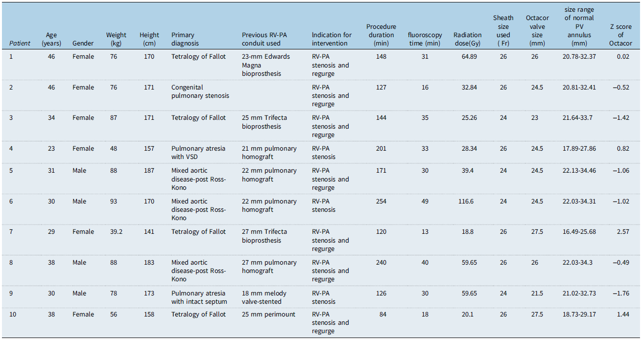
The demographic characteristics of the patients are presented in Table 1. Six patients were female and four were male. The mean age of the patients was 34.5 ± 7.4 years. The mean weight, height, and body surface area were 72.9 ± 18.6 kg, 168 ± 13.3 cm, and 1.82 ± 0.29 m2, respectively. The clinical characteristics of the patient cohort are summarised in Table 1. Primary diagnoses of the patients included tetralogy of Fallot in four patients and post-Ross-Kono procedure in three. Two patients had pulmonary atresia, and one had congenital pulmonary valve stenosis. With regard to the right ventricle to pulmonary artery conduit or pulmonary valve replacement used in the initial treatment of these patients, four received a pulmonary homograft, three had an implanted Trifecta bioprosthesis, one received the Edwards Magna bioprosthesis, one got the Melody valve TVP 18, and another had been treated with a Perimount valve. The median size of the original surgical conduits/pulmonary valve replacement was 24 mm Interquartile Range (IQR:21.75–25.5 mm). Patients presented with symptoms due to either isolated conduit stenosis (three patients) or a combination of stenosis and regurgitation (seven patients). Pre-stenting with a covered stent, CP stent (NuMed), prior to Myval™ Octacor implantation in four patients who had previous pulmonary homograft.
Demographics, clinical characteristics of the patients and procedural characteristics

BSA = body surface area; RV-PA = right ventricle to pulmonary artery; VSD = ventricular septal defect; Fr =French.
Procedural characteristics are summarised in Table 1. The median duration of the procedure was 146 minutes (IQR 124.5–210.75 min), with a median fluoroscopy time of 30.5 minutes (IQR 17.5–36.25 min). The median radiation dose was 36.12 Gy·cm2 (IQR 23.97–60.96 Gy·cm2). Four patients received a 24-French sheath, while six patients received a 26-French sheath. The median Z-score for the implanted Myval™ Octacor valves based on the body surface area of each patient was −0.50 (IQR −1.15 to + 0.97).
All patients underwent a baseline electrocardiogram. The median QRS duration was 135.5 milliseconds (ms) (IQR 115.7–171 ms).
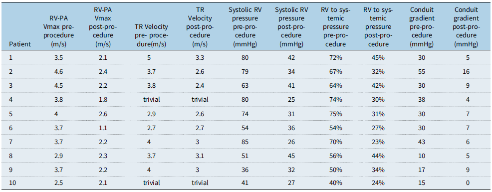
Table 2. summarises the echocardiographic measurements before and after the procedure. The median maximum flow velocity across the right ventricular outflow tract was 3.7 m/s (IQR 3.35–4.12 m/s) before the intervention and decreased to 2.2 m/s (IQR 2.0–2.32 m/s) following the intervention. Peak tricuspid regurgitation velocity was trivial and unquantifiable in two patients before the procedure; it remained unquantifiable after the procedure. The median tricuspid regurgitation velocity for the remaining eight patients decreased from 3.75 m/s (IQR 3.1–4.0 m/s) pre-intervention to 2.85 m/s (IQR 2.6–3.0 m/s) post-intervention. After valve implantation, all patients showed either trivial or no pulmonary regurgitation.
Echocardiographic findings and cardiac catheterisation haemodynamic data pre and post intervention

RV-PA = right ventricle to pulmonary artery; Vmax=maximum velocity; TR = tricuspid regurgitation; RV = right ventricle.
The haemodynamic data from cardiac catheterisation before and after valve implantation are displayed in Table 2. The median right ventricular systolic pressure decreased from 68.5 mmHg (IQR 48.5–80 mmHg) to 33 mmHg (IQR 26.75–41 mmHg) post-procedure.
Prior to valve implantation, six patients had right ventricular pressures greater than two-thirds of the systemic pressure, while four patients had pressures approximately half of the systemic pressure. These values are normalised by the end of the procedure. The procedure reduced the median right ventricular-to-systemic pressure ratio from 66% pre-procedure (IQR 53–72%) to 32% post-procedure (IQR 26–42.5%). The median peak instantaneous gradient across the right ventricular outflow tract or conduit decreased from 30 mmHg pre-procedure (IQR 16.5–39 mmHg) to 6.5 mmHg post-procedure (IQR 4.75–9 mmHg). There were no reported incidences of frame fracture, conduit rupture, device embolisation, coronary artery compression, or endocarditis in any of the cases. Significant symptomatic improvement was evident in all patients following the procedure.
Discussion
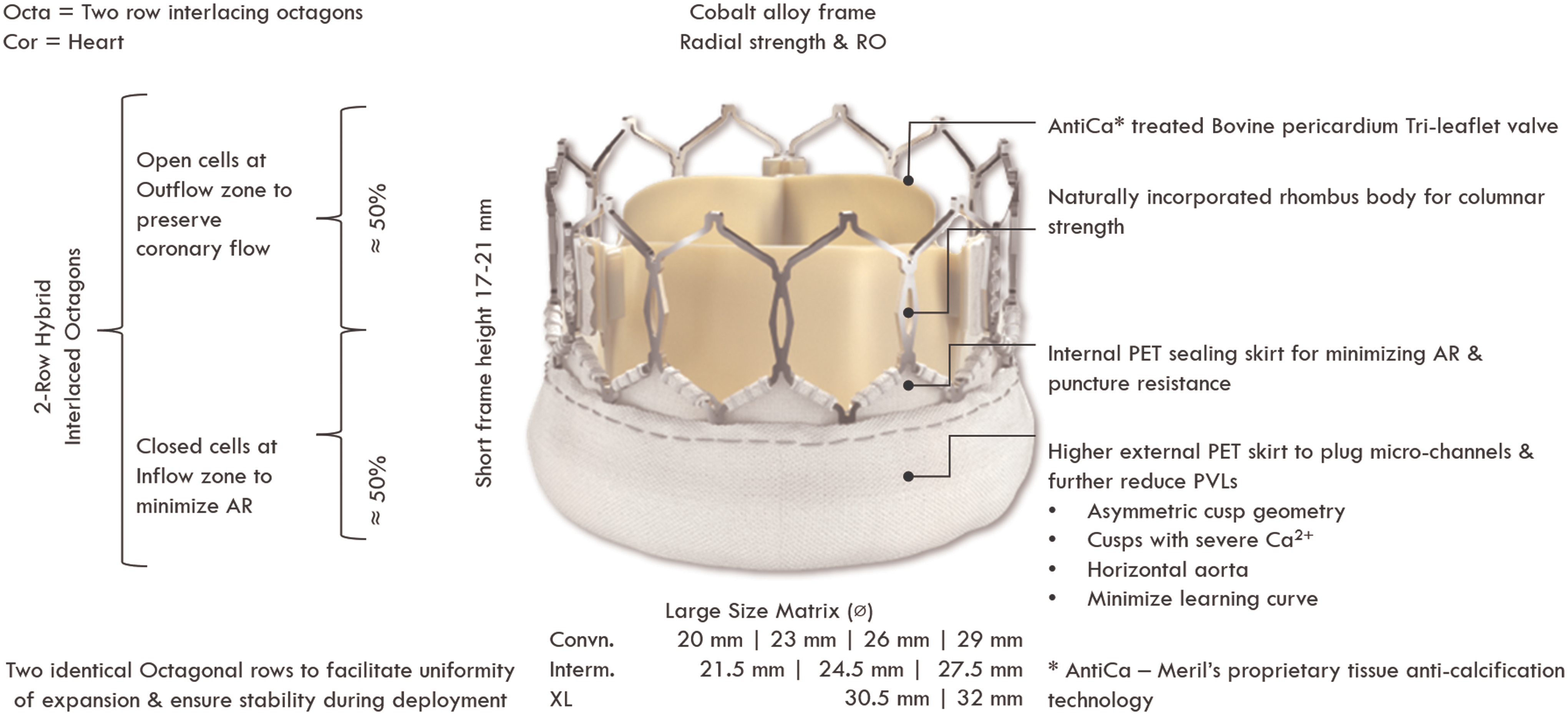
Off-label use of newer balloon-expandable valves has increasingly become common practice since the approval of the Melody valve and Edwards’ Sapien XT valve for use in the pulmonary position. Reference Driesen, Warmerdam and Sieswerda5 One of the new valves utilised in our patient cohort is the next-generation Myval™ Octacor valve. The Myval™ valve is manufactured from bovine pericardium, mounted on a nickel-cobalt alloy frame, and features a hybrid honeycomb structure with hexagonally arranged cells, as shown in Figure 2. Reference Al Nasef, Erdem and Aldudak10 The available sizes of the Myval™ valve range from 20 to 32 mm, in 1.5-mm increments. Reference Sivaprakasam, Reddy and Gunasekaran11 The Myval™ Octacor is an advanced version of the balloon-expandable Myval™ valve. The new design maintains a similar frame height to the previous Myval™ but incorporates only two layers of identical octagonal cells. This design minimises the extent of foreshortening during valve expansion and therefore provides sufficient deployment of the valve. The valve is also pre-mounted on its balloon delivery system, reducing the need for modifications during the procedure. Reference Montonati, Pellegrini and d’Atri8,Reference Jose, Mandalay and Cholenahally12 We routinely use the DrySeal sheath in all transcatheter pulmonary valve implantation cases as the standard of care in our centre. This provides protection to the tricuspid valve during the advancement of Myval™ and enables the precise positioning of the bioprosthetic valve in the right ventricular outflow tract.
Myval Octacor (Gen-2) transcatheter heart valve – design.

To the best of our knowledge, this is the first report on the use of the newer-generation Myval™ Octacor valve in the UK. Sivaprakasam et al. (2020) reported the first transcatheter pulmonary valve implantation using the Meril Myval™ valve in India. This procedure was performed on a symptomatic patient with severe dysfunction of the right ventricle-to-pulmonary artery conduit following the intracardiac repair of Tetralogy of Fallot. Reference Sivaprakasam, Arvind, Sridhar and Gunasekaran13 In 2021, Sivaprakasam et al. published their early multicentre experience involving seven patients with right ventricular outflow tract conduit stenosis, aged 17–60 years, who were treated with Myval™ valve implantation. All valves were implanted after pre-stenting of the outflow tract. During a median follow-up of 16 months, all patients were asymptomatic, with no significant valve-related adverse events. Only one patient had residual regurgitation. Reference Sivaprakasam, Reddy and Gunasekaran11 In our single-centre experience of a case series of 10 patients, we used the new generation Myval™ Octacor. Our patients were treated for either pulmonary stenosis or mixed-type pulmonary valve disease. The immediate and short-term results of the valve function were similar to those reported for the two approved valves for the pulmonary position, the Melody and the Edwards Sapien valves, respectively. Reference Nordmeyer, Ewert and Gewillig14,Reference Hascoët, Bentham and Giugno15 In our case series, only four patients underwent pre-stenting. They all had a pulmonary homograft in the pulmonary position, and stenting successfully alleviated the severe narrowing, creating a landing zone for the valve.
Al Nasef et al. reported the results of their multicentre experience of 53 patients suffering from dysfunctional pulmonary valves treated with the MyVal transcatheter heart valve implantation in the pulmonary position from Arabian Gulf countries, Turkey, and Spain. The resultant median peak instantaneous gradient across the right ventricular outflow tract was reduced from 23.5 mmHg pre-procedure to 10 mmHg post-procedure. MyVal transcatheter pulmonary valve implantation, therefore, became an option that showed procedural success with acceptable mid-term outcomes. Reference Al Nasef, Erdem and Aldudak10 In our study, there was a reduction in the median peak instantaneous gradient from 30 mmHg to 6.5 mmHg, which was similar to the previous report. In the report of Odemis and Yenidogan in 2022, the use of the Myval™ valve for treating severe pulmonary regurgitation in nine patients aged 8 to 34 years with native right ventricular outflow tract and conduit dysfunction was documented. It showed that the Myval™ valve was feasible and effective for pulmonary valve implantation. There was no pulmonary regurgitation observed post-procedure on echocardiography and angiography. They also reported normalisation of the right ventricle pressure after the implantation. Reference Odemis and Yenidogan16 In contrast, we did not implant any valves in isolated pulmonary regurgitation disease of the conduit. Our results were similar, as there was no pulmonary regurgitation and the right ventricular pressures normalised after the procedure in all patients. The immediate and short-term outcomes in our cohort showed no stent fracture, conduit rupture, coronary compression, or infective endocarditis.
This report does not undertake a direct comparison between the MyVal Octacor valve with the currently approved balloon-expandable valves—namely, the Melody and Sapien S3. Notably, it is known that Melody valve has limited size range up to a nominal diameter of 22 mm and off-label overexpansion for larger outflow tract up to 24 mm. Reference Shahanavaz, Qureshi and Levi17 Additionally, it has been associated with higher rates of infective endocarditis and stent fracture, as documented in various studies. Reference Malekzadeh-Milani, Ladouceur and Patel18,Reference Van Dijck, Budts and Cools19,Reference Cheatham, Hellenbrand and Zahn20,Reference Nordmeyer, Khambadkone and Coats21 In our case series, only one patient underwent a placement of a 21.5 mm valve, while eight patients received a valve between 24.5 and 27.5 mm, and one had a 23 mm valve. Thus, the majority of our cohort required valve sizes beyond the upper functional range of the Melody valve.
The Edwards Sapien S3 valve has a diameter range of 20, 23, 26, and 29 mm. Nominally intermediate sizes of the valves, such as the ones that our patients received, are typically accomplished in an off-label over-expansion of the nominal size of the valve. This off-label technique makes the valve function unpredictable, particularly with regard to leaflet coaptation and anchoring. Reference Patel, Levi and Cheatham22,Reference Shivaraju, Kodali and Thilo23,Reference Sellers, Sathananthan and Bouchareb24 Given this, we felt that precision would minimise malfunctioning of valve leaflets and reduce the risk of paravalvular leak.
The Myval™ Octacor valve offers a broader range of sizes, including intermediate diameters, allowing flexibility in valve selection without the need for off-label balloon modifications. Although the largest valve used in our series was 27.5 mm, the availability of larger sizes supported more accurate sizing for patients with dilated right ventricular outflow tracts. All valves were deployed using the manufacturer’s recommended balloon volumes, which helped ensure reliable leaflet function while reducing procedural variability and the risk for pulmonary regurgitation. These advantages are consistent with the findings of Houeijeh et al., who reported successful use of a 32 mm Octacor valve in a complex right ventricular outflow tract. Reference Houeijeh, Sudre, Juthier and Godart25
A comparative analysis between the Myval™ Octacor valve and other balloon-expandable valves may still be premature, primarily due to the limited number of off-label implantations using the Myval™ Octacor in comparison to the extensive clinical experience accrued over the years with the already approved valves.
Limitations of the study
1. Small Sample Size: The study involves only ten patients, which limits generalisability and the statistical power. A larger cohort is needed to evaluate the outcomes of the Myval™ Octacor valve more effectively.
2. Retrospective Design: This is a single-centre retrospective study. A multicentre prospective design could potentially yield stronger evidence regarding the safety and efficacy of the valve.
3. Lack of Long-term Follow-up: The study is based on short-term outcomes. Without long-term follow-up, the durability and long-term success of the valve remain questionable, which is critical for validating the long-term safety and performance of this device.
4. Lack of comparison with existing valves: The primary focus of the study is on the early outcomes and feasibility of the Myval™ Octacor valve, which makes any comparative analysis impractical. A head-to-head comparison with other balloon-expandable valves was beyond the scope of our current study. Further research—including comparative multicentre studies—is needed to better delineate the advantages and limitations of Myval™ Octacor relative to existing options.
Conclusion
This is the first report of the use of the Myval™ Octacor valve in the pulmonary position in the UK. All valve implantations were successful and safe based on our results. The short-term outcomes also demonstrated good valve function without significant stenosis or regurgitation. However, the identified limitations highlight the need for continued research, including larger, multicentre studies with extended follow-up to validate the findings and prove the efficiency of the valve function and freedom from re-interventions.
Acknowledgements
None
Financial support
This research received no specific grant from any funding agency, commercial, or not-for-profit sectors.
Competing interests
None
Ethical standards
The authors assert that all procedures contributing to this work comply with the ethical standards of the relevant national guidelines on human experimentation and with the Helsinki Declaration of 1975, as revised in 2008, and has been approved by the institutional committees. University Hospitals of Leicester (UHL) audit registration.