Introduction
Patients with major depressive disorder have been found to have structural changes in the prefrontal cortex, the subgenual medial orbital frontal cortex, the cingulate cortex, the amygdala, the hippocampus, the thalamus and the striatum (e.g. Drevets et al. Reference Drevets, Price, Simpson, Todd, Reich, Vannier and Raichle1997; Sheline, Reference Sheline2000; Davidson et al. Reference Davidson, Pizzagalli, Nitschke and Putnam2002b; Caetano et al. Reference Caetano, Kaur, Brambilla, Nicoletti, Hatch, Sassi, Mallinger, Keshavan, Kupfer, Frank and Soares2006). A model for depression has been proposed that involves frontal-limbic dysregulation among a dorsal-cortical compartment, a ventral-limbic compartment and the rostral cingulate (Mayberg, Reference Mayberg, Stuss and Knight2002; Tekin & Cummings, Reference Tekin and Cummings2002). The significant association between the frontal and subcortical structures with depression was further supported by the finding that an increased risk of depression was associated with lesions in the basal ganglia or in the left frontal region after a stroke (Morris et al. Reference Morris, Robinson, de Carvalho, Albert, Wells, Samuels, Eden-Fetzer and Price1996a, b; Lauterbach et al. Reference Lauterbach, Jackson, Price, Wilson, Kirsh and Dever1997; Carson et al. Reference Carson, MacHale, Kathryn, Lawrie, Dennis, House and Sharpe2000).
Depression has been found to be associated with selective attention biases towards negatively valenced, particularly sad, stimuli (Eizenman et al. Reference Eizenman, Yu, Grupp, Eizenman, Ellenbogen, Gemar and Levitan2003; Gotlib et al. Reference Gotlib, Krasnoperova, Yue and Joormann2004; Rinck & Becker, Reference Rinck and Becker2005). People with major depressive disorder seemed to be weak in inhibiting or disengaging attention from negative stimuli, for example on affective shifting tasks (Murphy et al. Reference Murphy, Sahakian, Rubinsztein, Michael, Rogers, Robbins and Paykel1999) and affective negative-priming tasks (Joormann, Reference Joormann2004; Gotlib et al. Reference Gotlib, Yue and Joormann2005; Goeleven et al. Reference Goeleven, De Raedt, Baert and Koster2006). On the contrary, attention to subsequent negative stimuli seemed to be facilitated by the preceding negative information, for example in solving anagrams of sad words (Rinck & Becker, Reference Rinck and Becker2005) and in the speed and rate of endorsement of subsequent negative-trait adjectives (Power et al. Reference Power, Cameron and Dalgleish1996). Consistent with the existing literature on the attention biases of depressed people, we also observed that their attention to negative stimuli, relative to their healthy counterparts, was more enhanced by preceding negative stimuli; and both depressed and control groups showed a similar extent of difficulty in inhibiting attention from previously ignored negative stimuli (Leung et al. Reference Leung, Lee, Yip, Li and Wong2007).
Studies have been conducted to identify the neural correlates of specific symptoms or the cognitive performance of depressed patients (e.g. Dolan et al. Reference Dolan, Bench, Brown, Scott and Frackowiak1994; Liotti & Mayberg, Reference Liotti and Mayberg2001; Goethals et al. Reference Goethals, Audenaert, Jacobs, Van de Wiele, Ham, Pyck, Vandierendonck, Van Heeringen and Dierckx2005). Some studies have focused on the attention biases towards negative information. For example, preferential increases in neural responses to sad but not happy facial expressions were noted in the left putamen, the left parahippocampal gyrus or the amygdala, and the right fusiform gyrus of depressed patients (Surguladze et al. Reference Surguladze, Brammer, Keedwell, Giampietro, Young, Travis, Williams and Phillips2005). However, depressed patients with left frontal hypo-activation were not found to show an attention bias towards valenced stimuli on an emotional Stroop task (Gotlib et al. Reference Gotlib, Ranganath and Rosenfeld1998). Despite these studies, there has been no investigation into the structural changes in depressed patients that are associated with the selective attention biases towards negative stimuli.
In the current study, voxel-based morphometry (VBM; Ashburner & Friston, Reference Ashburner and Friston2000) was used to investigate the regional gray-matter abnormality of depressed patients and to examine its relationship with attention facilitation and attention inhibition by negative information. Positive-priming (PP) and negative-priming (NP) tasks adopted from Tipper's priming paradigm (Reference Tipper1985) were used, which requires selective attention to one stimulus while simultaneously ignoring another stimulus. PP refers to the attention facilitation of the subsequent stimuli, particularly reflected by the reaction time (RT), once the same stimulus has been deliberately attended to on a preceding prime trial (Schacter & Buckner, Reference Schacter and Buckner1998). NP refers to the slowing down in RT to a stimulus that is the same as a distracting stimulus ignored on an immediately preceding trial (Tipper, Reference Tipper1985; Milliken et al. Reference Milliken, Joordens, Merikle and Seiffert1998), and is thought to reflect the inhibition of attention to the ignored distracting stimulus (Tipper, Reference Tipper2001). We also compared the priming performance and the structural correlates of the depressed participants with those of a matched group of healthy participants on a task that involved neutral stimuli and on one that involved negative stimuli. Thus, investigating the structural correlates of the priming effects that involve neutral and negative stimuli might help to improve our understanding of selective attention biases towards negative information in individuals with depression.
Method
Participants
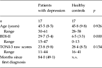
Seventeen female patients with major depressive disorder were recruited from an out-patient psychiatric clinic in Hong Kong when they visited the clinic for follow-up psychiatric consultation. Another 17 healthy female participants were recruited from the community through the personal network of the authors. All participants were right-handed. The depressed and healthy participants did not differ statistically in age or general intelligence as estimated using the abbreviated version of the Test of Nonverbal Intelligence Third Edition (TONI-3; Brown et al. Reference Brown, Sherbenou and Johnsen1997) (Table 1). The clinical participants were diagnosed by psychiatrists according to the ICD-10 criteria as suffering from major depressive disorder. They had been suffering from the illness for an average of 84·0 months (s.d.=49·1, maximum=217·1, minimum=20·7). Only patients who had received single depressive diagnostic labels and who had currently obtained a score on the Chinese version of the Beck Depression Inventory-II (BDI-II; Chinese Behavioral Sciences Society, 2000) above the recommended clinical cut-off score (Beck et al. Reference Beck, Steer and Brown1996) were included. All patients took medications that included selective serotonin reuptake inhibitors (SSRIs), tricyclic antidepressants, antipsychotics, benzodiazepine or hypnotics, or a combination of these. Both the patients and the healthy controls were interviewed using a checklist to exclude the possibility of another existing major psychiatric disease or a history of one, or the possibility of pre-existing neurological disease, mental retardation, or alcoholism. The healthy controls also completed the BDI-II and only those whose scores were below the clinical cut-off were included (Table 1).
Demographics of depressed and healthy participants

BDI-II, Beck Depression Inventory – II; TONI-3, Test of Nonverbal Intelligence Third Edition; n.a., not applicable.
Standard deviations are shown in parentheses.
The priming tasks
A typical trial of the priming task involved two consecutive displays: the first one was the prime and the subsequent one was the probe (Tipper, Reference Tipper1985). Each display consisted of two stimuli superimposed on each other, one of which was printed red and the other green. Throughout the experiment, the stimuli in red were the targets for the participants to identify whereas the stimuli in green were the distractors to be ignored. The relationships between the prime stimuli and the probe targets defined the conditions of interest. In the control condition, both the prime target and the distractor were different from the probe target and distractor. In the ignored repetition condition, the prime distractor had the same identity as the probe target. In the attended repetition condition, the prime target had the same identity as the probe target. The NP and the PP refer to the RT to the probe target in the ignored repetition condition and the attended repetition condition respectively relative to that in the control condition.
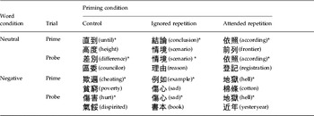
One hundred and forty neutral two-character Chinese compound words and 60 negative two-character Chinese compound words were used as stimuli in the priming task. The words were selected according to their low ratings on the pleasantness and arousal dimensions of emotion (Clark & Watson, Reference Clark and Watson1991; Clark & Steer, Reference Clark, Steer and Salkovskis1996) made by 36 Cantonese-speaking psychology undergraduates of The University of Hong Kong. The words were chosen to form one trial by matching their word frequency according to the Modern Chinese Word-Frequency Dictionary (Beijing yu yan xue yuan, Reference Beijing, yan and yu yan jiao1986). To test the valence effect, the negative words replaced the neutral words for the respective conditions of interest (Table 2). There were a total of 10 trials in each condition. The word pairs in the negative control condition were used as filler trials and were not included in the analysis. All 60 trials were randomly divided into six blocks among the participants.
Examples of the words used in the neutral-word and negative-word conditions in the priming task

* Words with asterisks were the targets to which the participants were asked to attend.
The priming task was programmed using e-prime (Schneider et al. Reference Schneider, Eschman and Zuccolotto2002), and was run on a notebook computer with a 60-Hz, 15-inch color screen. A typical trial started with a fixation cross being shown at the center of the computer screen for 500 ms, followed by a prime display, a fixation cross for 500 ms, a probe display, and then finally a blank screen for 500 ms. The participant was asked to press a key immediately after he/she had identified the red words in each display. A yellow mask appeared immediately on the screen to replace the word displayed. The participant was then asked to name the words so that the researcher could key in on a separate keypad the accuracy of the participant's response, and the next trial followed immediately. At the end of each block, the participant was reminded that he/she had to identify the words before pressing the key in order to minimize the possibility of an automatic response set that might have developed through the course of the task. Each participant finished six blocks before taking a short break. The same six blocks were then repeated with all the word trials being presented again randomly. After completing the priming tasks, the participants were given the BDI-II to check their level of depression and then proceeded to the scanning.
Magentic resonance imaging (MRI) acquisition and pre-processing
MRI was performed on all participants using a Signa 1.5 T imager (General Electric Medical Systems, Milwaukee, WI, USA) with a standard head coil. The following sequences were performed: axial spin–echo T1-weighted, fast spin–echo proton density and T2-weighted images, coronal fluid-attenuation inversion recovery sequences, and three-dimensional spoiled gradient-recalled (3DSPGR) images (slice thickness=3 mm with no gap for the whole brain, repetition time/echo time/inversion time=11·3/4·2/600 ms, acquisition matrix=256×256, flip angle=15°, field of view=23 cm).
All images were pre-processed and analyzed using the Statistical Parametric Mapping software (SPM2; Wellcome Department of Cognitive Neurology, University College London, UK) running on Matlab version 7.0.1.24704 (R14) Service Pack 1 (The MathWorks, Inc., Natick, MA, USA) and the extension VBM2 toolbox written by Christian Gaser from the University of Jena, Germany (http://dbm.neuro.uni-jena.de/vbm). Prior probability maps according to the Montreal Neurological Institute (MNI) template provided in SPM2 were used to segment and normalize all study images into gray matter, white matter, and cerebrospinal fluid. The resulting normalization parameters were then applied to the original whole-brain structural images. These images were segmented and spatially normalized again with the newly extracted individual brain mask to remove non-brain tissue. Spatial normalization of a 25-mm cut-off, medium regularization and 16 non-linear iterations were used. These segments were then modulated to correct for volume changes occurring during the spatial normalization, so that the resulting gray-matter values were referred to as gray-matter volume (GMV) whereas the unmodulated values were referred to as gray-matter concentration (GMC) (Mechelli et al. Reference Mechelli, Price, Friston and Ashburner2005; Eckert et al. Reference Eckert, Tenforde, Galaburda, Bellugi, Korenberg, Mills and Reiss2006). The images were smoothed using an isotropic Gaussian kernel of 12-mm full-width at half-maximum (FWHM) for unmodulated images and 8-mm FWHM for modulated images. A lower FWHM was applied to modulated images to obtain approximately the same resulting smoothness as the unmodulated images because modulation had additionally smoothed the data (Gaser, http://dbm.neuro.uni-jena.de/vbm/vbm2-for-spm2/statistical-analysis/). All images were resliced with 1×1×1 mm3 voxels.
Statistical analyses
RT on the probe trials of the priming tasks was used in the analyses, under the condition that the responses to both the prime and probe trials were correct. Extreme RT scores (<300 ms and >2000 ms) were considered outliers and were eliminated from the analyses (e.g. Joorman, Reference Joormann2004; Goeleven et al. Reference Goeleven, De Raedt, Baert and Koster2006). PP scores for the neutral words and negative words for each participant were computed separately by subtracting the RT in the control condition of the neutral words from the RT in the attended repetition condition of the respective word conditions. The lower the PP score was, the larger was the PP effect. Similarly, NP scores for neutral words and negative words were computed separately by subtracting the RT in the control condition of neutral words from the RT in the ignored repetition condition of the respective word conditions. The higher the NP score was, the larger was the NP effect. A 2×2 repeated-measures ANOVA was performed separately for the NP and PP scores, with the valence of words (neutral versus negative) being a within-subject variable, and the group (depressed versus control) being a between-subjects variable.
The processed images were analyzed with SPM2, using the framework of the general linear model (Friston et al. Reference Friston, Holmes, Worsley, Poline, Frith and Frackowiak1995). The primary analysis involved the identification of the GMC and GMV differences between the depressed patients and the healthy controls by applying one-tailed difference contrasts on the unmodulated and modulated gray-matter (GM) images respectively. Statistical tests on the modulated GM were performed with the global GMV as a covariate of no interest, so that disproportionate regional volume changes between the two groups relative to the overall GM size could be identified. We also performed ‘simple regression’ (correlation) analysis through the GMC and GMV maps and the BDI-II scores. In the secondary analysis, the gray-matter changes that were associated with the differences in priming effects of negative words from neutral words were examined. Differential PP and NP (DPP/DNP), denoting the differences between the negative words and the neutral words in PP and NP respectively, were calculated for each participant, by subtracting the priming scores of neutral words from the priming scores of negative words. The lower the DPP score, the larger was the enhancing effect of the negative words on the PP effect; and the lower the DNP score, the larger was the attenuating effect of the negative words on the NP. The voxel-based statistical tests used a covariate-only model that incorporated regressors including DPP and DNP, with the areas of GM changes between the depressed group and the control group that were identified in the primary analyses as a ‘mask’ to limit the regions of interest. The voxel-level statistical threshold was set at p<0·001, uncorrected, with an additional cluster extent threshold of eight voxels in the primary analyses. In the secondary analysis with the behavioral data of expected small effect size, this criterion was relaxed to an uncorrected p<0·005, with a cluster extent threshold of eight voxels.
Results
Behavioral measures
PP effects
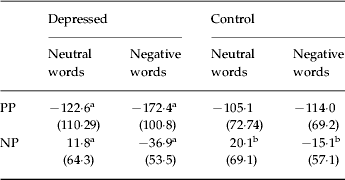
PP was greater for negative than for neutral words on all participants as a whole [F(1, 32)=8·50, p=0·006]. This effect was qualified by a trend towards significance on valence×group interaction [F(1, 32)=4·11, p=0·051]. Post-hoc paired-sample t tests showed that, in the depressed group, the PP effect for negative words was stronger than that for neutral words [t(16)=2·9, p=0·01], but in the control group, the difference between PP for neutral and negative words was not significant [t(16)=0·83, p=0·42]. To summarize, an enhancement of the PP effect by the negative words was found only in the depressed group (Table 3).
The mean positive priming (PP) and negative priming (NP) scores (in ms) and valence of words used as stimuli as a function of diagnostic group

Standard deviations are shown in parentheses.
a Significant difference between neutral words and negative words in the depressed group.
b Significant difference between neutral words and negative words in the healthy control group.
NP effects
NP was greater for neutral than for negative words [F(1, 32)=15·45, p<0·001] on all participants as a whole. Post-hoc paired-sample t tests showed that the NP effect was stronger for neutral words than for negative words in both the depressed group [t(16)=3·45, p=0·00] and the control group [t(16)=2·33, p=0·03]. However, the valence×group interaction was not statistically significant [F(1, 32)=0·04, p=0·85]. To summarize, attenuation of the NP effect by the negative words was found in both the clinical and control groups to a similar extent (Table 3).
VBM findings
Gray-matter differences between depressed and control groups
GMC
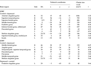
The depressed group showed reduced GMC in the right superior frontal gyrus [Brodmann area (BA) 8/9], the left middle frontal gyrus (BA 9), the right medial superior frontal gyrus (BA 32), the right orbital inferior frontal gyrus (BA 47), the right anterior cingulate gyrus (BA 32), the left median cingulate gyrus (BA 31), and the left insula (BA 13). Reduced GMC was also observed in the bilateral precentral gyrus (BA 6) and the right fusiform gyrus (BA 37; Table 4). There was a significant negative correlation between the BDI-II scores and the GMC at several of these regions, with the strongest correlation at the right anterior cingulate gyrus (BA 32), voxel local maxima r=−0·75.
Areas of differential gray-matter concentration (GMC) and gray-matter volume (GMV)

L, Left; R, right; BA, Brodmann area.
The threshold for between-group comparison (depressed group versus healthy control group) was set to p<0·001 uncorrected, the cluster extent threshold of eight voxels for all contrasts.
GMV
Decreased GMV was observed in depressed patients in the left median cingulate gyrus (BA 31/5), the left precuneus (BA 7), the left angular gyrus (BA 39), the left middle temporal gyrus (BA 21), the right temporal pole (BA 38), and the right precentral gyrus. However, depressed patients also showed increased GMV in the right middle frontal gyrus (BA 46) and the left posterior cingulate gyrus (BA 29; Table 4). There were both positive and negative correlations significant between BDI-II scores and GMV, with the strongest negative correlation at the right precuneus (BA 5), voxel local maxima r=−0·57, and the strongest positive correlation at the right middle frontal gyrus (BA 46), voxel local maxima r=0·58.
Structural correlates of DPP scores
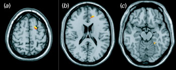
We performed the secondary analysis on DPP because only the depressed patients showed DPP, but not DNP, from that of the healthy controls. Among the GMC differences between the two groups, DPP was positively correlated with GMC in the right superior frontal gyrus (BA 8/9), the right anterior cingulate gyrus (BA 32), and the right fusiform gyrus (BA 37; Fig. 1). In other words, the more reduction in the concentration of the gray matter in these two regions relative to the whole brain, the stronger was the enhancing effect of the negative words on the PP effect. No statistically significant relationship was found between DPP and GMV.
Parametric t map showing regional gray-matter concentration (GMC) having a positive correlation with the differential positive priming (DPP) score for all participants. The analysis was performed with a ‘mask’ limiting the regions of interest derived from the structural differences between the depressed group and the healthy control group in the primary analysis. (a) Right superior frontal gyrus. (b) Right anterior cingulate. (c) Right fusiform gyrus.

Discussion
The findings show that specific structural changes in depression are associated with an attention bias towards negative information. We found that the facilitation of attention to repeated stimuli in PP was enhanced by the negative valence of the stimuli only in the depressed patients but not in the healthy controls. We further demonstrated that, among the structural differences identified between the depressed patients and the healthy controls in this study, such attention biases were associated with reduced GMC in the right superior frontal gyrus, the right subgenual anterior cingulate gyrus, and the right fusiform gyrus.
Our results concerning the right superior frontal gyrus and the right subgenual anterior cingulate gyrus are consistent with the role of the frontal and cingulate areas in attention regulation of emotion in humans. The right superior frontal gyrus has been considered part of the dorsal system of attention orienting (Posner & Rothbart, Reference Posner and Rothbart2007). It is involved in the goal-directed selection of visual stimuli and responses, and its activity is modulated by the detection of the stimuli (Corbetta & Shulman, Reference Corbetta and Shulman2002). Constituting the dorsolateral frontal area, the right superior frontal gyrus may also be involved in emotion regulation processes, particularly in the suppression of sadness (Lévesque et al. Reference Lévesque, Eugène, Joanette, Paquette, Mensour, Beaudoin, Leroux, Bourgouin and Beauregard2003). In fact, functional abnormalities in the right dorsolateral prefrontal cortex have been found in depressed patients when they ignored fearful stimuli while making a perceptual judgment task (Fales et al. Reference Fales, Barch, Rundle, Mintun, Snyder, Cohen, Mathews and Sheline2008). The role of the right dorsolateral prefrontal cortex in attentional modulation to emotional stimuli was corroborated by the finding that this region was hyperactive in depressed patients when they attended to emotional stimuli to make emotional judgment of the stimuli (Grimm et al. Reference Grimm, Beck, Schuepbach, Hell, Boesiger, Bermpohl, Niehaus, Boeker and Northoff2008). However, the subgenual anterior cingulate gyrus identified in this study corresponds to the ventral area (Bush et al. Reference Bush, Luu and Posner2000) or affective division (Pizzagalli et al. Reference Pizzagalli, Peccoralo, Davidson and Cohen2006) of the anterior cingulate cortex, which has extensive connections with the limbic and paralimbic regions. It is assumed to integrate salient affective and cognitive information, and subsequently to modulate the attention processes within the dorsal anterior cingulate and prefrontal areas (Mayberg et al. Reference Mayberg, Liotti, Brannan, McGinnis, Mahurin, Jerabek, Silva, Tekell, Martin, Lancaster and Fox1999). The role of the anterior cingulate in emotion regulation was further supported in a study that showed greater activation in this region associated with the difficulty in regulating sad emotion in patients with major depressive disorder (Beauregard et al. Reference Beauregard, Paquette and Lévesque2006). Given the top-down control exercised by the frontal region over the input from the ventral and subcortical regions during the process of selective attention towards valenced stimuli (Compton, Reference Compton2003), and the bottom-up contribution of the ventral anterior cingulate to frontal functions (Davidson et al. Reference Davidson, Lewis, Alloy, Amaral, Bush, Cohen, Drevets, Farah, Kagan, McClelland, Nolen-Hoekesema and Peterson2002a; Fales et al. Reference Fales, Barch, Rundle, Mintun, Snyder, Cohen, Mathews and Sheline2008), our speculation is that decreased GMC in the dorsal frontal and subgenual anterior cingulate regions is associated with the facilitation of the selective attention towards negative information. Although these two regions were found to be hyperactive during attention regulation of emotion in previous studies (Beauregard et al. Reference Beauregard, Paquette and Lévesque2006; Grimm et al. Reference Grimm, Beck, Schuepbach, Hell, Boesiger, Bermpohl, Niehaus, Boeker and Northoff2008), the structural change in the subgenual anterior cingulate and superior frontal gyrus of the depressed patients might be associated with a weakened ability to regulate or suppress the signals of the mood-congruent stimuli in our priming task. When a negative word was first detected, the valence of the word seemed to become the priority of the attentional goal registered in the dorsal frontal gyrus, so that attention to subsequent negative words was facilitated.
Only a few reported data support our findings that the right fusiform gyrus is involved in the attention bias towards negative information. Some of these data were contained in a functional MRI study showing increased activity in the right fusiform gyrus correlating with expressions of increasing sadness in the faces of people with depression (Surguladze et al. Reference Surguladze, Brammer, Keedwell, Giampietro, Young, Travis, Williams and Phillips2005). In the present study, we have shown that the reduced GMC in the right fusiform gyrus of the depressed participants, relative to the healthy controls, is associated with selective attention bias to negative words. In fact, some studies have also found that depressed patients showed deficits in facial emotion judgment (e.g. Walker et al. Reference Walker, McGuire and Bettes1984; Feinberg et al. Reference Feinberg, Rifkin, Schaffer and Walker1986), although others did not (e.g. Gaebel & Wölwer, Reference Gaebel and Wölwer1992). The role of fusiform gyrus in attention and evaluation of emotion might be one of the directions of future studies.
Regarding the overall structural differences between the depressed group and the healthy controls, our findings that depressed patients had lower GMC in their dorsolateral, medial and orbital frontal regions, and in the ventral anterior cingulate region, are consistent with previous neuroimaging and post-mortem studies showing that depressed people have structural or activity changes in these areas (Drevets et al. Reference Drevets, Price, Simpson, Todd, Reich, Vannier and Raichle1997; Marshall & Fox, Reference Marshall, Fox, Johnson, Hayes, Field, Schneiderman and McCabe2000; Bremner et al. Reference Bremner, Vythilingam, Vermetten, Nazeer, Adil, Khan, Staib and Charney2002; Davidson et al. Reference Davidson, Lewis, Alloy, Amaral, Bush, Cohen, Drevets, Farah, Kagan, McClelland, Nolen-Hoekesema and Peterson2002a, b; Harrison, Reference Harrison2002; Caetano et al. Reference Caetano, Kaur, Brambilla, Nicoletti, Hatch, Sassi, Mallinger, Keshavan, Kupfer, Frank and Soares2006). Moreover, the severity of depression measured by the BDI-II score was also found to be correlated with the reduction of the GMC in these regions. Contrary to our a priori prediction, we did not observe any structural differences between the depressed group and the healthy controls in the subcortical structures including the hippocampus and the amygdala. This might be related to the low power of the analysis, given the small sample size of this study. Nevertheless, it is also important to note that morphological changes are not required before significant cognitive abnormalities or improvements are detectable (MacQueen et al. Reference MacQueen, Campbell, McEwen, Macdonald, Amano, Joffe, Nahmias and Young2003; Vythilingam et al. Reference Vythilingam, Vermetten, Anderson, Luckenbaugh, Anderson, Snow, Staib, Charney and Bremner2004).
This study is not free from methodological limitations. The sample size is small and hence undermines the power of the statistical tests to discover structural differences and their relationship with the differential priming scores between the two groups of participants. The results should also be interpreted with caution because increase volume observed in VBM studies may relate to conformational changes in gyral morphometry. The findings of this study should also be considered as exploratory, particularly in those regions with small cluster size. Moreover, the results of this study should only be generalized to the female population with depression. According to Smith et al. (Reference Smith, Kyle, Forty, Cooper, Walters, Russell, Caesar, Farmer, McGuffin, Jones, Jones and Craddock2008), the depressive symptom profile is different between men and women. It is also thought that there is a volumetric difference between depressed men and depressed women, particularly in the amygdala and the anterior cingulate region (Hastings et al. Reference Hastings, Parsey, Oquendo, Arango and Mann2004), the orbitofrontal cortex (Lavretsky et al. Reference Lavretsky, Kurbanya, Ballmaier, Mintz, Toga and Kumar2004) and the hippocampus (Steffens et al. Reference Steffens, Byrum, McQuoid, Greenberg, Payne, Blitchington, MacFall and Krishnan2000; Vakili et al. Reference Vakili, Pillay, Lafer, Fava, Renshaw, Bonello-Cintron and Yurgelun-Todd2000).
In addition, for ethical reasons, psychotropic medication was not withdrawn from the depressed patients in this study. Normalization of the frontal and limbic metabolism and blow flow after treatment with antidepressants has been demonstrated (Mayberg, Reference Mayberg2003). In addition to affecting the RTs to the priming task, it is possible that taking antidepressants and other psychotropic drugs might also result in structural changes in the brain (Miguel-Hidalgo & Rajkowska, Reference Miguel-Hidalgo and Rajkowska2002). Thus, studies that use a larger sample size might replicate and extend these findings by using more defined depressive categories in terms of the age of the patients, the severity of symptoms, the subtypes of major depressive disorder, whether the depressive episodes are single or recurrent, and whether or not the patients are taking antidepressants. Similarly, the design of this study leaves open the possible interpretation that the association between the structural changes in depressed patients and the attention facilitation by negative stimuli is mediated by other factors. The question might be resolved with future imaging studies using a longitudinal approach, or using functional neuroimaging studies. In addition, we found a significant association of the enhancement of the PP effect by negative stimuli with GMC but not with GMV. Although the analysis of GMC and that of GMV are considered to detect different aspects of GM abnormalities (Good et al. Reference Good, Johnsrude, Ashburner, Henson, Friston and Frackowiak2001; Mechelli et al. Reference Mechelli, Price, Friston and Ashburner2005; Eckert et al. Reference Eckert, Tenforde, Galaburda, Bellugi, Korenberg, Mills and Reiss2006), the reasons for the different results from these two analyses need further investigation. Furthermore, even though we found an interesting association of the frontal region and the fusiform gyrus with the enhancement of the PP effect with negative stimuli in the depressed group, we did not correct the analyses for multiple statistical comparisons. This finding should be taken as preliminary, and future studies should involve further verification. Finally, our tasks involve repeated words as the stimuli, but real-life situations usually involve dynamic and semantically related visual information. Further studies are warranted to confirm the observed effect by using other stimuli such as faces or scenes.
This study complements and extends previous neuroimaging studies of attention biases towards negative information in major depressive disorder. Our findings suggest that neurostructural abnormalities in frontal and fusiform gyri are associated with attention biases to negative stimuli of depressed people relative to healthy controls. The study also highlights the importance of future longitudinal and imaging studies to identify the brain activities subserving the cognitive biases associated with the course and development of depressive disorder. This will help us to better understand the nature of attention biases in major depressive disorder, and will further validate the neural basis of the cognitive-emotional dysfunction of depression.
Acknowledgments
This project was supported by the National Natural Science Foundation of China (no. 30828012) as well as the Research Output Prize and the CRCG (ref. 200507176026) of The University of Hong Kong.
Declaration of Interest
None.