Introduction
The solitary lateral cystic neck mass in adults remains a diagnostic challenge for head and neck surgeons. The majority of published studies report a benign congenital cyst as the most common diagnosis in the absence of any other signs or symptoms of head and neck cancer; however, the rates of malignancy vary widely (9–72 per cent), with a direct positive correlation with increasing age.Reference Flanagan, Roland and Jones1–Reference Stefanicka, Gnojcakova, Kurinec and Profant8
Diagnostic uncertainty creates significant anxiety for patients, and reliance on inaccurate investigations may result in delayed or inappropriate management for concealed malignant disease in, for example, the oropharynx or thyroid.
A number of series have been published in an attempt to quantify the diagnostic accuracy of investigations, although these often focus on a single diagnostic modality. Ultrasound can be used to examine the thickness of the cyst wall and the presence of solid elements to help guide fine needle aspiration cytology (FNAC) or core needle biopsy.Reference Baatenburg de Jong, Rongen, Verwoerd, van Overhagen, Laméris and Knegt9 Computed tomography (CT) and magnetic resonance imaging (MRI) are useful to examine potential primary tumour sites and assess the anatomy surrounding the cystic mass for surgical planning, although their ability to differentiate benign and malignant neck masses is often limited.Reference Goyal, Zacharia and Goldenberg10,Reference Mittal, Malik, Sureka and Thukral11 Fluorine-18 fluorodeoxyglucose positron emission tomography-CT (PET-CT) is gaining popularity in the workup of cystic neck nodes,Reference Abadi, Johansen, Godballe, Gerke, Høilund-Carlsen and Thomassen12 but limitations include incidental false-positive findings in other organ systems in up to 33 per cent of head and neck cancer patients and significant doses of ionising radiation; a cost-benefit advantage has not been proven.Reference Britt, Maas, Kennedy and Hartig13 Fine needle aspiration cytology is useful in the diagnosis of solid neck masses but is often limited in lesions extensively affected by cystic degeneration because of a paucity of cellular material to target with resultant poor diagnostic accuracy.Reference Layfield, Esebua and Schmidt14 For cystic neck masses, the sensitivity and specificity yields improve as more viable solid material is obtained (open biopsy is better than core needle biopsy which is better than FNAC). The sensitivity for FNAC, core needle biopsy and frozen section has been quoted as 59 per cent, 83 per cent and 93 per cent, respectively, and specificity has been reported as 83 per cent, 100 per cent and 92 per cent, respectively.Reference Tabet, Saydy, Letourneau-Guillon, Gologan, Bissada and Ayad15 Although open biopsy obtains the greatest diagnostic accuracy, there are historical concerns regarding neck contamination and potential spillage or dissemination of cancer cells.Reference Martin and Romieu16,Reference McGuirt and McCabe17 However, evidence suggests that tumour seeding from the head and neck is a very rare occurrence.Reference Shinohara, Yamamoto, Tanabe, Maetani and Kim18,Reference Shyamala, Girish and Murgod19
We present our 10-year experience of investigating the solitary lateral cystic neck mass in adults, representing the largest published series of this clinical presentation to date. These numbers allow both a comparative analysis of diagnostic accuracy for a wide range of investigative modalities and regression analysis of variables that may increase the risk of a subsequent malignant diagnosis. Finally, we suggest a management algorithm to help stratify investigations for patients with these lesions and minimise time to diagnosis.
Materials and methods
Case identification and review
All patients presenting with a solitary lateral cystic neck mass to our tertiary academic head and neck centre between 2009 and 2019 inclusive were identified. We retrospectively reviewed operating theatre records, electronic patient records, and the head and neck cancer registry to collect demographic data, reports or outcome of investigations, histopathology reports, and final diagnosis.
Exclusion criteria included: less than 18 years of age, midline lesions, primary skin lesions (basal cell carcinoma, sebaceous cyst), multiple synchronous pathological lymphadenopathy and an obvious primary tumour on oral inspection or flexible endoscopy. Cases were also excluded if there was an obvious primary thyroid lesion on ultrasound that was later confirmed by thyroid excision histology. Human papillomavirus (HPV) testing of tissue potentially from primary oropharyngeal malignancies became routine midway through the study period and included in-situ hybridisation and immunohistochemistry for p16 protein; both were required for viral confirmation.
Literature review
In order to identify comparable case series, we searched the Medline electronic database for English-language articles between January 1990 to July 2020 using the following search terms: lateral neck cyst and lateral cervical cyst. We identified additional references by screening bibliographies of identified series.
Data analysis
Likelihood ratios predicting the presence of malignancy based on a positive result in each investigative modality were calculated, given a positive test result (sensitivity divided by 1-specificity) and a negative test result (1-sensitivity divided by specificity). Further, the accuracy of each test (i.e. the overall probability that a patient is correctly classified as having benign or malignant disease) was calculated (sensitivity × prevalence + specificity × (1 − prevalence). All analysis was performed using Microsoft Excel® and SPSS® statistical software (version 22).
Overview
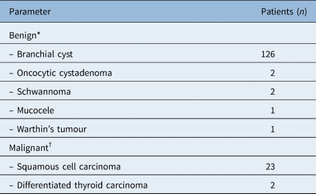
A total of 558 cases were identified from our initial case search. Following application of inclusion criteria and removal of duplicates, a total of 164 cases were identified with final histopathological diagnosis available for 157 patients (Table 1). This comprised 25 cases with a final diagnosis of malignancy and 132 benign cysts. The histopathological diagnosis was confirmed from either the primary tumour or cystic node. A total of 2 of 25 malignant cases had a diagnosis of differentiated thyroid carcinoma, both of which were diagnosed by cyst excision. The other 23 malignancies were squamous cell carcinoma (SCC). In these cases, a primary tumour was visualised on panendoscopy in nine cases. In the remaining 14 cases, clinicians performed tonsillectomies and guided biopsies of the high-risk sites (post-nasal space and tongue base) and identified a further 8 tumours. In the group with subsequently confirmed benign cysts (n = 132), rigid endoscopy was performed 24 times at the time of the initial excision, and biopsies of high-risk sites were performed in 16 cases including 5 tonsillectomies, all without yield of malignant tissue.
Final pathological diagnosis

*n = 132; †n = 25
For the SCC cases, the ipsilateral palatine tonsil was the most common site of primary tumour (13 of 23), followed by cancer of unknown primary disease (6 of 23). Base of tongue and hypopharynx were the site of primary disease in two cases each. No contralateral primary tumour to a cystic node was identified. Importantly, of 13 malignant cases tested for HPV, 11 were determined positive. The number of neck cysts presenting per year remained stable for the whole study period.
Age distribution
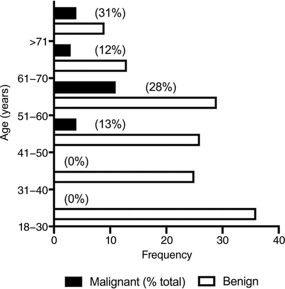
There were no cases of malignancy under the age of 40 years. The rate of malignancy increased with age, but did not rise above 31 per cent as a proportion of total cases per decade age group (Figure 1).
Squamous cell carcinoma and benign cases arranged by age group. The percentage of total malignant cases per age group are in brackets.

Risk factors
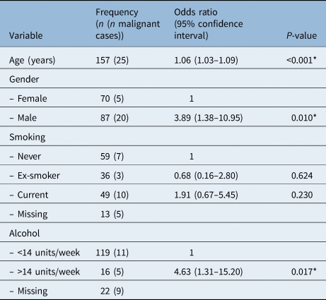
Univariate logistic regression was performed to identify variables associated with malignancy (Table 2). Three significant risk factors were identified: increasing age by year (odds ratio, 1.06, p ≤ 0.001), male sex (odds ratio, 3.89, p ≤ 0.010) and heavy alcohol intake (odds ratio, 4.63, p ≤ 0.017). Smoking was not an associated risk factor in this disease presentation as a whole; however, both patients discovered to have hypopharyngeal malignancy had heavy smoking and alcohol intake.
Model estimates for univariate logistic regression

*P < 0.05 indicates statistical significance
Investigations
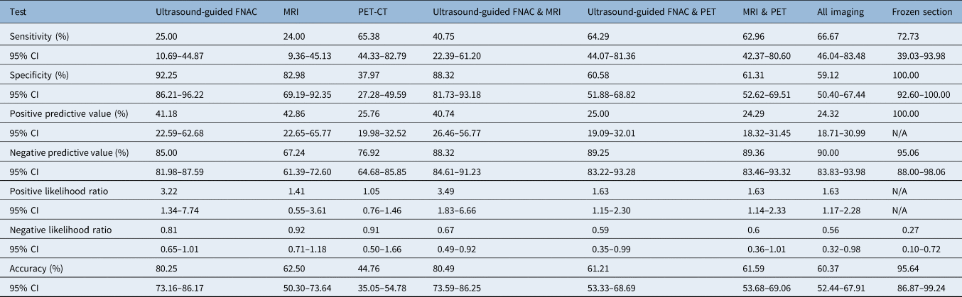
We performed over 740 investigations in our series, in addition to histopathology, although some investigations may have been performed prior to referral to our unit and were therefore not obtainable. Choice of investigation was dependent on primary clinician preference and departmental guidelines at the time of clinical review, although these were subject to numerous changes over the 10-year series. The diagnostic parameters of each investigation individually and in combination were analysed, and prior to open biopsy. As stand-alone investigations, ultrasound-guided FNAC and PET-CT demonstrated the highest specificity (92.25 per cent) and sensitivity (65.38 per cent), respectively (Table 3). At this initial stage in the management pathway, ultrasound-guided FNAC alone and the combination of ultrasound-guided FNAC with MRI provided the greatest accuracy (80.25 per cent and 80.49 per cent, respectively). However once proceeding to open biopsy, frozen section had a sensitivity and specificity of 72.73 per cent and 100 per cent, respectively, with an accuracy of 95.64 per cent.
Diagnostic test metrics

CT of the neck was only performed in two patients so was excluded from analysis. Positive predictive value, negative predictive value and accuracy are dependent on disease prevalence. FNAC = fine needle aspiration cytology; MRI = magnetic resonance imaging; PET = positron emission tomography; CT = computed tomography; CI = confidence interval; N/A = insufficient data to compute
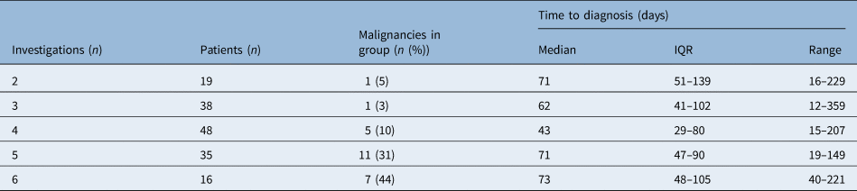
Further analysis on time to diagnosis was performed for all patients, grouping investigations by the number performed for each patient (Table 4). Interestingly, there was no significant correlation between number of investigations and time to diagnosis.
Summary table for time to diagnosis against number of investigations (excluding histopathology)

One patient's ‘time to diagnosis’ was classified as an outlier and removed from analysis. IQR = interquartile range
Outcome data
Four deaths were noted in the positive malignancy cases, with both hypopharyngeal SCC patients succumbing to disease within a year of diagnosis. Another patient died four years after treatment of metastatic recurrence, and the other patient died of breast cancer without receiving any treatment. The 5-year overall survival in the oropharyngeal primary and unknown primary group was 87.5 per cent (n = 21).
Published series
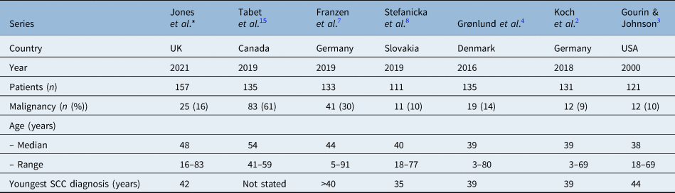
We have summarised important demographic data and comparisons between the present study and other large series in the literature (Table 5).
Comparison of published series on lateral cystic neck masses

*This article. SCC = squamous cell carcinoma
Discussion
In this series, we reported outcomes from a 10-year cohort of patients presenting to our tertiary head and neck centre with unilateral solitary cystic masses. The aim was to guide our stratification of investigations and provide tailored advice for our patients about their risk of malignancy. We failed to find any malignancies in patients under 40 years. Smoking was not a significant risk factor in this group, and the number of investigations performed did not appear to adversely lengthen the time to diagnosis. We also provide novel data for the accuracy of combinations of different investigations in solitary lateral cystic neck mass.
Despite routine p16 testing only being introduced midway through our series, 85 per cent of those tested were positive, demonstrating integrated HPV infection. This is likely to account for why the traditional risk factor of smoking was not of significance, and therefore we disagree with the approach of utilising this information in a risk-based approach to management.Reference Gillison, D'Souza, Westra, Sugar, Xiao and Begum20 Age over 40 years (odds ratio, 1.06 per year of age; p ≤ 0.001), male gender (odds ratio, 3.89; p ≤ 0.010) and alcohol intake (odds ratio, 4.63; p ≤ 0.0170) were significant variables on univariate analysis and should be considered when counselling patients, supporting several recent studies.Reference Franzen, Günzel, Buchali and Coordes7,Reference Stefanicka, Gnojcakova, Kurinec and Profant8,Reference Tabet, Saydy, Letourneau-Guillon, Gologan, Bissada and Ayad15 The youngest patient with malignancy in our cohort was 42 years.
The number of investigations performed did not appear to affect the timeframe of the patient pathway; in addition to resource considerations, this study provides important outcome data on individual and combined imaging modalities to help organise the most efficient and accurate test order. The highest overall accuracy (i.e. the weighted average of the sensitivity and specificity) is found with ultrasound-guided FNAC (80.25 per cent; 95 per cent confidence interval (CI): 73.16–86.17 per cent). Adding cross-sectional imaging in the form of an MRI to the ultrasound improves the sensitivity (from 25.00 to 40.75 per cent), increases the positive likelihood ratio (from 3.22 to 3.49), reduces the negative likelihood ratio (from 0.81 to 0.67) but does not significantly affect the accuracy (80.49 per cent; 95 per cent CI: 73.59–86.25 per cent). We therefore recommend an initial investigation order set of ultrasound-guided FNAC and MRI in these cases as a minimum prior to proceeding to an open biopsy. We can reassure patients that this combination of modalities gives the highest pre-biopsy accuracy, together with the highest chance of subsequently confirming disease in the event of a positive test.
FNAC sensitivity varies widely in the literature, and results obtained from cystic lesions demonstrate significantly lower accuracy. Tabet et al. performed a comprehensive cytopathological analysis of 135 cystic lesions, resulting in a positive predictive value of 92 per cent, and a sensitivity of 59 per cent,Reference Tabet, Saydy, Letourneau-Guillon, Gologan, Bissada and Ayad15 both significantly higher than the present series. However, with a malignancy rate in their studied cohort of 72 per cent, this represented a detection rate far in excess of other published series. Their reasoning that this may be because of the tertiary nature of their unit appears to be contradicted by our study but more likely represents the inclusion of partially necrotic lesions within their cohort. However, we believe inclusion of purely cystic lateral neck masses provides a more clinically relevant cohort to help guide management of this distinct clinic presentation group. Indeed, a previously published series of 2702 general head and neck aspirates from our unitReference Tandon, Shahab, Benton, Ghosh, Sheard and Jones21 reports sensitivity, specificity, positive predictive value, negative predictive value and accuracy rates of 89.5 per cent, 98.5 per cent, 97.3 per cent, 94.0 per cent and 95.1 per cent, respectively, demonstrating the difficulty of pre-biopsy tests when dealing with purely cystic lateral neck masses.
Adding PET-CT imaging to the ultrasound-guided FNAC and MRI combination significantly reduced the accuracy of the results in our series (60.74 per cent; 95 per cent CI: 52.80–68.29 per cent). However, PET-CT does have the highest sensitivity (70.83 per cent), albeit with the lowest specificity (37.97 per cent) and therefore a higher risk of false alarms and incidental findings as well as a high radiation dose (about 25 mSv compared with about 6 mSv for a CT of the thorax). These findings are consistent with other published series, the most comparable being the study by Sokoya et al.,Reference Sokoya, Chowdhury, Kadakia and Ducic22 who similarly published a 10-year retrospective series reporting a sensitivity and negative predictive value for detecting malignancy by PET-CT in unknown primary head and neck squamous cell carcinoma of 73.1 per cent and 68.9 per cent, respectively.
With its low specificity in this clinical scenario of solitary lateral cystic neck mass, we propose PET-CT should be reserved until after malignancy has been confirmed, optimising the use of this resource and minimising the risk of false alarms for patients who often then require further invasive investigations to rule out co-existing pathology (e.g. colonoscopy of the lower gastrointestinal tract). This timing will allow further imaging of the potential primary sites (if no evidence was found on primary examination) in keeping with international recommendations.Reference Civantos, Vermorken, Shah, Rinaldo, Suárez and Kowalski23
Should the above investigations fail to diagnose malignancy, open biopsy is the ‘gold standard’ for diagnosis. We emphasise that adding further pre-biopsy investigations to ultrasound-guided FNAC and MRI does not aid surgical planning and may actually reduce the accuracy of the diagnostic test battery. In the cases where frozen section was utilised, it was found to be a highly accurate adjunct during open biopsy. Sigel and Fisch eloquently demonstrated how operating on the neck can change the lymphatic drainage.Reference Sigel and Fisch24 This was important at a time when radical surgery was used as the sole treatment modality, which made frozen section especially important. Frozen section of a lymph node was popularised in the setting of cancer of unknown primary to confirm the diagnosis while the neck was open and to avoid delaying definitive surgery. In modern times, the use of frozen section in neck cysts remains controversial. In our series, the use of frozen section is insufficient to completely exclude malignancy (sensitivity 73 per cent), which led to false reassurance and delayed treatment in 3 of 11 subsequently confirmed cancer patients on paraffin histopathology, consistent with other published work.Reference Franzen, Günzel, Buchali and Coordes7
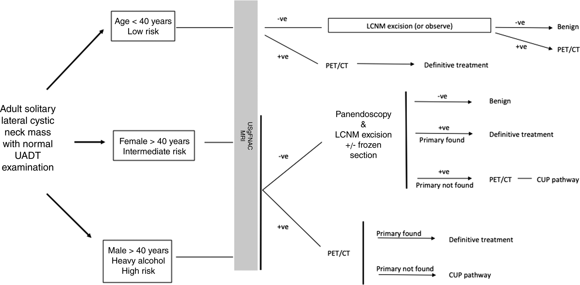
Based on our findings, we propose a diagnostic algorithm for the management of solitary lateral cystic neck mass (Figure 2). Several authors including Franzen et al. have suggested performing a panendoscopy, biopsies and tonsillectomy on anyone over the age of 40 years prior to cyst excision.Reference Franzen, Günzel, Buchali and Coordes7 In their series, they reported a malignancy rate of up to 80 per cent in the older age group, which justifies this approach. We found a much lower malignancy rate of 10 per cent for females over 40 years and 30 per cent for males over 40 years. We therefore believe the excess morbidity and time delay (25 per cent will remain cancer of unknown primary) does not justify a blanket biopsy approach in the absence of a clinically evident primary lesion. This would affect the utility of subsequent PET/CT imaging. From our series, half the patients with a subsequently confirmed malignant cystic node had an identifiable primary on rigid endoscopy. We therefore recommend a rigid endoscopy in patients over the age of 40 years at the time of cystic mass excision (intermediate to high risk). If a primary lesion is clinically evident at this more detailed mucosal examination, it is biopsied, and the planned cyst excision is abandoned. In the absence of a primary lesion but with subsequently confirmed malignancy and negative PET-CT, formal ipsilateral tonsillectomy and tongue base mucosectomy forms the second procedure as part of modern standard of care for cancer of unknown primary management. In our series, 50 per cent of the malignancies were in the ipsilateral palatine tonsil.
Proposed investigation pathway for management of adult lateral cystic neck masses. -ve = negative; +ve = positive; LCNM = lateral cystic neck mass; PET-CT = positron emission tomography-computed tomography; UADT = upper aerodigestive tract; USgFNAC MRI = ultrasound-guided fine needle aspiration cytology magnetic resonance imaging; +/- = with or without; CUP = cancer of unknown primary

• The solitary lateral neck cyst presents a diagnostic challenge
• No standardised investigation pathway exists
• Squamous cell carcinoma of the ipsilateral tonsil is the most common site of primary with human papillomavirus disease prevalent
• Age over 40 years, male sex and high alcohol intake (not smoking) were significant risk factors for malignancy
• Ultrasound-guided fine needle aspiration and magnetic resonance imaging in combination represent the most accurate pre-open biopsy tests
• Panendoscopy and biopsy of suspicious mucosal lesions (not blind biopsy) should be performed at the time of open biopsy in high-risk individuals
Several studies have demonstrated no impact on survival when excision biopsy is performed on a solitary neck node prior to radical radiotherapy.Reference Ellis, Mendenhall, Rao, McCarty, Parsons and Stringer25–Reference Zenga, Graboyes, Haughey, Paniello, Mehrad and Lewis27 Indeed, the combined 5-year overall survival in this series, including the oropharyngeal and unknown primary cases, was 87.5 per cent, comparing favourably with our published outcomes for all HPV positive and negative SCC (83 per cent and 53 per cent, respectively),Reference Dalton, Milinis, Houghton, Ridley, Davies and Williams28 which is reassuring. However, we hope that optimisation of the investigation strategy will help improve our pre-open biopsy diagnostic rate and potentially help to reduce the overall pathway time by reducing uncertainty regarding investigations.
Limitations
The retrospective nature of this series means that interpretation must be considered in this context. Recommended investigations were subject to intra-departmental changes and clinician preference over the course of the 10-year study period and may introduce selection bias for the investigations selected. Half of the 139 total MRI scans performed in our cohort were excluded from analysis because of indeterminate reports that failed to indicate a likelihood of malignancy from the cystic lesion appearance (when combined with a negative oropharynx for radiological primary lesion). We believe these omissions have only underscored the potential final test metrics for MRI, especially specificity and negative predictive value, and we still advocate this modality as an important anatomical baseline and oropharyngeal screen prior to cystic mass excision. Core needle biopsy has been advocated by a number of large series and guidelinesReference Civantos, Vermorken, Shah, Rinaldo, Suárez and Kowalski23,29 as a second line investigation but has not been included in this series because of the paucity of cases where it was utilised. Safety appears to be supported by published series and seeding risk is similarly rare.Reference Tandon, Shahab, Benton, Ghosh, Sheard and Jones21,Reference Shah and Ethunandan30 It remains a viable alternative second line investigation to fine needle aspiration, although in our experience the limitation of having very little solid material to target in these cystic lesions is not improved by use of a wider calibre core biopsy needle and should not delay progression through the diagnostic pathway to open biopsy. HPV-positive cases increased in the second half of the collection period, but this is likely because of increased testing rather than prevalence. The number of oropharynx SCC cases remained stable throughout the study period from 2008 to 2018.
Conclusion
The most common causes of a solitary lateral cystic neck mass are benign cysts of branchial origin. However, 16 per cent of patients with this presentation will harbour a malignancy (30 per cent males and 10 per cent females over 40), most commonly HPV-positive SCC from the ipsilateral oropharynx. Following a thorough history and physical examination, we recommend ultrasound-guided FNAC and MRI as initial investigations that provide the highest accuracy and then a risk-stratified approach based on age, gender and alcohol intake. The traditional head and neck risk factor of smoking is not indicative. With a low specificity and positive predictive value, PET-CT imaging should be reserved for confirmed malignant cases without an identified primary lesion to image potential high-risk sites. If a solitary, indeterminate high-risk cystic lesion is confirmed, we advise early excision of the mass for paraffin section histology, combined with an upper aerodigestive tract examination.
Competing interests
None declared