Introduction
Behçet's disease is a multisystem vasculitis, characterised by mucosal aphthous lesions, mainly in the oral and genital mucosa.Reference Akyol, Günbey, Karlı, Önem, Özgen and Sayarlıoğlu1 Akyol et al.Reference Akyol, Günbey, Karlı, Önem, Özgen and Sayarlıoğlu1 reported that olfactory dysfunction may denote neurological involvement in Behçet's disease.
The sense of smell is of great importance to people as a significant contributor to safety and quality of life. Olfactory deterioration is associated with a decrease in taste perception, and therefore, taste perception is greatly influenced by olfaction.Reference Landis, Scheibe, Weber, Berger, Bramerson and Bende2 Smell and taste play a role in stimulating gastric excretion in the context of normal digestive physiology, and at the same time serve as an early warning system against harmful substances.Reference Mattes3, Reference Samuels4
Early diagnostic methods, including olfactory evaluation tools, such as the University of Pennsylvania Smell Identification Test, have been previously used and reported.Reference Doty, Shaman and Dann5 Magnetic resonance imaging (MRI) can be usedReference Wattendorf, Welge-Lussen, Fiedler, Bilecen, Wolfensberger and Fuhr6–Reference Hummel, Witt, Reichmann, Welge-Luessen and Haehner9 for the reliable estimation of olfactory bulb volume and olfactory sulcus depth, in order to further identify the nature of the olfactory deficit.Reference Kim, Lee, Chung and Dhong7–Reference Mueller, Abolmaali, Hakimi, Gloeckler, Herting and Reichmann11
In the present study, olfactory bulb volume and olfactory sulcus depth were measured, using MRI, in patients with Behçet's disease and a control group, and the results were compared. No similar studies could be found in the literature related to olfactory bulb volume and olfactory sulcus depth values in Behçet's disease patients measured on MRI.
Materials and methods
All procedures of this retrospective study were applied according to the principles of the Declaration of Helsinki. Approval for the study was granted by the Clinical Research Ethics Committee of Adana City Training and Research Hospital (decision number 185; 27 March 2018). Cranial MRI scans were obtained from the Radiology Department of Kırıkkale University Faculty of Medicine hospital database.
Subjects
A retrospective screening was made of the cranial MRI scans in the hospital's picture archiving and communication system of 27 patients aged 18–60 years, diagnosed with Behçet's disease in the period from January 2015 to September 2017. The Behçet's disease diagnosis was confirmed by the dermatology department according to the International Study Group Criteria,Reference Chang and Kim12 and symptom duration (in years) was recorded.
The control group was formed of 27 non-Behçet's disease subjects, aged 18–60 years, with normal cranial MRI results documented in the picture archiving and communication system.
Subjects were excluded from the study if there was a history of trauma or surgery, sinonasal tumour, sinonasal infection, sinonasal polyposis, cerebrospinal fluid leakage, marked nasal septal deformity, marked facial deformity, multiple sclerosis, epilepsy, or Parkinson's disease.Reference Doğan, Bayar Muluk, Şahan, Asal, Inal and Ergün13
This information was obtained from the clinical data in the hospital files and could be a potential limitation of the study.
Cranial imaging measurements
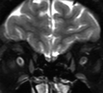
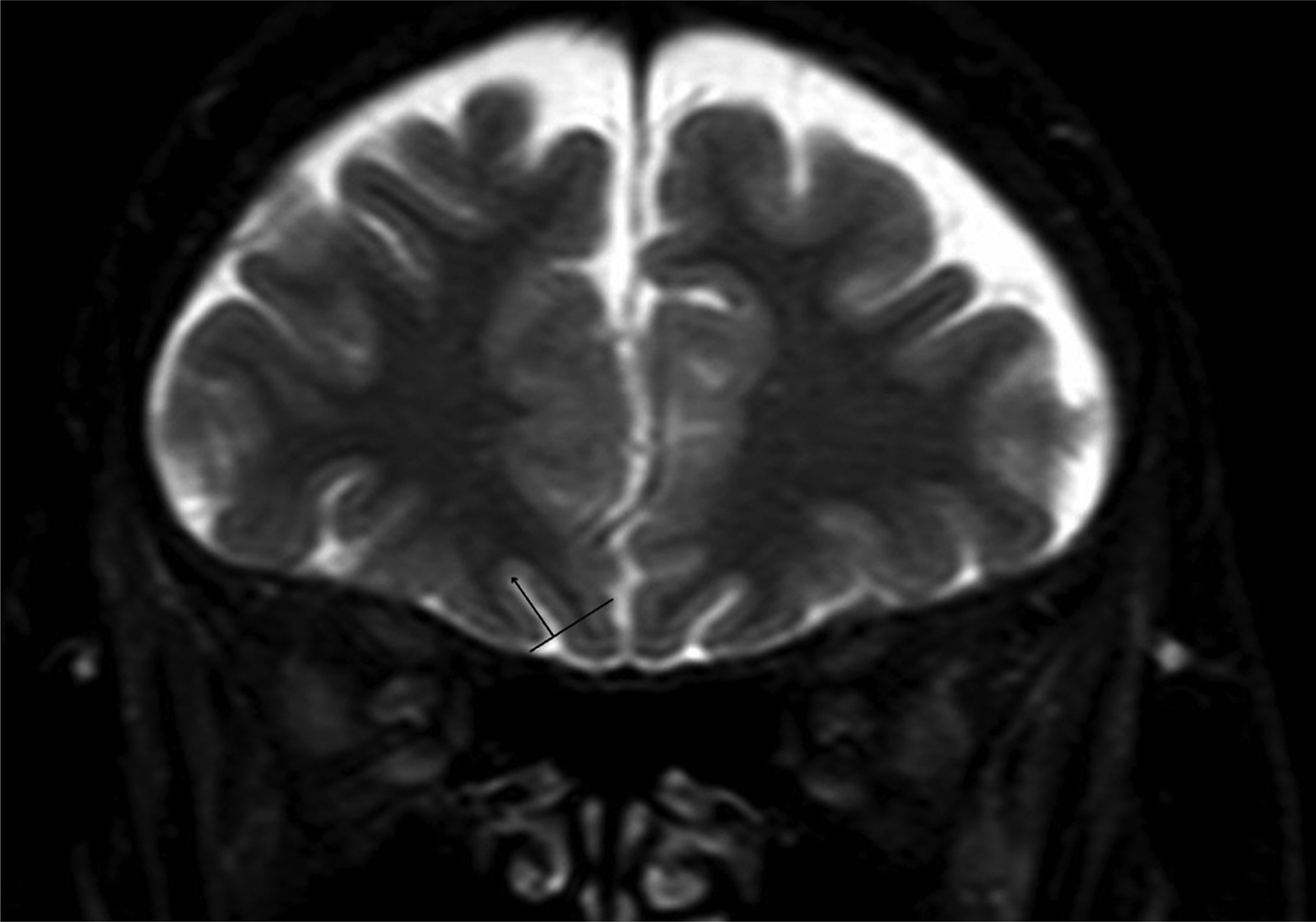
The MRI examinations were performed using a 1.5 Tesla MRI unit with a cranial coil (Philips MRI Systems, Achieva Release 3.2 (2013); Philips Medical Systems Nederland, Best, Netherlands). Fat-suppressed T2-weighted images in the coronal plane (repetition time/echo time = 6557/100 ms, field of view = 220 × 175 mm and matrix = 224 × 165 mm) were obtained using 5 mm slice thickness and 1 mm intersection gap. The measurements for standard olfactory bulb volume (Figure 1) and olfactory sulcus depth (Figure 2) were taken from coronal T2-weighted images.Reference Doğan, Bayar Muluk, Şahan, Asal, Inal and Ergün13–Reference Buschhüter, Smitka, Puschmann, Gerber, Witt and Abolmaali15 All MRI scans were obtained on the same scanner using the same protocol for measurement.
Olfactory bulb volume (outlined) on a coronal, T2-weighted, spectral pre-saturation with inversion recovery sequence magnetic resonance imaging scan of a 42-year-old female with Behçet's disease (disease duration: 4 years).

Olfactory sulcus depth (black lines) on a coronal, T2-weighted, spectral pre-saturation with inversion recovery sequence magnetic resonance imaging scan of a 45-year-old female with Behçet's disease (disease duration: 15 years).

Olfactory bulb volume was measured using a coronal T2-weighted spectral pre-saturation with inversion recovery (‘SPIR’) sequence. From anterior to posterior screening, the olfactory bulb can be clearly seen. The olfactory bulb surface was measured manually (in mm2) using an electronic cursor, and the volume was calculated in mm3 by multiplying this value by the slice thickness.Reference Doğan, Bayar Muluk, Şahan, Asal, Inal and Ergün13–Reference Buschhüter, Smitka, Puschmann, Gerber, Witt and Abolmaali15
Olfactory sulcus depth was measured on the coronal T2-weighted spectral pre-saturation with inversion recovery sequence by drawing a virtual tangent line from the inferior orbital gyrus to the gyrus recti in the posterior plane of the orbit. A perpendicular line was then drawn from this tangent line to the deepest point of the olfactory sulcus. The depth of this line provided the olfactory sulcus depth (in mm).Reference Doğan, Bayar Muluk, Şahan, Asal, Inal and Ergün13–Reference Buschhüter, Smitka, Puschmann, Gerber, Witt and Abolmaali15
The accuracy of measuring olfactory bulb volume and olfactory sulcus depth on MRI imaging is limited, which could be a shortcoming of the study.
Statistical analysis
Data obtained in the study were analysed using SPSS for Windows statistical software, version 16.0 (IBM, Chicago, Illinois, USA). The independent samples t-test, paired samples t-test, Pearson correlation test and Spearman's correlation rho efficient test were used in the analyses. No power analysis was performed because of the low number of Behçet's disease patients with cranial MRI scan data in the hospital database, which was a limitation of the study. A value of p < 0.05 was considered statistically significant.
Results
The Behçet's disease group was formed of 27 adult patients diagnosed with Behçet's disease, comprising 10 males and 17 females, with a mean age of 43.55 ± 7.98 years (range, 26–57 years). The control group was formed of 27 adults with no findings of Behçet's disease, comprising 10 males and 17 females, with a mean age of 43.40 ± 7.92 years (range, 26–57 years).
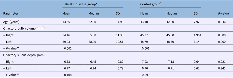
The olfactory bulb volume and olfactory sulcus depth measurements are shown in Table 1. Olfactory bulb volume on the right and left sides, and olfactory sulcus depth on the right side, were significantly lower in the Behçet's disease group than in the control group (p < 0.05) (Table 1).
Patient age and olfactory bulb volume and olfactory sulcus depth measurements

*n = 27; †n = 27. ‡Independent samples t-test result; **paired samples t-test result. SD = standard deviation
Olfactory bulb volume was significantly higher for the left side than the right side in both the Behçet's disease group and the control group (p < 0.05). No significant differences were determined between right- and left-sided olfactory sulcus depth values for both the Behçet's disease group and the control group (p > 0.05) (Table 1).
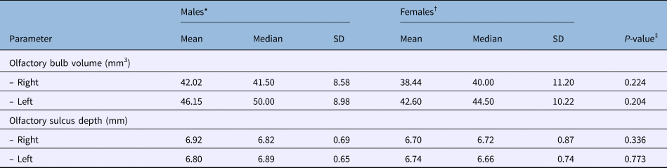
The olfactory bulb volume and olfactory sulcus depth values for the males and females of both groups are shown in Table 2. No significant differences were determined between right- and left-sided olfactory bulb volume and olfactory sulcus depth values according to gender (p > 0.05) (Table 2).
Olfactory bulb volume and olfactory sulcus depth in males and females in both groups

*n = 20; †n = 34. ‡Independent samples t-test result
Behçet's disease group correlations
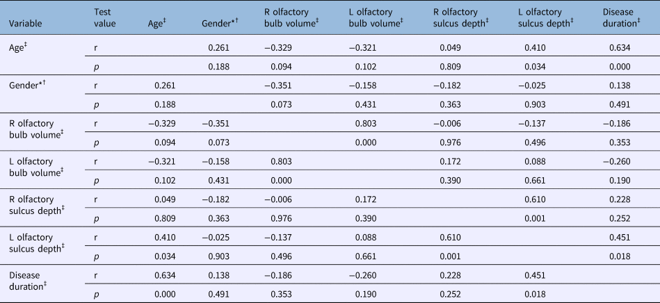
Positive correlations were determined between the right- and left-sided olfactory bulb volume values and between the right- and left-sided olfactory sulcus depth values (p < 0.05) (Table 3).
Correlation test results in Behçet's disease group*

*Code: male = 1, female = 2. †Spearman's correlation rho efficient test result; ‡Pearson correlation test result. R = right; L = left
As the duration of Behçet's disease increased, so too the left-sided olfactory sulcus depth values increased (p < 0.05) (Table 3). Behçet's disease duration was determined to be longer in older patients (p < 0.05) (Table 3).
Discussion
Behçet's disease is a chronic, recurrent condition that affects small vessels in many systems of the body. It is associated with a wide spectrum of clinical findings, affecting the vascular, ocular, mucocutaneous, gastrointestinal, musculoskeletal and central nervous systems.Reference Webb, Moots and Swift16 In patients with Behçet's disease, nasal mucociliary clearance time has been shown to be longer compared to control groups,Reference Ozbay, Kucur, Temizturk, Ozkan, Kahraman and Oghan17 but within the normal range of 20 minutes reported in literature.Reference Asai, Haruna, Otori, Yanagi, Fukami and Moriyama18, Reference Lale, Mason and Jones19
Veyseller et al.Reference Veyseller, Doğan, Ozücer, Aksoy, Meriç and Su20 evaluated nasal mucosal lesions in patients with Behçet's disease. The lesions were not specific to Behçet's disease and were mostly located in Little's zone and the medial surface of the lower concha, and less in the nasal septum. It was reported that nasal symptoms were associated with the presence of nasal lesions, but no correlations were determined between nasal findings and olfactory function.
In the present study, olfactory bulb volume and olfactory sulcus depth were evaluated in patients with Behçet's disease. A scan of the literature revealed studies that evaluated olfactory function in Behçet's disease using olfactory or smell tests.Reference Akyol, Günbey, Karlı, Önem, Özgen and Sayarlıoğlu1, Reference Lale, Mason and Jones19, Reference Veyseller, Doğan, Ozücer, Aksoy, Meriç and Su20 However, to the best of our knowledge, there has been no previous study of MRI measurements of olfactory bulb volume and olfactory sulcus depth in Behçet's disease.
The results of this study showed that the bilateral olfactory bulb volume and right-sided olfactory sulcus depth values were significantly lower in the Behçet's disease group compared to the control group. In addition, the left-sided olfactory sulcus depth increased with increasing Behçet's disease duration. In both the Behçet's disease group and the control group, olfactory bulb volume was significantly higher in the left side than in the right side. No differences were seen in olfactory bulb volume and olfactory sulcus depth according to gender. Positive correlations were determined between right- and left-sided olfactory bulb volume values, and between right- and left-sided olfactory sulcus depth values.
Buschhüter et al.Reference Buschhüter, Smitka, Puschmann, Gerber, Witt and Abolmaali15 measured the olfactory bulb on the MRI scans of 125 randomly selected subjects (58 males and 67 females, aged 19–79 years). Inter-individual variation in olfactory bulb volumes was relatively large, ranging from 41 mm3 to 97 mm3 for right-sided olfactory bulb volume and from 37 mm3 to 98 mm3 for left-sided olfactory bulb volume. In the present study, the median olfactory bulb volume values were 35.0 mm3 on the right side and 38.0 mm3 on the left side in the Behçet's disease group, and 45.0 mm3 on the right side and 48.5 mm3 on the left side in the control group. In the literature, olfactory bulb volume changes have been reported following post-viral, post-traumatic and sinonasal olfactory loss.Reference Mueller, Abolmaali, Hakimi, Gloeckler, Herting and Reichmann11, Reference Yousem, Geckle, Bilker, Kroger and Doty21–Reference Rombaux, Mouraux, Bertrand, Nicolas, Duprez and Hummel23 Hypoplasia of the olfactory bulb has been observed in patients with Alzheimer's diseaseReference Thomann, Dos Santos, Toro, Schönknecht, Essig and Schröder24 and schizophrenia.Reference Turetsky, Moberg, Yousem, Doty, Arnold and Gur25
Akyol et al.Reference Akyol, Günbey, Karlı, Önem, Özgen and Sayarlıoğlu1 investigated olfactory function in Behçet's disease using the Sniffin’ Sticks olfactory test. Odour identification and overall scores were lower in the Behçet's disease group than in the control group, but the differences were not statistically significant (p > 0.05). It was concluded that odour identification was more impaired in the Behçet's disease patients than in the healthy control subjects, but there was no group difference in respect of smell discrimination.
In Veyseller and colleagues’ study,Reference Veyseller, Doğan, Ozücer, Aksoy, Meriç and Su20 the effect of Behçet's disease on olfactory function was investigated using the Connecticut Chemosensory Clinical Research Center test in 30 patients with Behçet's disease compared to a control group. n-Butanol odour threshold and odour identification tests were also performed. The results showed that n-Butanol threshold test scores were 5.57 ± 1.0 for the Behçet's disease group and 6.47 ± 0.7 for the control group, on a scale of 7 (0 = worst score; 7 = best score). The identification test scores were 4.93 ± 1.3 for the Behçet's disease group and 6.15 ± 0.8 for the control group, and the mean Connecticut Chemosensory Clinical Research Center scores were 5.25 ± 1.0 for the Behçet's disease group and 6.31 ± 0.6 for the control group, with significant differences determined between the groups (p < 0.001). The significant difference in the Connecticut Chemosensory Clinical Research Center scores was reported to suggest an association between olfactory dysfunction and Behçet's disease. It was also stated that nasal symptoms are associated with the presence of nasal lesions, but there was no correlation between nasal findings and olfactory function.
In a recent study of olfactory dysfunction associated with neuro-Behçet's disease, by Doğan et al.,Reference Doğan, Ertaş, Özücer, Birday, Özturan and Veyseller26 the Connecticut Chemosensory Clinical Research Center olfactory test was used on 16 patients with neuro-Behçet's disease and compared with a healthy control group. The results showed that the mean Connecticut Chemosensory Clinical Research Center score of the neuro-Behçet's disease patients was 4.60 out of 7; this group was moderately hyposmic. The mean score of the control group was 6.5, with a statistically significant difference between the groups (p < 0.0001). The Connecticut Chemosensory Clinical Research Center scores of the neuro-Behçet's disease patients were significantly lower compared to both the healthy control subjects and those of Behçet's disease patients reported in literature. It was concluded that neuro-Behçet's disease presents with aggravated odour dysfunction compared to Behçet's disease. Neurological involvement, especially parenchymal involvement, appears to worsen olfactory dysfunction. The duration of the disease was found to be correlated with the severity of this dysfunction.
• Behçet's disease may cause a decrease in olfactory function
• Smaller olfactory bulb volume and olfactory sulcus depth values were found in Behçet's disease patients
• Behçet's disease patients should undergo regular olfactory function tests
To the best of our knowledge, this is the first study in literature to have measured olfactory bulb volume and olfactory sulcus depth in Behçet's disease patients. The results of the study demonstrated that the smaller olfactory bulb volume and olfactory sulcus depth values in the Behçet's disease group could be related to the main pathophysiological condition in Behçet's disease, as the disease is known to affect small vessels and to cause prolonged nasal mucociliary clearance time. Moreover, possible nasal mucosal lesions may lead to the delayed transport of the molecules in the nasal air system. Vascular problems may affect neural damage and, consequently, olfactory impairment may develop.
Conclusion
The results of this study indicate that Behçet's disease may cause a decrease in olfactory function related to lower olfactory bulb volume and olfactory sulcus depth. Follow up of Behçet's disease patients with regular olfactory function tests is recommended. If necessary, MRI examinations of the olfactory system should be conducted.
Competing interests
None declared