Introduction
Head and neck squamous cell carcinoma (SCC) is the sixth leading incident cancer in the world. It is likely that approximately 600 000 cases arise per year worldwide, and that only 40–50 per cent of patients with head and neck SCC survive for five years or more.Reference Leemans, Braakhuis and Brakenhoff1 Despite improvements in therapeutic and reconstructive modalities, the survival rates of head and neck SCC patients have improved little over the past 40 years.Reference Parkin, Bray, Ferlay and Pisani2 Treatment failures are determined by the development of local and/or regional recurrence, distant metastasis, or a second primary cancer. In patients with advanced-stage (i.e. stage III or IV) head and neck SCC, local recurrence rates are as high as 50 to 60 per cent, and 15 to 25 per cent of these patients will develop distant metastases.Reference Wirtschafter, Benninger, Moss, Umiel, Blazoff and Worsham3 Although locoregional recurrence is the most common source of treatment failure, up to one-third of deaths are due to distant metastases.Reference Alvi and Johnson4 Better prognostic markers are needed to improve prediction of locoregional recurrence and distant metastasis in head and neck SCC patients.
The process of epithelial cancer metastasis is complex and includes loss of adhesion, invasion, migration into and out of surrounding vascular and lymphatic structures, and survival and proliferation at other locations.Reference Pantel and Brakenhoff5 A large number of studies have demonstrated the critical role played by circulating tumour cells in the metastatic spread of most types of epithelial cancer.Reference Fidler6, Reference Valastyan and Weinberg7 Therefore, it is reasonable to hypothesise that detection and monitoring of circulating tumour cells might provide clinically important information relevant to diagnosis and treatment. The first report on tumour cells in the peripheral circulation has been attributed to Ashworth in 1869.Reference Hayes and Smerage8 Over the next 130 years, numerous attempts were made to develop reliable assays to detect circulating tumour cells in the peripheral blood. However, no one specific technology arose which could universally distinguish these circulating tumour cells. Over the past 20 years, prospects for the detection of circulating tumour cells have improved with the development of immunomagnetic separation technology, with its higher levels of sensitivity and specificity.
Now, a validated, standardised circulating tumour cell assay, the CellSearch system (Veridex, Raritan, New Jersey, USA), has been approved by the US Food and Drug Administration for clinical use.Reference Zhe, Cher and Bonfil9 This technology employs positive selection using magnetically labelled anti-epithelial cell adhesion molecule and immunocytochemical staining for cytokeratins 8, 18 and 19, in order to isolate and count circulating tumour cells. There is increasing evidence that circulating tumour cell number, as determined by the CellSearch system, correlates, as an independent predictor, with progression-free survival and overall survival in breast, colorectal and prostate cancer.Reference Miller, Doyle and Terstappen10 However, use of the CellSearch system to identify circulating tumour cells in patients with head and neck SCC has not previously been studied. If the CellSearch system could be shown to have similar utility in head and neck SCC patients, it could prove useful in patient monitoring and risk profile evaluation.
Therefore, we performed the current, preliminary study in order (1) to assess the feasibility of detecting circulating tumour cells in patients with advanced-stage head and neck SCC, and (2) to correlate our observations to clinical findings.
Materials and methods
Ethics
The study followed the principles established in the Declaration of Helsinki and was approved by the ethics committee of Beijing Tongren Hospital. All the patients involved gave written, informed consent to the publication of their case details.
Patients and peripheral blood sampling
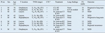
After giving informed consent, nine patients newly diagnosed with advanced-stage head and neck SCC (i.e. stage III or IV) were enrolled in the study between February and June 2011. All patients underwent imaging evaluation (including computed tomography scans of the head and neck and X-ray imaging of the chest) and endoscopy as part of routine clinical examination, before the initiation of therapy. The clinical characteristics of these patients are presented in Table I. Staging was carried out according to the Union of International Cancer Control tumour-node-metastasis (TNM) classification system (sixth edition).
Patient and circulating tumour cell data

*Circulating tumour cell (CTC) positivity (+) = more than 0 and up to 1 cell per 7.5 ml peripheral blood. Pt no = patient number; y = years; T = tumour; TNM = tumour-node-metastasis classification; FU (mth) = follow up (months); M = male; – = negative; CT = chemotherapy; RT = radiotherapy; mets = metastases; SD = stable disease; Sx = surgery; post-op = post-operative; NED = no evidence of disease; F = female
A total of 7.5 ml of peripheral blood was obtained using CellSave blood collection tubes (Veridex), prior to initiation of therapy. Blood samples were maintained at room temperature and processed using the CellSearch system within 72 hours.
Isolation and enumeration of circulating tumour cells
The CellSearch system, including the CellTracks AutoPrep, MagNest and CellSpotter Analyzer components, was used for isolation and enumeration of circulating tumour cells. The system prepared the sample in a semi-automatic fashion, enabling immunomagnetic selection, fluorescence staining, concentration and enrichment of circulating tumour cells. Sample processing was done as previously described.Reference Allard, Matera, Miller, Repollet, Connelly and Rao11 In brief, 7.5 ml of blood was mixed with 6.5 ml of dilution buffer, centrifuged and transferred into the CellTracks AutoPrep system. The remaining steps were performed automatically by the instrument, including all reagent addition, mixing, incubation and aspiration steps. Following aspiration of the plasma and dilution buffer layer, ferrofluids coated with antibodies to epithelial cell adhesion molecule were added, together with capture enhancement reagent. After incubation and magnetic separation, unbound cells and remaining plasma were removed, and ferrofluid-labelled cells were re-suspended in buffer, permeabilised, and fluorescently labelled using monoclonal antibodies specific for the relevant epithelial cells (i.e. cytokeratins 8, 18 and 19) and leukocytes (i.e. cluster of differentiation 45 protein). After incubation and repeated magnetic separation, unbound staining reagents were aspirated and residual cells were fixed.
The samples were transferred automatically to a cartridge then placed inside the MagNest cell presentation device. During an incubation of at least 20 minutes in the dark at room temperature, the immunomagnetically labelled cells were moved to the surface of the cartridge, due to the strong magnetic field of the MagNest device. They were then oriented for analysis using the CellSpotter Analyzer.
Identification of circulating tumour cells
The identification and enumeration of circulating tumour cells were performed by the CellSpotter Analyzer component. A fluorescence-based microscopy system generated reconstruction of cellular images. The criteria by which fluorescent images were identified as circulating tumour cells included round to oval morphology and a visible nucleus positive for 4′,6-diamidino-2-phenylindole, contained within cytokeratin-positive cytoplasm which stained negatively for cluster of differentiation 45 protein. Overlay of the 4′,6-diamidino-2-phenylindole nuclear staining pattern also showed that circulating tumour cells had a high nucleus-to-cytoplasm ratio, consistent with identification as malignant cells. The nucleus-to-cytoplasm ratios of the circulating tumour cells were greater than those of the surrounding white blood cells, which stained positive for cluster of differentiation 45 protein. These circulating tumour cells conformed to accepted criteria both for circulating tumour cells (i.e. the fluorescence criteria discussed by Fehm et al.)Reference Fehm, Solomayer, Meng, Tucker, Lane and Wang12 and for carcinoma cells (i.e. standard cytopathological criteria).Reference Atkinson13
Each sample was independently analysed by two trained readers without prior knowledge of the patient's clinical status.
Results
Circulating tumour cell detection
Using the CellSearch system, circulating tumour cells were successfully detected in three of the nine patients (33 per cent), i.e. more than 0 and up to 1 circulating tumour cells were detected per 7.5 ml peripheral blood. Figure 1 shows an example of circulating tumour cell detection. A summary of patient characteristics, including age, sex, tumour site, TNM stage, treatment and follow up, is provided in Table I.
Composite fluorescent images of circulating tumour cells detected by the CellSearch system. Cells staining positive for 4′,6-diamidino-2-phenylindole (DAPI) and cytokeratin (CK) and negative for cluster of differentiation 45 protein (CD45) are identified as circulating tumour cells. PE = phycoerythrin; APC = allophycocyanin

Circulating tumour cells and tumour localisation
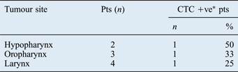
We assessed whether the frequency of circulating tumour cell detection was affected by the primary tumour site. Data are shown in Table II. The highest prevalence of circulating tumour cell positive cases was found amongst patients with hypopharyngeal SCC (one of two; 50 per cent), followed by oropharyngeal SCC (one of three; 33 per cent) and laryngeal SCC (one of four; 25 per cent).
Circulating tumour cell positivity by primary tumour site
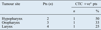
*See Table I for definition of circulating tumour cell positivity (CTC +ve). Pts = patients
Circulating tumour cells and regional metastasis
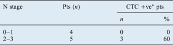
We also assessed whether the presence of circulating tumour cells correlated with lymph node metastasis. Patients were grouped according to their nodal stage and the frequency of circulating tumour cell positive cases in these groups was compared. Of the four N0–N1 cases, none was positive for circulating tumour cells (0 per cent); in contrast, of the five N2–N3 cases, three were positive for circulating tumour cells (60 per cent). However, there was no statistically significant association between the presence of circulating tumour cells and the presence of nodal metastasis (p = 0.167, Fisher's exact test; Table III).
Circulating tumour cell positivity by node stage
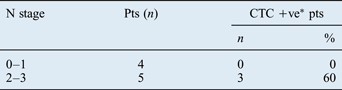
P = 0.167, circulating tumour cell positivity (CTC +ve) vs nodal stage, Fisher's exact test. *See Table I for definition of CTC +ve. N = node; Pts = patients
Circulating tumour cells and disease progression and recurrence
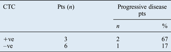
We then evaluated the correlation between the presence of circulating tumour cells and the extent of disease progression. After a mean follow-up time of 14 months (range, 12–16 months), only one of the six patients (17 per cent) who were negative for circulating tumour cells had locoregional recurrent or progressive disease; in contrast, two of the three patients (67 per cent) who were positive for circulating tumour cells had progressive disease (Table IV). However, no statistically significant association was found between the presence of detectable circulating tumour cells and the presence of locoregional recurrent or progressive disease (p = 0.15, log-rank test; Table IV).
Disease progression by circulating tumour cell status
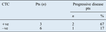
P = 0.15, disease progression vs circulating tumour cell positivity (CTC +ve), log-rank test. *See Table I for definition of CTC +ve. Pts = patients; –ve = negative
Discussion
Over the past few decades, advances in imaging techniques and treatment modalities have resulted in improved local control for patients with head and neck SCC. However, these improvements have not translated into significantly improved overall survival in all studies.Reference Cognetti, Weber and Lai14 This has been due mainly to distant metastases, which occur in up to 20 per cent of head and neck SCC patients.Reference Yilmaz, Hoşal, Ozyar, Akyol and Gürsel15–Reference Senft, de Bree, Hoekstra, Kuik, Golding and Oyen17 Since there is currently no successful curative treatment for metastatic disease and the expected survival time for these patients is poor, a reliable blood test which could identify patients at high risk of disseminated disease prior to the development of clinically detectable metastasis would have clear clinical relevance.
Circulating tumour cells are shed by both primary and metastatic cancers, and are thought to mediate the haematogenous spread of cancer to distant sites.Reference Pantel and Brakenhoff5 Circulating tumour cells may even represent an earlier and more reproducible indication of disease status than current imaging criteria.Reference Budd, Cristofanilli, Ellis, Stopeck, Borden and Miller18 In head and neck SCC patients, the presence of circulating tumour cells is associated with poorer outcomes.Reference Jatana, Lang and Chalmers19 Moreover, recent findings have indicated an additional role for circulating tumour cells: they seem to be able to efficiently ‘re-seed' the primary tumour.Reference Kim, Oskarsson, Acharyya, Nguyen, Zhang and Norton20 Therefore, circulating tumour cells may contribute not only to distant metastasis but also to locoregional recurrence.
Over the past 40 years, several methods of detecting circulating tumour cells have been developed, including detection by physical characteristics (e.g. size or weight) and by biological characteristics (including immunocytochemistry, flow cytometry and reverse transcriptase polymerase chain reaction).Reference Hayes and Smerage8 A few studies using these techniques have revealed the presence of circulating tumour cells in the peripheral blood of head and neck SCC patients. Hristozova et al. demonstrated that 18 of 42 (43 per cent) patients with inoperable head and neck SCC with regional metastasis had detectable circulating tumour cells, using flow cytometric analysis of epithelial cell adhesion molecule and cytokeratins.Reference Hristozova, Konschak, Stromberger, Fusi, Liu and Weichert21 They found a significant association between circulating tumour cell prevalence and N stage, but not T stage. Jatana et al. prospectively studied 48 patients with head and neck SCC, using a negative depletion process based on depletion of normal cells; they found that 71 per cent of patients had circulating tumour cells present, and that these patients had a statistically significantly worse disease-free survival time compared with patients with no circulating tumour cells.Reference Jatana, Balasubramanian, Lang, Yang, Jatana and White22 In a previous study, our group used reverse transcriptase polymerase chain reaction to identify cytokeratin 19 messenger RNA as a marker of disseminated tumour cells, and found this marker to be present in 15 of 32 (47 per cent) patients with laryngeal or laryngopharyngeal carcinoma; we also found an increased risk of local recurrence and a reduced disease-free survival time in patients positive for this marker.Reference Li, Yu, Huang, Fang, Ni and Wang23 However, at this time there was no one detection technique that was reliably reproducible in different laboratories.
In this study, we used the CellSearch system, a new, immunomagnetic circulating tumour cell assay, to detect circulating tumour cells in the peripheral blood of head and neck SCC patients. This system is the first automated, standardised, clinically approved system for the detection and quantification of circulating tumour cells in peripheral blood, and has been used clinically in cases of metastatic breast, colorectal and prostate cancer.Reference Miller, Doyle and Terstappen10, Reference Cristofanilli, Budd, Ellis, Stopeck, Matera and Miller24 The CellSearch system uses magnetically labelled anti-epithelial cell adhesion molecule microbeads and immunocytochemical staining for cytokeratins 8, 18 and 19, in order to isolate and count circulating tumour cells in peripheral blood. Both the markers employed in the system have been previously proven suitable for detecting head and neck SCC cells.Reference Chaubal, Wollenberg, Kastenbauer and Zeidler25, Reference Ziober, Mauk, Falls, Chen and Ziober26 The CellSearch detection technique is reproducible across different laboratories and can identify circulating tumour cells in patients with different cancer types.Reference Miller, Doyle and Terstappen10
Allard et al. have published findings from a multicentre study designed to determine the accuracy of the CellSearch system in healthy subjects and patients with nonmalignant disease.Reference Allard, Matera, Miller, Repollet, Connelly and Rao11 These authors reported that 321 of 344 (93.3 per cent) tested samples contained no circulating tumour cells in 7.5 ml of blood, while only 1 of 344 (0.3 per cent) samples contained more than 1 circulating tumour cell. The mean number ± standard deviation of circulating tumour cells in healthy subjects and patients with nonmalignant disease was 0.1 ± 0.2 per 7.5 ml blood. Allard and colleagues' data demonstrated that the CellSearch system provides an accurate and reproducible assay that can count circulating tumour cells reliably across laboratories; these data also confirmed the findings of Riethdorf et al. Reference Riethdorf, Fritsche, Müller, Rau, Schindlbeck and Racket27 To date, the CellSearch system is the only validated and standardised clinical assay of circulating tumour cells.
The primary goal of our pilot study was to determine whether the CellSearch system could be used to detect circulating tumour cells in patients with head and neck SCC. Our data indicated that the CellSearch system successfully detected circulating tumour cells in the peripheral blood of three of nine (33 per cent) advanced-stage head and neck SCC patients. The highest prevalence of circulating tumour cell positive patients was found among those with hypopharyngeal cancer, followed by those with oropharyngeal and laryngeal cancer. Our preliminary data showed a greater proportion of circulating tumour cell positive patients in the N2–N3 group (60 per cent) compared with the N0–N1 group (0 per cent). Our data also suggested a lower prevalence of progressive disease among circulating tumour cell negative patients (17 per cent) compared with positive patients (67 per cent). Although there was no significant correlation between the presence of circulating tumour cells and progression-free survival, this negative result may have been due to the small number of patients (nine) enrolled in this pilot study. It appears that circulating tumour cells may represent a novel prognostic indicator in head and neck SCC. Further studies, including greater patient numbers and longer follow-up times, are needed.
• The presence of circulating tumour cells in peripheral blood is a marker of invasive cancer
• This study used the CellSearch automated assay to detect circulating tumour cells in advanced-stage head and neck squamous cell cancer patients
• The system successfully identified circulating tumour cells in peripheral blood
One possible limitation regarding the sensitivity of the CellSearch system is the requirement that circulating tumour cells express both epithelial cell adhesion molecule (for capture) and cytokeratins (for analysis). The vast majority of epithelial tumours express epithelial cell adhesion molecule.Reference Went, Lugli, Meier, Bundi, Mirlacher and Sauter28 Furthermore, cancer cells may stop expressing epithelial cell adhesion molecule during epithelial-to-mesenchymal transition (the process by which tumour cells acquire a more invasive and migratory phenotype).Reference Frederick, Helfrich, Coldren, Zheng, Chan and Bunn29, Reference Yang and Weinberg30 Theoretically, it is also possible that circulating tumour cells that express epithelial cell adhesion molecule do not express cytokeratins, and are thus undetectable by the CellSearch system. In addition, cytokeratin down-regulation has been observed as a characteristic feature of epithelial-to-mesenchymal transition in head and neck SCC.Reference Bleijerveld, Brakenhoff, Schaaij-Visser, Damen, Altelaar and Heck31 This may be one reason why our study detected a low frequency of circulating tumour cell positivity. More research is needed to further elucidate the biological mechanisms underlying circulating tumour cells and tumour-specific phenotypes in head and neck SCC.
Conclusion
The data from this study indicate that circulating tumour cells can be successfully isolated in patients with advanced-stage head and neck SCC, using the CellSearch system. Further studies on a larger patient population are needed to confirm our findings and to evaluate the clinical benefit of using circulating tumour cell number as a diagnostic and therapeutic indicator in head and neck SCC.
Acknowledgements
This work was supported by Beijing Municipal Natural Science Grant number 7122039 and Beijing Municipal Health Bureau Grant number 2009208. The funders played no role in study design, data collection, data analysis or manuscript preparation.