While airway foreign body is a common life-threatening accidental airway obstruction in children, negative pressure pulmonary oedema due to airway obstruction by foreign bodies in children is uncommon. Negative pressure pulmonary oedema occurs when airway obstruction-induced negative intrathoracic pressure to pull fluid from the pulmonary capillaries into the alveoli or interstitial tissue. Reference Tami, Chu, Wildes and Kaplan1–Reference Lemyze and Mallat3 It has been estimated that the incidence of NPPE was up to 12% in the patients with upper airway obstruction. Reference Tami, Chu, Wildes and Kaplan1 However, many NPPE may have been misdiagnosed because of a lack of familiarity with the syndrome, and thus, true incidence of NPPE might be higher than estimated.
In normal lung, pulmonary fluid homeostasis depends on net difference between hydrostatic and colloid osmotic pressures, as well as on the permeability of the capillary membrane (Starling’s law). Reference Lemyze and Mallat3 Under normal conditions, most of the fluid that transfer across the pulmonary capillaries is removed from the interstitium through lymphatic system to the systemic circulation. Reference Matthay, Folkesson and Clerici4 In addition, intercellular tight junctions of alveolar epithelium prevent fluid intrusion from interstitium into alveolar spaces. However, when the hydrostatic pressure in the pulmonary capillary bed increases while negative lung interstitial pressure occurs, the rate of fluid transfer across the capillary walls rises, causing oedema in the peri-microvascular interstitial spaces followed by alveolar flooding if the interstitial oedema reached a critical quantity. Reference Matthay, Folkesson and Clerici4,Reference Ware and Matthay5
Two possible mechanisms have been proposed to explain the pathogenesis of NPPE caused by upper airway obstruction. First is that NPPE is developed by substantial fluid movement from the pulmonary capillaries into the interstitium and alveolar space due to negative intrathoracic pressure, which is generated by deep inspiratory efforts against upper airway obstruction. Reference Lemyze and Mallat3,Reference Oswalt, Gates and Holmstrom6 The second suggested mechanism is that the mechanical stress developed from respiration against an obstructed upper airway may induce breaks in the alveolar epithelial tight junctions and pulmonary microvascular membranes, which results in increased pulmonary capillary permeability and protein-rich pulmonary oedema. Reference Lemyze and Mallat3,Reference Ware and Matthay5,Reference West, Tsukimoto, Mathieu-Costello and Prediletto7–Reference Koh, Hsu and Eng9
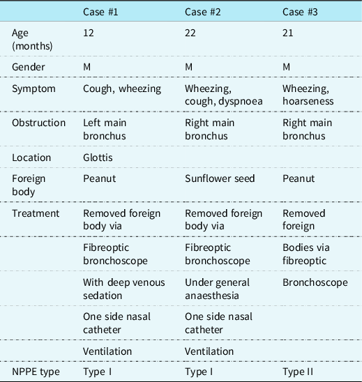
NPPE is classified into two types, that is, type I and II. Reference Bisinotto, Cardoso Rde and Abud10 Type I occurs, generally in adult, when the patient has increased negative intrathoracic pressure due to inspiration against a closed glottis, while type II is common in children who have chronic upper airway obstruction and sudden drop of intrathoracic pressure with acute removal of the chronic airway obstruction. Reference Bisinotto, Cardoso Rde and Abud10 In the past, while type II NPPE in the children caused by airway foreign bodies has been reported as case reports, type I NPPE in the children has not been reported. In this article, two cases of type I NPPE and one case of type II NPPE in the children (Table 1), and the difference in pathogenesis as well as location of the foreign bodies in the two types were discussed.
Characteristics of the three cases with negative pressure pulmonary oedema

Case report
Case 1
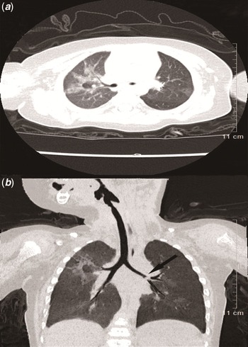
A 1-year-old boy visited our hospital with an irritating cough followed by wheezing one day after eating “peanut porridge.” On arrival, his respiratory rate was 46 breaths/minute, oxygen saturation was 94%. He was in consciousness, shortness of breath and in use of accessory muscles, but no cyanosis in the lips. Wheezing sound in the left lung and blistering sound in the right lung were auscultated. Chest CT showed blockage of the distal airways in the left main bronchus, obstructive emphysema in the left lung and multiple patchy high-density shadows in the right lung. He was diagnosed as foreign body obstruction in the left main bronchus complicated with type I NPPE. A piece of peanut fragment was successfully removed from the lower lobe via fibreoptic bronchoscope with deep venous sedation of propofol and one side nasal catheter ventilation, which resulted in opening of the distal left main bronchus. After the procedure, the wheezing sound disappeared immediately and his breathing was stable. The blistering sound in the right lung, however, was not disappeared till 24 hours after the procedure. He was discharged 48 hours after the procedure without any respiratory symptoms or signs (Fig. 1).
Chest CT of the Case #1 before removal of the foreign bodies. Pulmonary emphysema caused by hyperventilation in the left lung was observed and patchy high-density exudation was observed in the right lung in ( a ). The location of the foreign body indicated by the arrow in ( b ).

Case 2
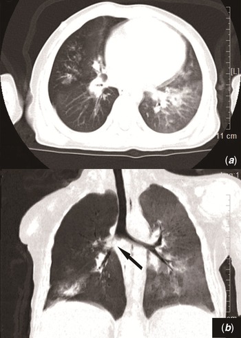
A 22-month-old boy was admitted to hospital with wheezing. Four hours before the admission, he suddenly had irritating cough, flushed face and dyspnoea when he ate sunflower seeds. His parents took out a small piece of sunflower seed from his mouth before coming to the hospital. When he arrived at the hospital, his respiratory rate was 48 breaths/minute and oxygen saturation was 93%. He was in consciousness, laboured breathing and in use of accessory muscles. Auscultation of wheezing and blistering sounds in both lungs was noticed. Chest CT indicated a foreign body in the right main bronchus and right middle lobe bronchus, obstructive emphysema in the right lung and multiple patchy and high-density shadows in both lungs. He was diagnosed as foreign body in right bronchus and type I NPPE. Under general anaesthesia and one side nasal catheter ventilation, a sunflower seed located inside the trachea below the glottis and a fragment of sunflower seed located at the beginning of the right main bronchus were removed via fibreoptic bronchoscope. The child was in stable breathing after the procedure. The blistering sound in both lungs disappeared 24 hours later and chest X-rays taken 48 hours later showed normal lung image (Fig. 2).
Chest CT of the Case 2. ( a and b ) showed the foreign bodies. Obstructive emphysema in the right lung, and high-density exudation in the lower lobe of the left lung were observed.

Case 3
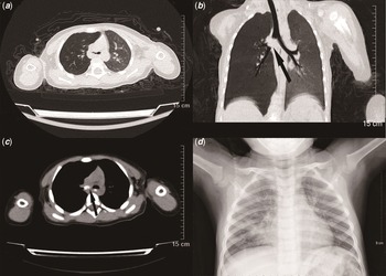
A 21-month-old boy had wheezing and hoarseness after eating peanuts half month ago. The symptoms were worsened for the last 24 hours before he was taken to the hospital. Physical examination showed that he had a laboured breathing and decreased respiratory sound in the right lung. Chest CT indicated a foreign body lodged in the right main bronchus causing emphysema in the right lung. Peanut was removed from the glottis and the opening area of right middle bronchus, respectively, via fibreoptic bronchoscope. After the procedure, the boy’s breathing was stable without wheezing. However, he developed shortness of breath after a few minutes of the procedure, and blistering sound was auscultated in both lungs, especially in the right lung. Chest X-ray showed patchy and high-density infiltration shadows in both lungs, especially in the right lung. Diagnosis of pulmonary oedema was considered, and thus, continuous positive airway pressure ventilation and furosemide were applied. After 24 hours of treatment, the boy was fully recovered and type II NPPE was confirmed (Fig. 3).
Chest CT and X-ray images of the Case #3. ( a ) showed obstructive emphysema in the right lung. ( b and c ) showed the location of foreign body in the right main bronchus indicated by the arrows. ( d ) showed high-density exudation in both lungs after removal of the foreign body, which was taken 24 hours after treatment.

Discussion
NPPE is one of the non-cardiogenic pulmonary oedemas seen in clinic, and it may be classified into two types. Reference Bisinotto, Cardoso Rde and Abud10,Reference Bhattacharya, Kallet, Ware and Matthay11 Type I NPPE occurs, commonly seen in adult, when the patient has increased negative intrathoracic pressure due to inspiration against a closed glottis. Type I NPPE often occurs immediately after acute airway obstruction caused by acute laryngitis, epiglottitis or respiratory foreign body. In contrast, type II NPPE is common in children who have chronic upper airway obstruction and development of more positive intrathoracic pressure by creating positive pressures at the end of expiration. When the chronic obstruction is acutely removed, the intrathoracic pressure suddenly drops and this negativity of the pressure is transmitted to the pulmonary capillaries, resulting in extravasation of fluid into the interstitial tissue of the lung. Reference Bisinotto, Cardoso Rde and Abud10 Type II NPPE usually occurs after relief of chronic airway obstruction, for example, after the operation of tonsillar hypertrophy, adenoid hypertrophy and upper respiratory tract tumours. Reference Toukan, Gur and Bentur12,Reference Masuda, Asano and Tsuzuku13
The incidence of NPPE is between 0.1 and 11%, Reference Bhattacharya, Kallet, Ware and Matthay11,Reference Tebay, Bouti and Tebay14,Reference Xiong and Sun15 and it has been reported that the incidence of NPPE in China is as high as 9.6%. Reference Chen, Wang and Ma16 The most common cause of NPPE in adults is the laryngospasm after extubation under general anaesthesia, with an incidence of 3% Reference Bhattacharya, Kallet, Ware and Matthay11,Reference Toukan, Gur and Bentur12 ; but the cause of NPPE in children is different from that in adults, which is mainly due to airway obstruction. While the reported incidence of NPPE in the literature is largely different, through careful observation and analysis on the cases of airway obstruction by foreign bodies in children who admitted to our hospital in the past decades, we found that pulmonary oedema existed before the removal of foreign bodies as evidenced by chest X-ray although they had atypical respiratory symptoms, suggesting type I NPPE might be a common complication for children with airway foreign body obstruction.
The pathogenesis of NPPE is mainly related to the increase of intrathoracic negative pressure. However, there are obvious differences in pathogenesis and location between type I and type II NPPE. In the type I NPPE, due to acute airway obstruction, the human body inhales hard to relieve hypoxia, resulting in a significant increase in negative intrathoracic pressure, which could reach −24∼−50 cmH2O in children. The increase of negative intrathoracic pressure leads to the raise of blood volume reflux in the right ventricle, the right ventricular end-diastolic volume, the pulmonary blood volume and the hydrostatic pressure of pulmonary capillary, thereby disrupting the balance of Starling’s law, which causes the pulmonary capillary fluid leakage into the pulmonary interstitial tissues and alveoli, resulting in type I NPPE. Reference Masuda, Asano and Tsuzuku13 Respiratory obstruction not only increases negative intrathoracic pressure but also increases respiratory frequency. A large amount of gas is stored in the alveoli, that is, an increase in the end-expiratory lung volume induces a mild auto positive end-expiratory pressure, which offsets the excessive hydrostatic pressure of pulmonary capillary caused by the initial increase in negative intrathoracic pressure. Reference Masuda, Asano and Tsuzuku13 When the foreign body is removed rapidly, the pressure in the alveoli suddenly drops, and the auto positive end-expiratory pressure disappears. Excessive hydrostatic pressure of pulmonary capillary promotes pulmonary oedema, which results in type II NPPE. Reference Masuda, Asano and Tsuzuku13 However, this mechanism of the auto positive end-expiratory pressure is not suitable for atelectasis caused by complete obstruction of foreign bodies. The Case #3 of the current study showed that pulmonary oedema occurred after the removal of foreign body, which was consistent with the pathogenesis of type II NPPE as discussed above.
Previous studies have not discussed on the NPPE occurrence location, specifically, the difference of occurring places between the type I and type II NPPE. Here, we found that type I NPPE was prone to occur on the opposite side of the obstruction as well as the non-obstructive lung segments on the same side of obstruction, and that, in contrast to the type I, type II NPPE was prone to occur on both sides with more severe oedema on the obstructive side. In this regard, for the Case #1 of this study, an obstruction by foreign body occurred in the left main bronchus, but type I NPPE occurred in the right lung. The mechanism of type I NPPE occurrence at the non-obstructive side of the lung may attribute to the excessive venous reflux with increased negative pressure in the thoracic cavity resulting in flowing to the left and right pulmonary arteries with lacking of automatic positive end-expiratory pressure but increasing of the hydrostatic pressure in the pulmonary capillaries on the healthy side. Similar pathogenic mechanism may attribute to the occurrence of more severe type I NPPE on the obstructive side in the Case #2 of this study. In the Case #2, bilateral pulmonary oedema resulted from the foreign bodies located in right main bronchus and right middle lobe bronchus. Oedema on the left lung was diffuse and uniform, while it was focal on the right lung, which was possibly due to insufficient local automatic positive end-expiratory pressure that counteracts the high capillary hydrostatic pressure.
It has been reported that hypoxia, inflammation and high hydrostatic pressure of pulmonary capillaries may contribute to the rupture of alveolar-capillary membrane and impairment of the barrier function, the increase of permeability and the occurrence of pulmonary oedema. Reference Bhattacharya, Kallet, Ware and Matthay11,Reference Toukan, Gur and Bentur12,Reference Bhaskar and Fraser17 Consistently, in type I NPPE as seen in the Case #1 of this study, lung of the obstructive side is overventilated, end-expiratory volume increased, and the ventilatory blood flow is out of proportion. The pathologic alteration of ventilation versus blood flow in the lung led to more severe hypoxia and increased capillary permeability in the contralateral side which resulted in pulmonary oedema in the lung of the contralateral side.
Diagnosis of NPPE mainly depends on the history of foreign body inhalation, clinical manifestation and imaging features of pulmonary oedema. Reference Toukan, Gur and Bentur12,Reference Masuda, Asano and Tsuzuku13,Reference Xiong and Sun15,Reference Bhaskar and Fraser17 Differential diagnosis between NPPE and the following diseases should be in consideration: cardiogenic pulmonary oedema caused by cardiac disease, low colloidal osmotic pulmonary oedema caused by hypoproteinemia, neurogenic pulmonary oedema caused by craniocerebral trauma or virus infection, allergic pulmonary oedema and aspiration pneumonia. None of the three children in this study had aforementioned diseases and were in good health before the airway foreign body obstruction.
Treatment for NPPE includes oxygen inhalation, CPAP ventilation, endotracheal intubation ventilator-assisted ventilation and, if necessary, diuretics to reduce pulmonary oedema. But application of diuretics and glucocorticoids has been reportedly controversial, Reference Toukan, Gur and Bentur12,Reference Xiong and Sun15 and thus, neither diuretics nor glucocorticoids was used in the current study. Prognosis of the disease is good and it is generally relieved within 12–24 hours after treatment.
Taken together, the incidence of type I NPPE is common, while type II NPPE is indeed rare, in children with airway foreign bodies. Pathogenesis and location of the two types of NPPE are different. In clinical practice, paediatricians should be aware of the risk of potential complications of type I and II NPPE when they treat and remove the foreign bodies from airways in children.
Acknowledgements
We appreciate the valuable support of Wei-Ran Dong in image data supply.
Financial support
This study was supported by Medical Science Research Project of Hebei Province (No. 20211000).
Author contributions
QL collected the data, draughted, and edited the manuscript. LZ analysed the images and designed the study. All the authors reviewed and approved the final manuscript.
Conflict of interest
The authors have no conflict of interests to disclose.
Ethical standards
The study was approved by the Ethics Committee of Children’s Hospital of Hebei Province (No. 187).