1. INTRODUCTION
Pulsed laser irradiation of a solid target generates transient plasma of very short temporal existence with the characteristic parameters that depend on the irradiation conditions such as laser fluence, wavelength, pulse duration, and ambient conditions. After production plasma expands at supersonic velocity and its parameters vary drastically with axial and radial distance from the target surface. Besides several applications in various research fields such as material growth and processing, synthesis of nano-particles, elemental analysis of multi-component materials (Braren et al., Reference Braren, Bukoski and Norton1993), laser produced plasma is also a potential source of highly charged ions for accelerators and ion implanters (Sharkov & Scrivens, Reference Sharkov and Scrivens2005). Several laser based ion sources have been designed and extensive experimental work has been reported to optimize their performance (Kashiwagi et al., Reference Kashiwagi, Hattori, Hayashizaki, Yamamato, Takahashi and Hata2004; Yeates et al., Reference Yeates, Costello and Kennedy2010; Alekseev et al., Reference Alekseev, Balabaev, Vasilyev, Satov, Savin, Sharkov, Shumshurov and Roerich2012; Zhao et al., Reference Zhao, Zhang, Jin, Liu, Wang, Sun, Zhang and Zhao2016). Laser ion source has the capability to vaporize and ionize any solid material and can provide large peak ion current. But its major drawback is large ion energy spread (Burdt et al., Reference Burdt, Tao, Tillack, Yuspeh, Shaikh, Flaxer and Najmabadi2010), which restrict the possibility of further ion acceleration by application of the electric field. The externally applied magnetic field may modify the characteristics of ions emitted from the laser plasma and help to establish better understanding of the ionization and acceleration mechanisms. In addition, the study of magnetic field interaction with expanding plasma is relevant to the pulsed laser deposition (Qingrun & Gao, Reference Qingrun and Gao1997; Tsui et al., Reference Tsui, Minami, Vick and Fedosejevs2002), extreme ultraviolet lithography (Harilal et al., Reference Harilal, Tillack, Tao, O'Shay, Paguio and Nikroo2006; Roy et al., Reference Roy, Hassan, Harilal, Endo, Mocek and Hassanein2014), laser induced breakdown spectroscopy (Shen et al., Reference Shen, Lu, Gebre, Ling and Han2006), inertial fusion (Lindl, Reference Lindl1995), and astrophysical plasmas (Pant, Reference Pant1994).
In recent years, numerous studies of laser–plasma interaction with applied radial and axial magnetic field have been reported. Wolowski et al. (Reference Wolowski, Badziak, Ivanova-Stanik, Parys, Stepniewski and Woryna2004) reported that the current density of W ions close to the magnetic field axis significantly increases by application of 1.2 T axial magnetic field. Kondo et al. (Reference Kondo, Kanesue, Tamura, Dabrowski and Okamura2010) demonstrated that the total ion current and yield of highly charged Ag ions increases with the application of 0.42 T axial magnetic at the target surface. Ikeda et al. (Reference Ikeda, Takahashi, Okamura and Horioka2016) investigated the behavior of Fe plasma in solenoidal magnetic field and showed that total ion charge increases with the increase of magnetic field. The spectroscopic measurements revealed that the presence of 0.3 T transverse magnetic field at the surface of Cu target leads to the significant increase of the intensity of some neutral and ion emission lines (Pagano et al., Reference Pagano, Hafeez and Lunney2009). Harilal et al. (Reference Harilal, Tillack, O'Shay, Bindhu and Najmabadi2004) observed changes in Al plume dynamics, electron temperature, electron density, and enhancement in the ionization and spectral emission due to the presence of 0.64 T transverse magnetic field. Roy et al. (Reference Roy, Harilal, Hassan, Endo, Mocek and Hassanein2015) applied 0.5 T axial magnetic field at the surface of Sn target and noticed plasma plume focusing, increase of electron temperature, and decrease of electron density as a function of the distance from the target surface. Torrisi et al. (Reference Torrisi, Margarone, Gammino and Ando2007) passed expanding metallic (Al, Cu, Ta) plasmas through a cylindrical magnet and observed enhancement in the total ion yield, ion charge state and average ion energy. In this experimental arrangement a big cylindrical magnet (length = 14 cm, inner dia = 15 cm) was placed at a distance of 6.5 cm from the target surface. As a result the magnetic field at and close to the target surface, where all activity (ionization, acceleration, recombination, etc) takes place, was quite weak. Moreover, ion energy analyzer (IEA) measurement showing explicit evidence of ion charge state and ion energy enhancement of individual ion charge states were not reported.
In this work, we measured properties of Cu ions emitted from the laser produced plasma without and with axial magnetic field applied in the vicinity of the target surface. Experiments are conducted in wide range of laser fluence (2–24 J/cm2) and the integrated ion yield, average ion energy, ion charge state distribution, and energy distribution of individual ion charge states are measured with the help of ion collector (IC) and time-of-flight (TOF) IEA. We observed significant effect of the magnetic field on these ion parameters. The initial plasma parameters estimated using experimentally measured ion properties are used to explain the effect of applied magnetic field on the ion charge and energy enhancement.
2. EXPERIMENTAL SETUP
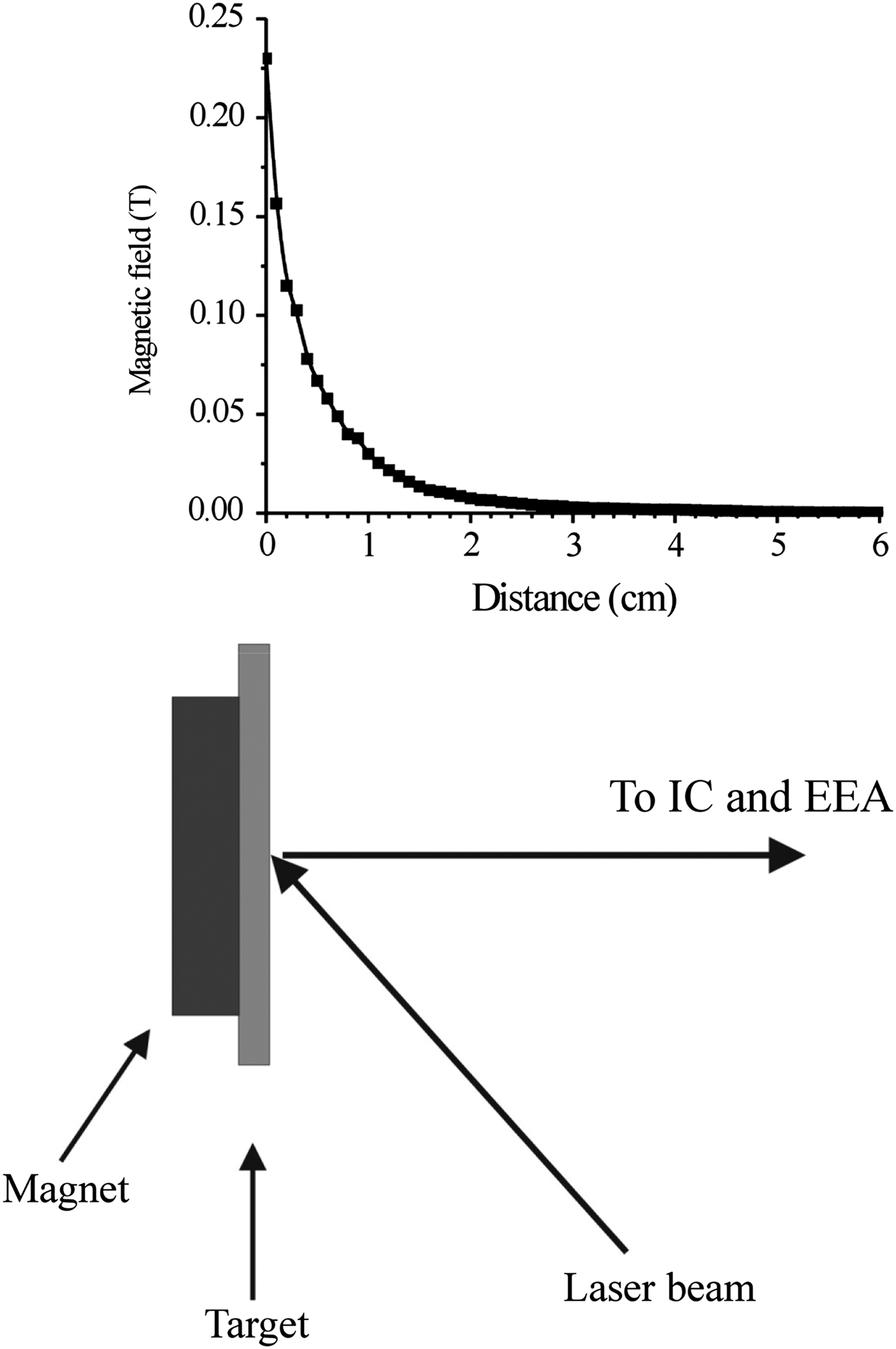
A block type NdFeB permanent magnet (1 × 1 × 0.5 cm3) was placed behind 2 mm thick 99.99% pure Cu target. Magnetic field at the target surface was 0.23 T, which exponentially decreases along the surface normal and drops down to zero at a distance of about 3 cm (see Fig. 1). The experimental setup used for the generation and diagnostics of laser generated ions is described in detail elsewhere (Ilyas et al., Reference Ilyas, Dogar, Ullah and Qayyum2011, Reference Ilyas, Dogar, Ullah, Mahmood and Qayyum2012). The experimental setup consists mainly of an Nd:YAG laser with associated beam delivery optics, ablation chamber, IC, TOF-IEA, ion detection, and data acquisition systems. The pulsed radiation from an Nd:YAG laser (wavelength = 1064 nm, pulse duration = 6 ns) is focused by an optical lens (focal length f = 50 cm) onto the surface of a Cu target. Laser beam enters the vacuum system through a quartz window and impinges on the target at an angle of 45° with respect to the target surface normal. The target can be moved in vertical direction with the help of a vacuum feedthrough that permits to change the laser spot position at the target. The area (7.85 × 10⁻³ cm2) of slightly elliptical laser spot on the target surface was inferred by analysis of the crater using optical microscope. The laser fluence at the target was varied in the range of 2–24 J/cm2 by changing the laser energy.
Schematic of the target-magnet arrangement and measured magnetic field profile along the target surface normal.

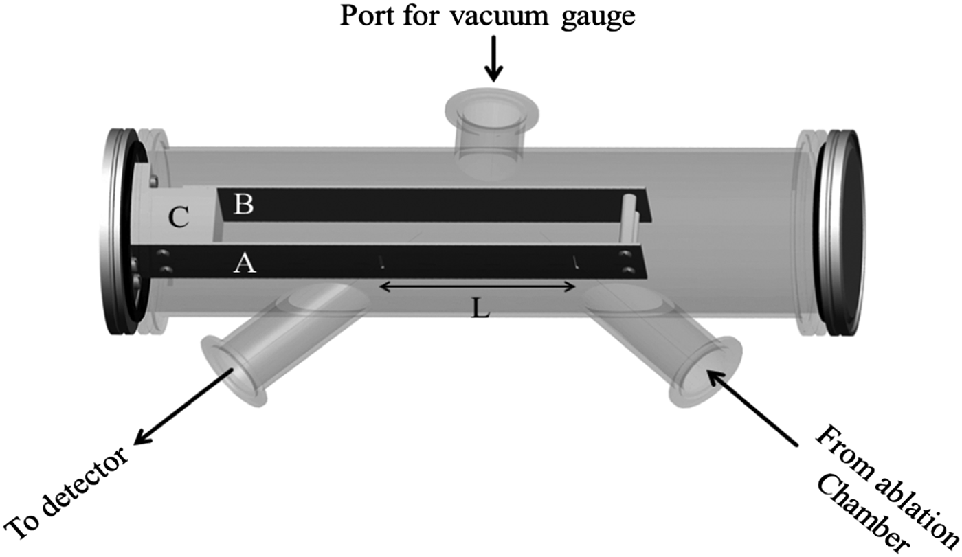
A removable cylindrical IC was placed along the target surface normal at a distance of 6.5 cm from the target. The IC has a circular aperture of 6 mm in diameter. While a grounded aperture of the same diameter was placed before the IC (Ilyas et al., Reference Ilyas, Dogar and Qayyum2013). The IC is biased with −50 V to repel electrons emitted from the laser plasma. The IC signal was determined from the voltage drop across a resistor with the help of a fast digital storage oscilloscope (1 GHz bandwidth). The IC measures a time-resolved ion signals during the plasma expansion (see Fig. 2), which was used to measure total ion charge and average ion energy. Also an electrostatic IEA was placed at a distance of 45 cm from target along surface normal. The sketch of IEA is shown in Figure 2. It consists of two parallel plates, the front plate was grounded and back plate was at an applied potential V a. Ions were admitted into IEA through a 2 mm wide entrance slit with some initial energy and charge state E i/q i e and subsequently decelerated by the constant electric field of −V a/d between the plates, where d the distance between the plates was 35 mm. In this configuration, only ions with a particular energy and charge state will leave the device through a 2 mm wide exit slit. The distance L between the entrance and exit slit of this device is 103 mm. The energy resolution of this device is given as (Hofer et al., Reference Hofer, Hass and Gallimore1999):
 $$\displaystyle{{\Delta E_{\rm i}} \over {E_{\rm i}}} \approx \displaystyle{{\sqrt 2 W} \over {2L}} = 1.4\% $$
where W is the width of entrance and exit slits, and L is the distance between entrance and exit slits. IEA was calibrated with variable (0.5–5 keV) energy Ar+ ion beam extracted from the duoplasmatron ion source and focused by an Einzel lens on the entrance slit of IEA. Technical details of duoplasmatron and ion current measuring setup is given elsewhere (Qayyum & Ahmad, Reference Qayyum and Ahmad1994). The ions were detected by means of a Channel Electron Multiplier (CEM) (Model KBL5RS, manufactured by Dr Sjuts optotechnik GmbH), whose signal was acquired by a fast digital storage oscilloscope (1 GHz bandwidth). A fast photodiode was used to trigger the oscilloscope at the time laser hits the target. The flight distance of ions from target to the CEM was 1.31 m. This whole arrangement provides energy resolved TOF mass spectra of the positively charged ions ablated by a laser shot. The Einzel lenses placed before and after the IEA are used to manipulate or optimize the signal of a specific ion charge state. But these Einzel lenses are placed at the ground potential during this study. The experiments were conducted in vacuum below 10−6 torr to minimize the charge exchange with the ambient residual gas.
Sketch of the ion energy analyzer (IEA). Here ‘A’ is front plate, ‘B’ is back plate, ‘C’ is ceramic insulating block and ‘L’ is distance between the centers of 2 mm wide entrance and exit slits.

3. RESULTS AND DISCUSSION
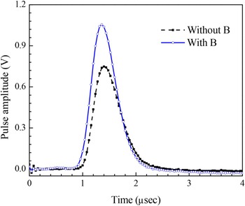
Typical TOF Cu ion spectra measured by IC without and with application of the axial magnetic field are shown in Figure 3. For this measurement, Cu target was ablated with the laser fluence of 5.48 J/cm2 and each spectra is the average of IC signal measured for three consecutive laser shots. Two very clear effects of the magnetic field on the ion signal are: (1) The peak ion signal increases up to about 33% when magnetic field is applied, and (2) the peak position and leading edge of the ion pulse is slightly shifted to earlier times when the magnetic field is applied. Similar ion spectra without and with application of magnetic field were measured for the range of laser fluence from 2 to 24 J/cm2. The total ion charge per pulse Q for various values of laser fluence was evaluated by integrating the corresponding TOF ion spectra as
 $$Q = \displaystyle{{\int_{t_{\rm i}}^{t_{\rm f}} {V(t){\rm d}t}} \over R}$$
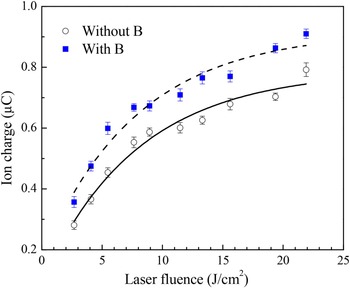
where V(t) is instantaneous voltage developed across the load resistance R, while t i and t f respectively are the time values corresponding to start and end of the ion signal. The total ion charge so obtained without and with application of magnetic field is plotted as a function of the laser fluence in Figure 4. The error bars indicate statistical error of total ion charge measured for three consecutive laser shots. The smooth curves are polynomial fit to the experimental data points. In agreement with the previous studies (Baraldi et al., Reference Baraldi, Perea and Afonso2011; Ilyas et al., Reference Ilyas, Dogar, Ullah and Qayyum2011), initially there is a rather fast rise of ion charge with laser fluence up to ~7 J/cm2, afterwards the increase in ion charge with laser fluence is rather slow and seems to reach saturation at higher values of the laser fluence. The saturation of ion charge at high-fluence values is presumably due to the reflection and/or absorption of laser radiation by the plasma itself. Plasma produced by leading edge of the laser pulse prevents laser radiation from reaching the target. Consequently, bulk of the energy in remaining part of the laser pulse will be reflected and/or absorbed by the ablated material plasma in front of the target surface. Therefore, the given amount of laser energy delivered at higher values of the laser fluence is less effective in causing vaporization as compared with the energy delivered at lower values of the laser fluence. However, the important result in present context is that the ion charge with magnetic field is constantly higher than the case when magnetic field is not applied.
TOF ion spectra measured by ion collector without and with application of magnetic field for the laser fluence of 5.48 J/cm2.

Integrated ion charge as a function of laser fluence without and with application of magnetic field.

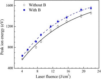
The shifting of peak position and leading edge of the ion pulse to earlier time indicates that the energy of ions that constitute leading edge of the ion pulse is increased by application of the magnetic field (see Fig. 3). The ion energies corresponding to signal onset, 50% of the rising edge and maxima of ion pulse were obtained from the TOF for both cases. The ion energy increase of 146, 71, and 42 eV was found for the ion corresponding to the signal onset, 50% of the rising edge and maxima of the ion pulse respectively. Previously, the contribution of ion charge states to various sections of the ion pulse has been obtained by the deconvolution procedure using the Coulomb-Boltzmann-shifted function (Caridi et al., Reference Caridi, Torrisi and Giuffrida2010, Reference Caridi, Torrisi and Cutroneo2013). These studies showed that the leading part of ion pulse comprises highly charged ion, whereas tail is mostly due to singly charged ions. Our results indicate that the Cu ions in leading part of the ion pulse, which are highly charged ions, have relatively higher energy rise as compared with the low charge state ions in trailing part of the ion pulse. The peak ion energy as a function of the laser fluence obtained from the ion pulses generated without and with application of the magnetic field is shown in Figure 5. The peak ion energy with magnetic field is constantly high throughout the laser fluence range investigated here.
The peak ion energy as a function of laser fluence obtained from time-of-flight (TOF) ion collector spectra without and with application of magnetic field.

The TOF-IEA measurements were performed to investigate the effect of axial magnetic field on the ion charge state distribution and charge state resolved ion energy distributions. Figure 6 shows typical TOF-IEA spectra recorded without and with magnetic field for five similar values of the laser fluence. The filtering criterion of the IEA was fixed at E/q = 502 eV/charge. The individual peaks represent the arrival of Cu ions of various charge states at detector meeting the filtering criterion of IEA. Let us first consider TOF-IEA spectra recorded without magnetic field (see first column of Fig. 6). The measurement was started from the laser fluence value of 2 J/cm2 but Cu1+ appeared at 2.68 J/cm2, which seems to be the appearance threshold of Cu1+ without magnetic field. As the laser fluence further increases highly charged Cu ions emerged and maximum attainable ion charge state gradually increases with the laser fluence. For the laser fluence of 2.68, 7.64, 11.46, 19.36, and 23.69 J/cm2, the number of detected positive Cu ion charge state were 1, 3, 5, 6, and 7 respectively. These results clearly indicate that, firstly, the laser fluence threshold for appearance of a particular charged state can be obtained by fine tuning of the laser fluence, and secondly, the ion charge state distribution of laser plasma can be changed by variation of the laser fluence. These observations are in agreement with our previously reported work on Ti and W targets (Ilyas et al., Reference Ilyas, Dogar, Ullah, Mahmood and Qayyum2012; Abbasi et al., Reference Abbasi, Hussain, Ilyas, Rafique, Dogar and Qayyum2015).
Time-of-flight ion energy analyzer spectra obtained by irradiating a Cu target with various values of the laser fluence. The ion spectra shown in first and second column are generated without and with application of magnetic field respectively. For these measurements filtering criterion of the ion energy analyzer was fixed at E/q = 502 eV/charge.

The TOF-IEA spectra recorded with magnetic field are shown in the second column of Figure 6. By application of magnetic field the appearance threshold of Cu1+ decreases down to 2.21 J/cm2. At the laser fluence of 2.68 J/cm2, which is the appearance threshold of Cu1+ without magnetic field, the intensity of Cu1+ with magnetic field is about 1.8 times higher than that of the case without magnetic field (see first row of Fig. 6). For the measurement performed with magnetic field at the laser fluence of 7.64, 11.46, 19.36, and 23.69 J/cm2, the number of detected positive Cu ion charge state were 4, 6, 7, and 9 respectively, which indicate that the number of detected ion charge states was increased when magnetic field is applied. The average ion charge state
 $\bar Z$
of Cu plasma was estimated from TOF-IEA spectra as (Burdt et al., Reference Burdt, Tao, Tillack, Yuspeh, Shaikh, Flaxer and Najmabadi2010),
 $$\bar Z = \displaystyle{{\mathop \sum \nolimits_n nI_n} \over {\mathop \sum \nolimits_n I_n}}$$
where n is the charge state and I
n
is the intensity of ions in respective charge state. For the laser fluence of 23.69 J/cm2,
 $\bar Z$
found to increase from 2.9 to 3.4 with the application of magnetic field. This finding is in line with the IC measurements that total ion charge increases by application of the magnetic field (see Fig. 4).
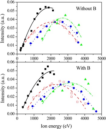
The charge state resolved ion energy distributions of first four charge states of Cu ions were measured by using the procedure reported elsewhere (Woryna et al., Reference Woryna, Parys, Wolowski and Mroz1996). Plenty of TOF-IEA spectra were recorded in a wide range of filtering criteria E/q = 70–1850 eV/charge by varying the back plate voltage of IEA. Laser fluence was kept fixed at 23.69 J/cm2 during this experiment. These spectra permit to obtain a plot between intensity of ion signal and ion energy for different charge states recognized in TOF-IEA spectra. Figure 7 shows plots of the ion intensity as a function of ion energy for first four charge states of Cu ions measured without and with magnetic field. In both cases, there is a regular shift of ion energy distributions toward higher values as the charge state increases. In addition, the energy distributions of individual charge states became broader and peaks of ion distributions moved toward higher energy values with the application of magnetic field.
Energy distributions of Cu1+ (■), Cu2+ ( ), Cu3+ (
 ), and Cu4+ (
 ) measured at the laser fluence of 23.69 J/cm2 without and with application of magnetic field.

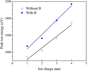
According to electrostatic model (Demtröder & Jantz, Reference Demtröder and Jantz1970; Margarone, Reference Margarone2007), which is normally used to explain ion acceleration in laser plasmas, when high-intensity laser beam interacts with a solid target a plume of high density ionized gas (plasma) is formed at the surface of the super-heated target in a time period shorter than the laser pulse duration. Afterward, plasma expands along target surface normal due to thermal and adiabatic processes. For the times slightly higher than the laser pulse duration (few tens of nanosecond in this case) plasma remains inside the Knudsen layer of the target and satisfy conditions of local thermal equilibrium. The plasma particles (electrons and ions) have similar temperatures and hence similar energies but due to huge mass difference electron velocities are much higher than that of ions. As a result two plasma regions of the opposite charges are created inside the Knudsen layer. This charge separation establishes an equivalent potential, V 0 that is applied over a distance of the order of Debye length and accelerates ions in the direction of plasma expansion. The peak ion energies, obtained by fitting of the data shown in Figure 7, are plotted versus charge state in Figure 8. The peak ion energy rise proportional to ion charge state is due to the existence of equivalent potential V 0 inside the non-equilibrium plasma, which accelerate ions proportional to their charge state. The equivalent potentials obtained from slopes of the straight lines, shown in Figure 8, were 273 and 341 V for the case without and with magnetic field respectively. Another important observation from Figure 8 is that the energy of singly charge Cu ions is much larger than the equivalent accelerating potential that exits inside the plasma, which indicates that ions possess energy in addition to that acquired during Coulomb acceleration. The additional ion energy gain is presumably due to thermal and adiabatic processes. It has been reported (Kelly, Reference Kelly1992; Margarone, Reference Margarone2007) that ion energy at a large distance from the target (or at a time much longer than laser pulse duration) is given as E ion = E C + E T + E A, where E C, E T, and E A is coulomb, thermal and adiabatic energy of the ions respectively. Our results indicate that the sum of thermal and adiabatic energies (E T + EA) of single charge ions is quite high as compared with their coulomb energy. But to the best of our knowledge, the quantitative information about E T and E A is not available for comparison.
Peak ion energies of first four charge states of Cu ions as a function of the charge state measured without and with application of magnetic field at the laser fluence of 23.69 J/cm2.

The Debye length λD can be calculated using the expression valid under local thermal equilibrium (LTE) condition as (Doggett & Lunny, Reference Doggett and Lunny2009),
 $${\rm {\rm \lambda}} _{\rm d}(m) = \sqrt {\displaystyle{{{\rm \varepsilon} _0kT_{\rm e}} \over {e^2n_{\rm e}}}} $$
where T e is the electron temperature, n e is the electron density of laser generated plasma, ε0 is the permittivity of free space and e is the elementary charge. According to hydrodynamic model of laser–plasma interactions (Puell, Reference Puell1970), T e can be estimated from the ion properties through the following expression derived for the similar parameter regime,
 $$E_{\rm a} = C(\bar Z + 1)T_{\rm e}$$
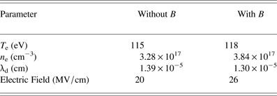
where E a is the average ion energy far from the laser plasma source and factor C depends on the nature of plasma expansion (for isothermal expansion C = 5). As discussed in recent literature (Burdt et al., Reference Burdt, Tao, Tillack, Yuspeh, Shaikh, Flaxer and Najmabadi2010), E a can be obtained by taking average of the peak ion energies of various charge states shown in Figure 7. The electron temperatures estimated using Eq. (5) were 115 and 118 eV for the plasma without and with magnetic field respectively. The plasma ion density at the position of IC can be estimated as (Pagano et al., Reference Pagano, Hafeez and Lunney2009),
 $$n_{\rm i}(t) = \displaystyle{{I(t)} \over {Aev}}$$
where I is the ion current measured by IC, v is the ion velocity and A is the area of IC. Ion velocity was calculated by dividing the target-IC distance with TOF. At the laser fluence of 23.69 J/cm2, the measured ion velocities corresponding to the time of maximum ion flux were 6.66 × 106 cm/s and 6.87 × 106 cm/s for the case without and with magnetic field respectively. Considering 3D self-similar expansion of the plasma and neglecting ion recombination, the plasma ion density at any distance dʹ along target surface normal is given as (Pagano et al., Reference Pagano, Hafeez and Lunney2009),
 $${n}^{\prime}_{\rm i}(t{\rm ^{\prime}}) = n_{\rm i}(t)\left( {\displaystyle{d \over {d{\rm ^{\prime}}}}} \right)^3$$
where d is the distance of IC from target, which was 6.5 cm for this experiment. The calculated plasma ion densities at the distance of 1 mm from the target were 3.28 × 1017 cm−3 and 3.84 × 1017 cm−3 for the case without and with magnetic field respectively. Plasma can be assumed in quasi-neutral state because spatial scale length of the plasma is much larger than the characteristic length over which charges are electrostatically shielded. The condition of quasi-neutrality implies that n i ≈ n e. These values of electron densities are in good agreement with the literature for laser plasma produced under similar conditions (Harilal et al., Reference Harilal, Bindhu, Issac, Nampoori and Vallabhan1997; Torrisi et al., Reference Torrisi, Caridi, Margarone and Borrielli2008a ). According to McWhirter's criterion plasma will be in local thermal equilibrium (LTE) (McWhirter, Reference McWhirter, Huddlestone and Leonard1965) if
 $$n_{\rm e} \ge 1.6 \times 10^{12}T_{\rm e}^{1/2} (\Delta E)^3$$
where n
e (cm−3) is electron density, T
e (K) is electron temperature and
 $\Delta E\;(eV)$
is the maximum energy difference between the atomic (ionic) states expected to be in LTE conditions. Here we verify that plasma is in LTE: For the case without magnetic field T
e = 115 eV [estimated using Eq. (5)] and taking ΔE = 4.89 eV for CuI transition at 353.67 nm, for these values of T
e and ΔE Eq. (8) gives the critical electron density of 2.16 × 1017 cm−3. While the calculated electron densities at the distance of 1 mm from the target are 3.28 × 1017 cm−3 and 3.84 × 1017 cm−3 for the case without and with magnetic field respectively (see Table 1). This shows that McWhirter's criterion is satisfied for the plasma at and very close to the target surface.
Plasma parameters without and with magnetic field at the laser fluence of 23.69 J/cm2

The calculated values of plasma electron temperature, electron density, Debye shielding length and transient electric field along surface normal for both cases (without and with magnetic field) at the laser fluence of 23.69 J/cm2 are given in Table 1. The resultant electric fields inside the plasma without and with application of magnetic field were about 20 and 26 MV/cm respectively. These electric field values are only approximate because of the transient nature of the laser plasma. Nevertheless, our results indicate that the electric field developed in the laser plasma increases with the application of magnetic field. Previously, electric field has been estimated for 1064 nm laser produced plasma of various materials in the comparable laser fluence range as: 16 MV/cm for Al plasma at 23 J/cm2 (Caridi et al., Reference Caridi, Torrisi and Giuffrida2010), 32 MV/cm for Si plasma at 30 J/cm2 (Torrisi et al., Reference Torrisi, Caridi, Margarone and Borrielli2008a ) and 25 MV/cm for Ni plasma at 20 J/cm2 (Torrisi et al., Reference Torrisi, Caridi, Margarone and Giuffrida2008b ).
The influence of magnetic field on the ion trajectory can be estimated by comparing the cyclotron period of the ions with the time it spent to pass the magnetic field region. For instance the cyclotron period of Cu ions, at the magnetic field of 0.1 T, is about 36 µs. The average ion velocity measured by IC at the laser fluence of 23.69 J/cm2 is 6.87 × 106 cm/s. It shows that the ions will pass the magnetic field region in just 0.3 µs. This means that magnetic field has hardly any effect on the ions trajectories. Therefore, the increase of ion yield with application of magnetic field cannot be due to the radial magnetic focusing of the ions. On the other hand due to very short electron cyclotron period, the electrons are trapped in magnetic field nearby the target surface. The electron path length and consequently number of electron–ion interactions close to the target surface are significantly increased. The rise in number of electron–ion interactions boosts up the ionization processes, which is responsible for the observed increase in ion yields and ion charge states. As it is indicated above, the ion acceleration along target surface is due to the presence of self-generated electric field produced by the separation of electrons and ions clouds. The applied magnetic field most probably modifies the densities of electrons and ions clouds and their separation in such a way that self-generated electric field inside the plasma increases. This enhanced electric field is responsible for the increment of ion energy observed by application of the magnetic field.
4. SUMMARY OF RESULTS
The ion flux emitted from Cu plasma produced by 1064 nm Nd:YAG laser, without and with axial magnetic at the target surface, have been investigated in wide range of the laser fluence. We have shown that the presence of magnetic field at the target surface has strong influence on the emitted ion flux. The major experimental results of this study can be summarized as: (1) The integrated ion yield increases with the application of magnetic field; (2) threshold laser fluence for the appearance of Cu1+ decreases from 2.68 to 2.21 J/cm2 by application of the magnetic field; (3) for a given laser fluence the number of detected ion charge states increases with the application magnetic field; (4) the average ion energy increases and energy distributions of individual ion charge states become broader and shift toward higher energy values. The plasma parameters estimated from the experimental results have been used to draw rather clear picture of the ion flux and ion energy enhancement mechanism by magnetic confinement of the plasma. These results are quite important for fundamental understanding of the laser produced plasmas and may help to improve the characteristics of multiply charged laser based ion sources. In future, similar experiments will be performed with the application of variable magnetic field at the target surface.