Enlarged bronchial arteries or systemic-to-pulmonary collateral arteries have been described in a significant number of patients with cyanotic congenital heart disease.Reference Andrade and Leipsic 1 – Reference Kaufmann, Kan, Mitchell, Flaherty and White 4 In infancy, they do not cause clinical symptoms, although there are well-documented cases with significant left-to-right shunting resulting in pulmonary hyperperfusion, congestive left heart failure, and respiratory distress.Reference Andrade and Leipsic 1 , Reference Knauth, Marshall, Geva, Jonas and Marx 5 , Reference Snir, de Leval, Elliott and Stark 6 Haemoptysis, resulting from enlarged bronchial arteries or anomalous collateral arteriesa is a well-known late problem in the natural history of complex cyanotic cardiac malformations or chronic pulmonary disease. However, in infancy this complication is extremely uncommon.Reference Aghaji, Friedberg, Burlingame and Litwin 2 , Reference Knauth, Marshall, Geva, Jonas and Marx 5 , Reference Rauch, Rauch and Koch 7 We describe the case of a 6-month-old girl who developed pulmonary haemorrhage owing to systemic pulmonary collateral arteries following corrective surgery for tetralogy of Fallot with absent pulmonary valve.
Case report
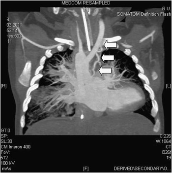
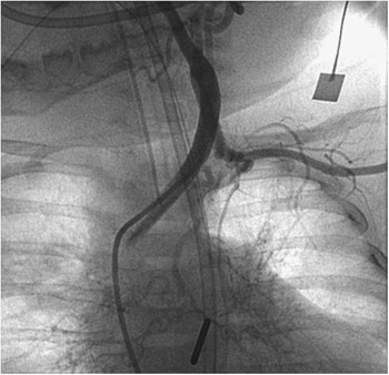
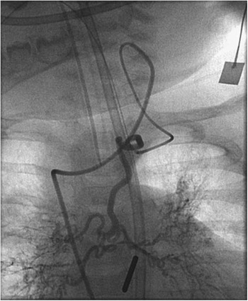
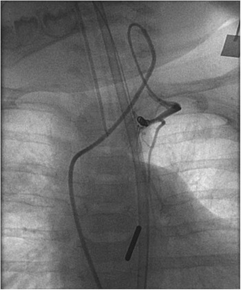
In our female patient, tetralogy of Fallot with absent pulmonary valve syndrome was diagnosed prenatally. Postnatal echocardiography revealed additional right aortic arch with cervical origin of the left subclavian artery. Genetic testing confirmed microdeletion 22q11.2. Since she was haemodynamically and respiratory stable, surgery was postponed. At the age of 6 months, she was admitted with severe respiratory distress requiring mechanical ventilation. Oxygen saturation (SaO2) at room air was 88%. High-resolution computed tomography revealed typical features of absent pulmonary valve syndrome with long-distance compression of left and right main bronchus by massive dilatation of both central pulmonary arteries. Following clinical improvement, surgical repair was performed including transatrial ventricular septal defect patch closure, patch enlargement of the right ventricular outflow tract, and reduction of right and left pulmonary arteries. Post-operatively, oxygenation (SaO2 96%) and ventilation improved dramatically. Echocardiography showed second-degree pulmonary regurgitation with half systemic right ventricular systolic pressure. Starting from post-operative day 11, there was progressive deterioration of the respiratory situation with varying atelectasis of both upper lobes and expectoration of increasing amounts of blood, which were first misinterpreted as being caused by possible upper gastrointestinal bleeding. After 4 weeks, she required reintubation and mechanical ventilation. Flexible bronchoscopy localised haemorrhage to the left upper lobe. Repeat high-resolution computed tomography demonstrated a good cardiosurgical result with significant reduction in diameter of both central pulmonary arteries, resulting in effective relief of bronchial compression. Furthermore, a significant systemic collateral artery originating from the left internal mammary artery perfusing large areas of both lungs (Fig 1), had increased dramatically in size as compared with the pre-operative high-resolution computed tomography. Arterial catheterisation was performed and the left innominate artery was entered from the right aortic arch. Owing to the cervical origin of the left subclavian artery, the origin of this vessel was shifted cranially close to the bifurcation of the carotid artery where the large systemic collateral artery originated from the left internal mammary artery supplying parts of both lungs (Fig 2). Using the 4-French catheter as a guiding catheter, selective catheterisation of this vessel was achieved with a 2.9-French microcatheter (GlidecathTM, Terumo Corp®, Tokyo, Japan; Fig 3) followed by successful coil occlusion with two platinum coils (MWCE18s-2.0-2-NESTER, Cook Medical Inc., Bloomington, USA) (Fig 4). The following day, she was extubated and discharged home after 3 weeks with oxygen saturations of 95%. Following an uneventful period, she was readmitted at the age of 9 months with severe respiratory tract infection requiring prolonged mechanical ventilation.
By flash computed tomography and contrast with three-dimensional reconstruction, the systemico-pulmonary collateral from the left subclavian artery to the left upper lobe is detected. Note the right aortic arch with cervical origin of the left subclavian artery.

Angiography into the right innominate artery shows cervical origin of the left subclavian artery.

Selective injection demonstrates a large systemic collateral artery originating from the left internal mammary artery supplying parts of both lungs.

Selective injection demonstrates complete occlusion of collateral vessel (Figure 3) following placement of 2 platinum coils.

After 18 days of respirator therapy, she relapsed with recurrent pulmonary haemorrhage originating again from the left upper lobe, as confirmed by bronchoscopy. Repeat cardiac catheterisation demonstrated two additional aortopulmonary collateral arteries originating from the left internal mammary and the left subclavian artery, both having increased in size from very tiny vessels at the first examination. The vessels were entered in the previous technique and subsequently occluded by four platinum coils each (MWCE 18s 2.0-2 hilal, Cook Medical Inc., Bloomington, USA). Pulmonary haemorrhage ended abruptly and the infant was extubated 2 days later.
Discussion
Tetralogy of Fallot with absent pulmonary valve is characterised by rudimentary pulmonary valve tissue, resulting in severe pulmonary stenosis and regurgitation with subsequent aneurysmal dilatation of the main pulmonary arteries.Reference Siwik, Preminger and Patel 8 Severe respiratory problems due to bronchial obstruction by dilatated and tortuous pulmonary arteries are frequent.Reference Knauth, Marshall, Geva, Jonas and Marx 5 , Reference Snir, de Leval, Elliott and Stark 6 , Reference Siwik, Preminger and Patel 8 However, significant systemic-to-pulmonary collateral arteries may represent an underappreciated additional cause of cardiorespiratory compromise.Reference Knauth, Marshall, Geva, Jonas and Marx 5 A large retrospective analysis described collateral arteries in 7 out of 50 patients with tetralogy of Fallot and absent pulmonary valve, adding to respiratory compromise by increased pulmonary blood flow with pulmonary congestion and left atrial enlargement and bronchial compression.Reference Knauth, Marshall, Geva, Jonas and Marx 5 In our patient, pulmonary haemorrhage contributed significantly to the secondary post-operative respiratory deterioration. Pulmonary haemorrhage, well described as a serious complication in the natural history of older patients with cyanotic congenital heart disease,Reference Andrade and Leipsic 1 , Reference Aghaji, Friedberg, Burlingame and Litwin 2 , Reference Hofbeck, Wild and Singer 3 , Reference Kaufmann, Kan, Mitchell, Flaherty and White 4 , Reference Snir, de Leval, Elliott and Stark 6 , Reference Rauch, Rauch and Koch 7 may present either with numerous small or with large systemic-to-pulmonary collateral arteries.Reference Aghaji, Friedberg, Burlingame and Litwin 2 , Reference Hofbeck, Wild and Singer 3 , Reference Rauch, Rauch and Koch 7 To the best of our knowledge, pulmonary haemorrhage has not been described yet as a complication of systemic-to-pulmonary collateral arteries in infancy. In our patient, interventional occlusion of the collateral vessels successfully terminated the haemorrhage on two occasions, as has been described in older patients.Reference Aghaji, Friedberg, Burlingame and Litwin 2 , Reference Veshti, Vida, Padalino and Stellin 9
Selective catheterisation of the collateral vessel was technically demanding because of the cervical origin of the left subclavian artery, an anomaly described exclusively in microdeletion 22q11.2, with the origin of the subclavian artery dislocated to the neck close to the bifurcation of the carotid arteries.Reference Rauch, Rauch and Koch 7 , Reference Yoon, Kim, Kim and Chung 10 Catheterisation of this retrograde vessel was achieved with a 2.3-French catheter (GlidecathTM, Terumo®) using a 4-French catheter (GlidecathTM, Terumo®) as a guiding catheter. The combination of these catheters allowed safe placement of several 0.018 inch platinum wires, despite the tortuosity of the collateral vessels.
The course of our patient confirms previous experience that in infants with cyanotic congenital heart disease the possibility of systemico-pulmonary collateral arteries should be taken into consideration, if a patient develops unexpected respiratory problems in the post-operative period.Reference Knauth, Marshall, Geva, Jonas and Marx 5 Whether earlier postnatal surgical repair may have prevented development of those clinically relevant collaterals remains speculative. Interventional coil occlusion is the treatment of choice, resulting in prompt clinical improvement of the patients.Reference Aghaji, Friedberg, Burlingame and Litwin 2 , Reference Kaufmann, Kan, Mitchell, Flaherty and White 4 , Reference Yoon, Kim, Kim and Chung 10 On the basis of the described experience, we would recommend performing careful screening by high-resolution computed tomography of the thorax in patients with absent pulmonary valve syndrome, if there is any suspicion of the presence of abnormal collateral arteries.