Introduction
In our experience, collectors often pass over lichens where a conspicuous fruiting structure is absent, possibly believing a reliable identification to be difficult. This view is shared in papers by other authors, especially those concerning genera such as Cryptothecia Stirton, Stirtonia A. L. Smith and some species of Herpothallon Tobler where asci are either absent, dispersed indiscriminately across the thallus or often in cryptic ascigerous zones (Makhija & Patwardhan Reference Makhija and Patwardhan1998; Cáceres Reference Cáceres2007; Wolseley & Aptroot Reference Wolseley and Aptroot2009). In the case of Stirtonia, prior to the publication of a world key (Aptroot Reference Aptroot2009), there had been a paucity of publications in existence that recorded Stirtonia species (Aptroot Reference Aptroot2009; Wolseley & Aproot Reference Wolseley and Aptroot2009), possibly for the above reason. The publication of a world key to any given genus frequently precipitates a flow of new species into the genus (Aptroot et al. Reference Aptroot, Sipman and Cáceres2013; Seavey & Seavey Reference Seavey and Seavey2014). However, in the case of Stirtonia, only one new addition to the genus has been described over a span of five years (Kalb et al. Reference Kalb, Buaruang, Mongkolsuk and Boonpragob2012). Although this may indicate an intrinsic rarity of species within the genus, it seems more likely to us that the genus is a victim of undercollecting due to the cryptic nature of its fruiting characteristics and/or thallus. This argument is strengthened by the finding of three new additions to the genus within a four hectare forest (this paper), when only 14 species are known worldwide.
After the transfer of described foliicolous Stirtonia to different genera (Lücking Reference Lücking2008), the genus was believed to be restricted to the Asian tropics (Makhija & Patwardhan Reference Makhija and Patwardhan1998). However, in more recent times S. neotropica Aptroot and S. schummii Aptroot have been described from the Netherland Antilles and the Seychelles, respectively (Aptroot Reference Aptroot2009), and two previously named species were discovered in south Florida, USA (Lücking et al. Reference Lücking, Seavey, Common, Beeching, Breuss, Buck, Crane, Hodges, Hodkinson and Lay2011). These, coupled with the three new species described from Everglades National Park in this paper, suggest the genus can be considered pantropical.
All three of the new species described herein were found in coastal forested habitat and, with one exception, within less than 200 m from the shore of Florida Bay. While Stirtonia byssoidea has also been collected from other locations and habitats, S. coei and S. latispora are currently known only from multiple collections at the site given above. By Florida standards, where periodic hurricanes rarely allow tree species to attain their full stature, the site has a moderately large mature arboreal flora but without a closed canopy which allows a large degree of filtered sunlight to penetrate. Understorey shrubs and forbs are essentially absent. The bark of Krugiodendron ferreum (Vahl) Urban (Rhamnaceae) was the favoured substratum (8 out of 27 aggregate Stirtonia collections), although 23 other arboreal species were present. That tree has smooth to furrowed bark which has a neutral or very slightly basic pH (F. Seavey & J. Seavey, unpublished data).
Materials and Methods
All collections were examined using standard stereoscopic and light microscope techniques. A Leica DFC295 compound microscope and a Leica S8APO inspection microscope were used to view hand-cut sections and thalline superficial structures, respectively. All macroscopic and microscopic images were captured via computer using Leica Application Suite V4.2.0 28 software. Ascigerous zone dimensions were determined from computer images using the software's measuring program. Measurements of internal structures were obtained from untreated water mounts. The software's automatic setting was employed and may have enhanced some of the images. No additional enhancement was used unless otherwise noted. Thin-layer chromatography (TLC) was carried out in accordance with Orange et al. (Reference Orange, James and White2001), using solvents A and C. Controls used were atranorin (Lecanora leprosa Fée), norstictic acid (Graphis librata C. Knight) and perlatolic acid [Canoparmelia caroliniana (Nyl.) Elix & Hale]. Spot test abbreviations used are C (sodium hypochlorite), K (potassium hydroxide), I (Lugol's solution 1%) and P (para-phenylenediamine). All collections were made by the authors within Everglades National Park and will be curated at the South Florida Collections Management Center (FNPS). They are corticolous unless otherwise noted.
The New Species
Individual colour images of the new species are available at: http://www.seaveyfieldguides.com/Lichens/
Stirtonia byssoidea F. Seavey & J. Seavey sp. nov.
MycoBank No.: 809720
Similar to Stirtonia alba but differing by its byssoid, thicker thallus and smaller ascospores.
Type: USA, Florida, Everglades National Park, Monroe County, Key Largo Ranger Station, 25°05′N, 80°27′W, corticolous on Guapira discolor in coastal hammock forest, 8 April 2014, F. Seavey & J. Seavey 12934E (FNPS—holotype; FLAS—isotype).
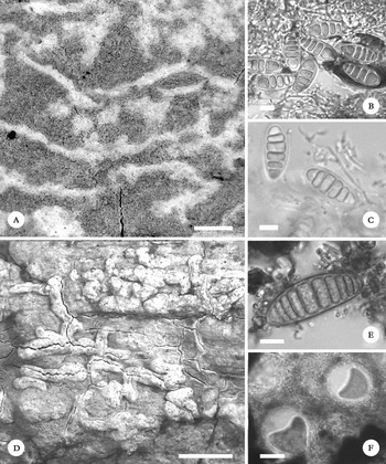
A–C, Stirtonia byssoidea; A, byssoid nature of the thallus and white ascigerous zones; B, megacephalic ascospores; C, rarely observed ascospores where the enlarged upper cell subdivides. D–F, S. coei. D, thallus with ascigerous zones raised significantly above the thallus surface; E, typical ascospore stained with Lugol's; F, asci showing the more or less globose configuration stained with Lugol's. Scales: A & D=1 mm; B, C & E=10 μm; F=100 μm.

Thallus corticolous, ecorticate, endophloeodal, byssoid, continuous, up to 8 cm diam., greenish white to greyish green, 100–200 µm thick, inspersed with calcium oxalate crystals 15–20 µm diam., I+ blue, often appearing pruinose due to projecting greyish hyphae, usually with a distinct white prothallus. Photobiont Trentepohlia.
Ascigerous zones indicated by white pruinose-like lines level or slightly raised above the thallus surface, branched or unbranched, 0·5–4·0×0·08–0·35 mm, I+ blue. Asci subglobose to globose, fissitunicate, 8-spored, not visible in surface view. Ascospores obovoid, 22–28×10–12 µm, 5–6-celled, upper cell often larger, occasionally subdividing into 6–7 cells.
Chemistry
K−, KC−, C−, P−. UV− but white ascigerous zones UV+ white. Perlatolic acid by TLC.
Etymology
The specific epithet refers to the distinct byssoid thalline surface.
Habitat and distribution
Stirtonia byssoidea is widespread in the south-east quadrant of Everglades National Park especially close to water, including Florida Bay. It is common and better developed in the coastal hammock discussed above, appearing on the bark of at least 11 different tree species. No host specificity was observed and the bark of the 11 species ranged from smooth to rough and somewhat furrowed.
Discussion
In the field, Stirtonia byssoidea can be recognized by its distinct greenish grey byssoid thallus usually appearing wispy with a hand lens. The white ascigerous zones may be barely discernible or encompass over one half of the thallus in larger well-developed specimens. Nearly all of the ascospores obtained from 19 collections of this taxon were 5-celled, with an enlarged upper cell in apparent full maturity (Fig. 1B). However, in one collection we observed the upper cell subdividing, creating 6–7 cells of roughly equal size (Fig 1C). Although this seems a rare event, the key below includes both configurations. Stirtonia alba Groenh. ex Makhija & Patw. also contains perlatolic acid and has ascospores with enlarged upper cells, but the latter are much larger (38–60×14–22 µm) while its thallus lacks the distinct byssoid nature. Stirtonia dubia A. L. Sm. has similar size ascospores but also lacks the byssoid thallus and contains no substances.
Selected additional specimens examined. USA: Florida: Miami-Dade Co., Goodrich Hammock, on Salix caroliniana, 2013, 6351; south of Old Tamiami Trail, on Salix caroliniana, 2005, 5961; Monroe Co., Key Largo Hammock, on bark of dead tree, 2014, 6347; Key Largo Hammock, on Gymnanthes lucida, 2014, 6348; Key Largo Hammock, on Sideroxylon salicifolium, 2014, 6349; Key Largo Hammock, on Sideroxylon celastrinum, 2014, 6350.
Stirtonia coei F. Seavey & J. Seavey sp. nov.
MycoBank No.: 809721
Similar to Stirtonia neotropica and S. curvata Aptroot but differing from both by containing no substances, lacking a more or less shiny thalline surface, strongly raised ascigerous zones and having an I+ blue reacting thallus.
Type: USA, Florida, Everglades National Park, Monroe County, Key Largo Ranger Station, 25°05′N, 80°27′W, corticolous on Krugiodendron ferreum in coastal hammock forest, 8 April 2014, F. Seavey & J. Seavey 12915E (FNPS—holotype; FLAS—isotype).
Thallus corticolous, ecorticate, thin, endophloeodal, matt to somewhat shiny, continuous, 8–12 cm diam., grey to greenish grey, 30–60 µm thick, inspersed with small calcium oxalate crystals 10–15 µm wide, I+ blue, with brownish white prothallus. Photobiont Trentepohlia.
Ascigerous zones indicated by white thick pruinose-like lines strongly raised above the thallus surface, branched or unbranched, 0·60–3·60×0·25–0·65 mm, I+ blue. Asci subglobose to globose, fissitunicate, 8-spored, not visible in surface view. Ascospores 40–45×15–16 µm, 10–12-celled, cells isolocular or mid cells slightly larger.
Chemistry
K−, KC−, C−, P−. UV−. No substances by TLC.
Etymology
The name commemorates the untiring efforts of Ernest F. ‘Tom’ Coe, who first advocated the creation of Everglades National Park in 1928 and then fought valiantly until its fruition in 1947. He is recognized as the single most important driving force for the park's establishment.
Habitat and distribution
Stirtonia coei is common in the coastal hammock mentioned above. It occurs in sunnier locations than the above species and well back from the shoreline. Although locally common, it is currently known only from this location but the same comments given for S. latispora (below) could apply here also. Its position in the hammock might indicate an intolerance of even the finest salt spray.
Discussion
Stirtonia coei is an easily overlooked species as the grey thallus blends in cryptically with its substratum. In many cases it appears as a grey stain on grey bark and, unless the white ascigerous lines are abundant, it will likely be missed. In the Aptroot key it would key out at couplet 12 that includes S. neotropica and S. curvata. It differs from the latter by its I+ blue thallus, wider, more strongly raised ascigerous zones, and lack of substances. Stirtonia neotropica has smaller ascospores, an I− ecrystallate thallus lacking raised ascigerous zones and contains gyrophoric acid. Stirtonia latispora, described below, also contains no substances but has larger and much wider ascospores, immersed white ascigerous zones and a distinct green epiphloeodal thallus containing larger calcium oxalate crystals. Stirtonia ramosa Makhija & Patw., described from the Andaman Islands, lacks thalline oxalate crystals, has ascospores with fewer septa and differs further by containing 2′-O-methylnorsuperphyllinic and 4′-O-demethylsuperconfluentic acids.
Selected additional specimens examined. USA: Florida: Monroe Co., Key Largo Hammock, on Krugiodendron ferreum, 2014, 6343E; Key Largo Hammock, on Reynosia septentrionalis, 2014, 6344E.
Stirtonia latispora F. Seavey & J. Seavey sp. nov.
MycoBank No.: 809722
Similar to Stirtonia curvata but differs by its larger ascospores with an enlarged mid cell, an I+ blue thallus and lack of perlatolic acid.
Type: USA, Florida, Everglades National Park, Monroe County, Key Largo Ranger Station, 25°05′N, 80°27′W, corticolous on Krugiodendron ferreum in coastal hammock forest, 25 April 2014, F. Seavey & J. Seavey 12933E (FNPS—holotype).
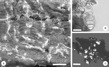
A–C, Stirtonia latispora; A, white ascigerous zones contrasting sharply with bright green thallus; B, ascospore showing the large middle cell; C, large calcium oxalate crystals restricted to non-ascigerous parts of the thallus. Scales: A=1 mm; B=20 μm; C=100 μm.

Thallus corticolous, ecorticate, epiphloeodal, matt to somewhat shiny, continuous, 2·5–3·5 cm diam., greyish green, 50–80 µm thick, inspersed with large calcium oxalate crystals 25–30 µm diam., I+ blue. Prothallus absent. Photobiont Trentepohlia.
Ascigerous zones in the form of white pruinose-like lines immersed or level with the thallus surface, irregularly branched, 0·65–2·75×0·15–0·30 mm, I+ blue. Asci globose, fissitunicate, 8-spored, but rarely more than 2–4 maturing; often visible in surface view as dark spots. Ascospores ellipsoid, 52–55×20–23 µm, 10–12-celled, one mid cell much larger.
Chemistry
K−, KC−, C−, P−. UV−. No substances by TLC.
Etymology
The specific epithet refers to the broad ascospores of this species.
Habitat and distribution
Currently known from three corticolous collections on Krugiodendron ferreum, Reynosia septentrionalis Urb. and Piscidia piscipula (L.) Sarg., all from the coastal hammock mentioned above. Several other similar coastal forests exist in the park, also within a few metres of the bay, but they are remote and difficult to access either by foot or boat. These may well be fertile areas for other populations of this species.
Discussion
This species is quite easy to recognize in the field as the thin bright white lines of the ascigerous areas stand out in sharp contrast to the greyish green thallus. The white of the ascigerous zones is a mix of asci, hyphae surrounding the asci and crystalline structures. It is only the hyphae which react I+ blue leaving the non-hyphal interspaces I−. The ascospore ontogeny of this species appears to us to be taxonomically significant. Multiple sections encountering several dozen ascospores show that the septation begins with a mid-septum followed rapidly by two additional septa subdividing each half leaving four more or less equal cells. Septation then continues from the ends toward the middle. Curiously, however, septation ceases in one of the mid cells while the other continues subdividing until as many as 12(13) cells are produced. Even post-mature ascospores retain the enlarged mid cell. As we observed no deviation from this pattern, the trait is included in both the protologue above and the key below. In the worldwide key of Aptroot, Stirtonia latispora would key out at couplet 12 along with S. curvata and S. neotropica. However, the former contains perlatolic acid and has ascospores that are shorter, narrower and isolocular. The latter contains gyrophoric acid and much smaller, narrower ascospores. The recently described S. rhizophorae Kalb & Mongkolsuk (Kalb et al. Reference Kalb, Buaruang, Mongkolsuk and Boonpragob2012) is also somewhat similar but has much larger ascospores with a larger number of septa and contains confluentic acid.
Selected additional specimens examined. USA: Florida: Monroe Co., Key Largo Hammock, on Piscidia piscipula, 2014, 6540E; Key Largo Hammock, on Krugiodendron ferreum, 2014, 12935E.
Key to Stirtonia species known from the Neotropics
-
1 Thallus without secondary metabolites ... 2
Thallus containing perlatolic, gyrophoric or 2′-O-methylperlatolic acids ... 4
-
2(1) Ascospores<35 µm long, with 8 cells or less ... S. dubia A. L. Sm.
Ascospores>40 µm long, with 10 cells or more ... 3
-
3(2) Ascospores 40–45×15–16 µm, cells isolocular, ascigerous zones strongly raised ... S. coei F. Seavey & J. Seavey
Ascospores 52–55×20–23 µm, mid cell distinctly enlarged, ascigerous zones immersed ... S. latispora F. Seavey & J. Seavey
-
4(1) Ascospores 22–28×10–12 µm, with enlarged upper cell, thallus containing perlatolic acid ... S. byssoidea F. Seavey & J. Seavey
Ascospores without enlarged upper cell ... 5
-
5(4) Ascospores 22–28×10–12 µm, 6–7-celled, thallus containing perlatolic acid ... S. byssoidea F. Seavey & J. Seavey
Ascospores>30 µm long, perlatolic acid absent ... 6
-
6(5) Thallus containing 2′-O-methylperlatolic acid, ascospores 50–110 µm long, 12–17-celled ... S. macrocarpa Makhija & Patw.
Thallus containing gyrophoric acid, ascospores 35–38 µm long, 11–12-celled ... S. neotropica Aptroot
We thank Dr James Lendemer and an anonymous reviewer for their insightful comments and suggestions. Everglades National Park provided support for this project.