Atrial septal defect is a common form of congenital heart disease, accounting for nearly 5–10% of all congenital cardiac defects in children. Reference Mitchell, Korones and Berendes1 Transcatheter closure of secundum atrial septal defects is considered the best intervention whenever applicable; it is regarded as superior to surgical atrial septal defect closure, especially in terms of patient morbidity. It shows fewer complications, entails a shorter hospitalisation period, requires less blood products and, in most countries, incurs considerably lower treatment costs. Reference Butera, Carminati and Chessa2,Reference Kim and Hijazi3 The Amplatzer septal occluder and similar self-centering devices are the most commonly used for transcatheter closure of large atrial septal defects. Reference Dalvi, Pinto and Gupta4
Whereas large atrial septal defects in adults are defined as an atrial septal defect larger than 20–25 mm, Reference Romanelli, Harper and Mottram5,Reference Varma, Benson and Candice Silversides6 there is no accepted definition for large atrial septal defect in children. There are also no conclusive results as to whether transcatheter closure is safe and feasible for large atrial septal defects in children. Reference Pinto, Jain and Dalvi7 For instance, Ohno et al. considered an atrial septal defect device/weight ratio larger than 1.5 to be a contraindication for transcatheter closure. Reference Ohno, Chaturvedi, Lee and Benson8 Conversely, Houeijeh et al. published a series of transcatheter closure of large atrial septal defects in young children with device/weight ratio more than 1.5. Cardiac erosion is a known complication after large atrial septal defect device closure. Reference Houeijeh, Hascoëtb and Bouvaistc9 The mortality associated with erosion is 19.6%; deficient aortic rim (present in 89% of the erosion cases) and a large device size to upstretched atrial septal defect ratio were identified as risk factors for erosion. Reference Amin10,Reference Di Bernardo, Fasnacht and Berger11
Moreover, quantitative evaluation of right ventricular performance post-transcatheter closure remains challenging due to right ventricle complex anatomy and structure. Reference Sheehan and Bolson12 Two standard modalities for better assessment of right ventricle function and volume are four-dimensional echocardiography and cardiac MRI. Four-dimensional echocardiography can overcome two-dimensional limitations by neglecting geometric assumptions and by utilising multiple images to reconstruct the right ventricle chamber. Reference Mor-Avi, Sugeng and Lang13 Myocardial strain and strain rate are more accurate than velocities as indices of ventricular contractility; by eliminating translational artefact, strain rate values are more dependent on pressure overload than volume overload. Reference Vitarelli, Sardella and Roma14 Four-dimensional echocardiography is a potential useful tool in studying the atrial septal defect device and its points of contact or pressure especially in relation to the aorta, aortic-mitral plane, pulmonary, and systemic veins. Reference El-Saiedi, Agha and Shaltoot15
Cardiac MRI is the gold standard for non-invasive measurements of right ventricle size and function. Reference Geva16 It is also used to assess the size and location of atrial septal defects, to guide the real-time positioning of an atrial septal defect device, to depict the position of the atrial septal defect device with respect to adjacent structures (the right pulmonary veins, the superior and inferior venae cavae, the coronary sinus, the mitral valve, the aortic valve and root) and finally, to assess any early sign of erosion. Reference Fratz, Chung and Greil17,Reference Lapierre, Raboisson, Miró, Dahdah and Guérin18
Aim of the study
The main aim of this study is to evaluate outcome of transcatheter closure of very large atrial septal defects by using two-dimensional conventional echocardiography, tissue Doppler imaging, four-dimensional speckle tracking imaging and compare these results with age-matched control group of 40 healthy children group (2).
The secondary aim is to compare these results to cardiac MRI values of left ventricle and right ventricle volumes and function, to evaluate the capability of cardiac MRI to accurately depict the position of the large atrial septal defect devices with respect to adjacent structures, and to assess the safety and efficacy of transcatheter closure of very large atrial septal defects in young children at mid-term follow-up.
Patients and methods
This is a case–control study of 80 children divided into two groups:
Group 1 – Forty children post-transcatheter closure of atrial septal defect (device/weight ratio more than 1.5 and mid-term follow-up of at least 12 months) at paediatric cardiology clinics in two centres, Cairo University Children Hospital and Beni Suef University Hospital.
Group 2 – Control group of 40 healthy age-matched children who only have the echocardiographic studies.
The study was approved by both universities’ Institutional Review Board, and all parents provided written informed consent.
Group (1) had the routine clinic follow-up. Echocardiography and Doppler examination were done in supine and left lateral positions using Philips EPIQ 7 C machine with probe X5-1, S8-3, or X7-2 MHz (multifrequency transducer) according to the age of the patient. Examinations consisted of:
-
(a) Conventional echo-Doppler: different Doppler velocity measurements of blood flow at the heart valves including M-mode measurements, two-dimensional, pulsed, continuous wave, and colour.
-
(b) Tissue Doppler: pulsed wave tissue Doppler imaging measures included systolic (S′) and diastolic (E′, A′, E′/A′ ratio), as well as the calculation of the global myocardial performance index (Tei index) of both right ventricle and left ventricle according to the guidelines of the American Society of Echocardiography. Reference Lang, Badano and Mor‐Avi19
-
(c) Speckle tracking imaging: four-dimensional images were obtained. Offline speckle tracking analysis performed using 4D Tom-Tec software analysis programme. Reference Baur20
Group (1) had cardiac MRI using a Philips Achieva, the Netherlands (1.5 T) and Siemens Aera, Germany (1.5 T); patients were scanned in supine position using a phased array cardiac coil. Children below 6 years old were sedated using chloral hydrate under supervision of a senior anaesthetist. The examination was explained to patients more than 6 years old. First, a survey reference scan was taken. This was followed by steady-state free precession sequence with parallel imaging: balanced fast field echo was acquired in two-chamber, four-chamber, short axis, and axial zero angle scans in 30 cardiac phases covering the whole heart. Coronal oblique views were done to assess the pulmonary veins and venae cavae. The mitral valve was evaluated by a four-chamber view. Imaging of the left atrial roof was acquired with a sagittal oblique view in the intersection with the middle of the atrial septum. For the aortic annulus and root, the sequence was applied twice – first in the coronal oblique plane along the axis of the proximal ascending aorta, followed by an axial oblique plane parallel to the axis of the valve. The goal was to show the relationship of the device to all the venous openings to the atria, as well as to the aortic valve’s annulus and root, the mitral valve, and the atrial roof. Reference Fratz, Chung and Greil17
Cardiac MRI interpretation: Images were reviewed independently by two senior radiologists with no prior access to the echo results. Measuring the right ventricular volumes was done by manually tracing the endocardial and epicardial borders of the ventricle in axial zero angle views, while the left ventricular volumes were measured by tracing the short axis images with recheck on the right ventricle. Trans-aortic and transpulmonary flow assessment (Q-flow) and the assessment of associated congenital anomalies and complications were also done. Reference Fratz, Chung and Greil17 We also looked for any evidence of protrusion of the device disks into the opening of the systemic or the pulmonary veins in the atria during the cardiac cycle. Finally, we attempted to identify the presence of contact between the device and both, the mitral valve (anterior leaflet or annulus or both) and the left atrial roof. Reference Lapierre, Raboisson, Miró, Dahdah and Guérin18
Statistical tests
Data were statistically described in terms of range, mean ± standard deviation, median, frequencies (number of cases), and percentages. To compare categorical data, Chi square test was performed. Student t test was used when comparing between two groups, while ANOVA test was used to compare between all groups. Finally, an exact test was used instead whenever the expected frequency was less than 5. p Values less than 0.05 were considered statistically significant.
Results
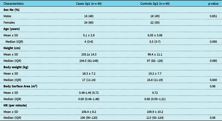
There was no statistical difference between the two groups as regard to sex, age, weight, height, body surface area, and heart rate (not significant p value) (Table 1).
Baseline characteristics of patients Gp1 and controls Gp2 at FU

Gp: group, FU: follow up, No: number, cm: centimeters, kg: kilograms, HR: heart rate, IQR: interquartile range.
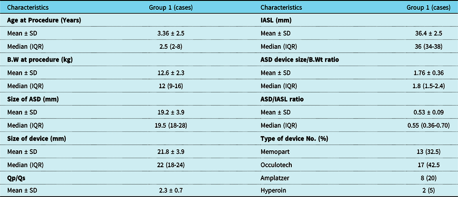
Group (1) data at the time of the procedure are summarised in Table 2, which includes patient age, weight, interatrial septum length, device type and size, and device size/weight and device size/interatrial septum length and Qp/Qs ratios.
Baseline characteristics of group (1)

ASD: Atrial Septal Defect, B.W: Body Weight, IASL: Interatrial Septal Length, No: number, mm: millimeters, HR: heart rate, Kg: kilograms, Qp: pulmonary flow, Qs, systemic flow, SD: standard deviation, IQR: interquartile range.
Group (1) follow-up at the time of investigations ranged from 1.4 to 10 years with a mean of 2.7 years and a median of 2.2 years.
Both groups were subject to two-dimensional conventional (echo-Doppler) as well as advanced echocardiography (tissue Doppler and speckle tracking imaging).
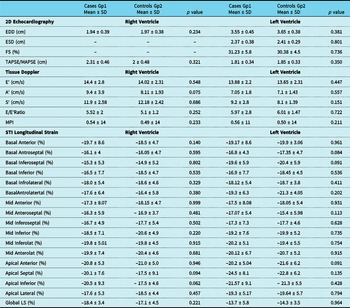
There was no statistical difference between the three methods as regards to both right ventricle and left ventricle parameters (Table 3).
Comparison of 2D echocardiography, TD and STI measurements of RV and LV between groups (1) and (2).

Gp: Group, 2D: two-dimensional, TD: tissue Doppler, LS: longitudinal strain, EDD: End diastolic diameter, ESD: End systolic diameter, TAPSE/MAPSE: Tricuspid/Mitral annular plane systolic excursion, MPI: Myocardial Performance Index, STI: Speckle tracking Imaging. p-value is considered significant <0.05.
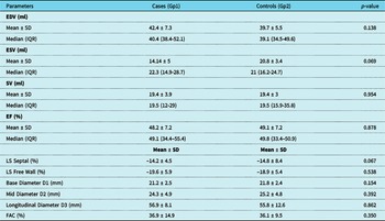
Four-dimensional echocardiography assessment of right ventricle volumes, ejection fraction, and longitudinal strain using Tom-Tec software analysis programme in both groups showed no significant difference between both groups. p Value is not significant for all studied parameters (Table 4).
4D Echocardiography of RV volumes, function, longitudinal strain, and dimensions comparison between the cases Gp1 and controls Gp2

Gp: group, RV: right ventricle, EDV: end diastolic volume, ESV: end systolic volume, SV: stroke volume, EF: ejection fraction, LS: longitudinal strain, FAC: fractional area change, IQR: interquartile range.
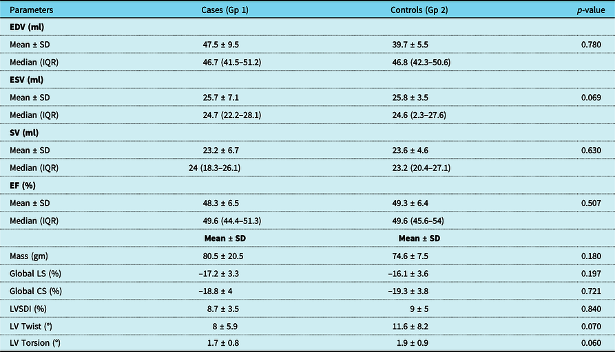
Four-dimensional echocardiography assessment of left ventricle volumes, ejection fraction, mass, strain, twist, and torsion using Tom-Tec software analysis programme in both groups showed no significant difference between both groups. p Value is not significant for all studied parameters (Table 5).
4D Echocardiography of LV volumes, function, mass strain, twist and torsion comparison between the groups 1 and 2

Gp: group, SD: standard deviation, IQR: interquartile range, EDV: end diastolic volume, ESV: end systolic volume, SV: stroke volume, EF: ejection fraction, LS: longitudinal strain, CS: circumferential strain, SDI: strain delay index.
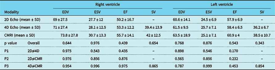
Group (1) had cardiac MRI to detect left ventricle and right ventricle volume and function, early signs of complications, and relation of the large atrial septal defect devices to adjacent structures. Table 6 shows comparison between two-dimensional echocardiography, four-dimensional echocardiography, and cardiac MRI with regard to end diastolic and end systolic volumes, ejection fraction, and stroke volume of both left ventricle and right ventricle. There was no significant difference between the three modalities’ measurements. p Value is not significant (Table 6).
Comparison of 2DE, 4DE, CMRI measurements between RV and LV.

E=echocardiography; RV=right ventricle; LV=left ventricle; EDV=end diastolic volume; ESV=end systolic volume; SV=stroke volume; EF=ejection fraction; CMRI=cardiac MRI.
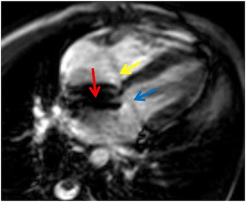
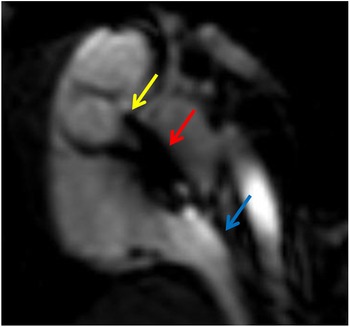
Cardiac MRI image analysis showed no encroachment on pulmonary or systemic veins. The devices touched the anterior mitral leaflet in five patients (Fig 1), but there was no effect on movement and no mitral regurgitation. There was extrinsic contact of the device with the aortic annulus in all examined patients (Fig 2). There was no device deformity nor pericardial effusion (as sign of early erosion) in any patient.
Steady state free precision (SSFP) Four chamber view showing large ASD device in place (red arrow). The ASD device is seen touching the anterior mitral leaflet (blue arrow), the yellow arrow refers to the tricuspid valve.

Steady state free precision (SSFP) oblique short axis view: showing the ASD device in place with no deformity (red arrow), the yellow arrow shows extrinsic contact of the large ASD device with the aortic annulus. The blue arrow points to the inferior venae cavae (IVC) opening into the the right atrium.

Discussion
Transcatheter closure of secundum atrial septal defects is now considered the first choice whenever applicable. It is superior to surgical atrial septal defect closure with regard to patient morbidity; it is characterised by fewer complications, shorter hospitalisation period, a reduced need of blood products, and in most countries considerably lower treatment costs. Reference Butera, Carminati and Chessa2,Reference Kim and Hijazi3 With progressive experience in transcatheter closure of atrial septal defects, device size is gaining more close attention, as excessively large devices are prone to mushroom deformities, encroaching on cardiac structures and possible serious complications such as cardiac erosions. Reference Dalvi, Pinto and Gupta4 However, Houeijeh et al. published a series of transcatheter closure of large atrial septal defects in young children with device/body weight ratio more than 1.5. This ratio of 1.5 was previously considered a contraindication for transcatheter closure of atrial septal defects in children. Reference Ohno, Chaturvedi, Lee and Benson8,Reference Houeijeh, Hascoëtb and Bouvaistc9
Our goal is to assess the safety and efficacy of transcatheter closure of large secundum atrial septal defects in children and to compare the patients on mid-term follow-up to matched healthy controls using multiple measurement modalities including two-dimensional, advanced four-dimensional, and cardiac MRI. There are different definitions of large atrial septal defects in children, Reference Dalvi, Pinto and Gupta4,Reference Pinto, Jain and Dalvi7,Reference Houeijeh, Hascoëtb and Bouvaistc9 and few reports of transcatheter closure of large atrial septal defects in infants and children. Reference Wood, Holzer and Texter21 The device/body weight ratio in previous series of transcatheter closure of secundum atrial septal defects in small children was less than 1.5. Reference Abu-Tair, Wiethoff, Kehr, Kuroczynski and Kampmann22,Reference Wyss, Quandt and Weber23 In our study, analogous to Houeijeh et al., the atrial septal defect size mean was 19.2 ( ±3.9) and ranged from 12 to 30 mm, and the device/body weight ratio mean was 1.76 (±0.36) with a range of 1.5–2.5 . Reference Houeijeh, Hascoëtb and Bouvaistc9 Our study was at mean follow-up period of 2.7 years, a range of 1.4–10, and a median 2.2 years. This is the first report to our knowledge of mid-term follow-up of transcatheter closure of such large atrial septal defects by different modalities.
There were different device types in our study: Memopart (32.5%), Occlutech (42.5%), Amplatzer (20%), and Hyperion (5%) (Table 2), these devices are all self-centering Amplatzer like. The choice of the device was dependent on the availability at the time of the procedure. There was no difference between any of the devices used on FU, in accordance with Faccini and Butera study which showed no difference between different device in transcatheter closure of atrial septal defects. Reference Butera and Faccini24
Two-dimensional, tissue Doppler, and speckle tracking imaging echocardiography of right ventricle and left ventricle showed no difference between patients at mid-term follow-up and healthy matched controls as shown in Tables 3 and 4. There was no significant difference as regard two-dimensional echocardiography measurements, fraction shortening, tricuspid annular plane systolic excursion, mitral annular plane systolic excursion, E, A, S (waves), E/E’ ratio, myocardial performance index, longitudinal strain (septal, mid, basal), and global strain. This proves return of right ventricle and left ventricle to normal relatively early despite large atrial septal defects in young children. This is in accordance with Ozturk et al who noted significant decrease in right ventricle diameter and significant increase in right ventricle myocardial performance index and right ventricle longitudinal strain and no significant change in left ventricle size 1 month following transcatheter closure of atrial septal defects. Reference Ozturk, Ozturk and Zilkif25
In our study, four-dimensional echocardiography assessment of left ventricle volumes, ejection fraction, mass, strain, twist, and torsion using Tom-Tec software analysis programme in both patients and healthy control groups showed no significant difference (p value not significant) for all studied parameters as shown in Table 5, despite large Qp/Qs and volume overload prior to intervention. This is in accordance with Teo et al. who showed a significant reduction in right ventricle volumes at 6 months post-atrial septal defect closure (p < 0.0001) and right ventricle ejection fraction was significantly increased (p = 0.025). Reference Teo, Dundon and Molaee26 There was a significant increase in the left ventricular volumes (p = 0.003 and p = 0.016), that is, return of both right ventricle and left ventricle volumes and function to normal. Reference Teo, Dundon and Molaee26 In contrast to our study, in Veldtman et al.’s study of 40 patients aged 20–71 years (median 38), only a third of the patients demonstrated persistent right ventricle enlargement at 1 year follow-up. Reference Veldtman, Razack and Siu27 This emphasises that late device closure in adult patients leads to incomplete remodelling and confirms the need for early closure of atrial septal defects in childhood.
Cardiac MRI is a valuable imaging modality for precise morphologic and functional information before and after transcatheter closure of atrial septal defects. It detects contact of the device with adjacent structures, early pericardial effusion plus the volume and function of right ventricle and left ventricle. Reference Lapierre, Hugues, Dahdah, Déry, Raboisson and Miró28,Reference Weber, Weber and Ekinci29 There are many reports that oversizing of the atrial septal defect device is the culprit, leading to erosion and perforation of the atrial wall. Reference Amin10,Reference Divekar, Gaamangwe, Shaikh, Raabe and Ducas30 Large atrial septal defect devices in young children lead to significant protrusion and contact with the adjacent structures, especially the left atrial roof and the aortic root which were observed in 76 and 100% of patients, respectively, in Lapierre et al study. Reference Lapierre, Hugues, Dahdah, Déry, Raboisson and Miró28 Most of the complications from erosion occur within 1 year of device closure, but more than 10% of the cases of erosion occurred more than 1 year after device closure. Reference McElhinney, Quartermain, Kenny, Alboliras and Amin31 Our study median follow-up is 2.2 years which ensured that there are no signs of late erosion.
In our study, there was no significant difference between cardiac MRI assessment of right ventricle and left ventricle volumes and function and assessment by two-dimensional- and four-dimensional echocardiography, in accordance with Sarwar et al. Reference Sarwar, Shapiro, Abbara and Cury32 This is different than Van der Zwaan et al. who proved that correlations between right ventricle volumes obtained by three-dimensionall echo and cardiac MRI (r = 0.71–0.97) were significantly better than the two-dimensional echo-derived correlations (p < 0.001). Reference van der Zwaan, Helbing and Boersma33 In our study, cardiac MRI showed no encroachment on pulmonary or systemic veins, the devices touched the anterior mitral leaflet in five patients (Fig 1), but there was no effect on movement and no mitral regurgitation. This is in accordance with Lapierre et al. who found contact of atrial septal defect devices with mitral valve in three patients, 9 years post-atrial septal defect device closure. Reference Lapierre, Hugues, Dahdah, Déry, Raboisson and Miró28 Our study also echoed a similar result that there was no encroachment on pulmonary or systemic veins as Lapierre et al. Reference Lapierre, Hugues, Dahdah, Déry, Raboisson and Miró28
In large atrial septal defects, there is typically deficient or absent retro-aortic rim. Reference Divekar, Gaamangwe, Shaikh, Raabe and Ducas30 The original design of the self-centering Amplatzer-like devices required that the disks of the device circumferentially straddle the rims of the septal defect. The use of the device in large atrial septal defects, where the rims of the defect are typically deficient in the retro-aortic region, required the straddling of the disks. Reference Lapierre, Hugues, Dahdah, Déry, Raboisson and Miró28 This explains the extrinsic contact of the device with the aortic annulus by cardiac MRI in all patients group 1 (Fig 2). Cardiac MRI also showed no device deformity and no pericardial effusion (as sign of early erosion) in any patient. Erosion is rare in children and this is explained by growth of the atria which gradually reduces the contact of the edges of the device with the free atrial walls and the aortic root providing better protection against erosion compared with adults. Reference Weber, Weber and Ekinci29 In contrast, a recent study has shown that the absence of the aortic rim, poor posterior rim consistency, septal malalignment, and dynamic morphology of the atrial septal defect in echocardiographic findings can significantly increase the risk of erosion. Reference McElhinney, Quartermain, Kenny, Alboliras and Amin31 This study further emphasise the importance of long-term follow-up as erosion risk is higher in cases of large atrial septal defects device closure like our series.
Limitations
The definition of a “large atrial septal defect” remains controversial. In fact, the larger diameter of the atrial septal defect is probably not accurate as the shape of the defect is often not round but rather oval or crescentic. However, this diameter criterion is widely used in most of the published studies. Reference Butera and Faccini24, The other limitation is the non-availability of pre-catheter advanced echo data.
Conclusion
Mid-term follow-up of children with very large atrial septal defect devices showed that right ventricle and left ventricle volumes and function returned to normal values. This was confirmed by comparing results with control group of healthy children with matched ages. There were no complications detected by echo or by cardiac MRI post-large atrial septal defect device closure, which proves safety of large devices in young children. There was also no significant difference (by p value) between four-dimensional and cardiac MRI in assessment of both right ventricle and left ventricle volumes and function. This favours the usage of 4-dimensional Echo in the assessment of right ventricle and left ventricle volumes and functions as it proves to be a less invasive, less time consuming, and more affordable method of reaching an accurate assessment of cardiac functions.
Acknowledgments
We would like to thank all participating patients and their families.
Financial support
This research received no specific grant from any funding agency, commercial or not-for-profit sectors.
Conflict of interest
None.
Ethical standards
The authors assert that all procedures contributing to this work complied with the Helsinki declaration of 1975, as revised in 2008. The Study was approved by Ethics Review Board in both of Cairo University and Beni Suef university.