Introduction
Domestic livestock and humans share many similar pathogens (Haydon et al., Reference Haydon, Cleaveland, Taylor and Laurenson2002). Pathogens that originate in domestic or wild animals and cause infections in humans are known as zoonotic organisms; pathogens that originate with humans and cause disease in animals are termed as anthroponotic organisms. Zoonotic diseases account for about 61% of all the pathogens infecting humans and therefore are an important public-health issue worldwide. Furthermore, as the global human population continues to increase, anthroponosis is now an emerging issue. Some human diseases currently pose conservation risks to wildlife (Taylor et al., Reference Taylor, Latham and Woolhouse2001). A high percentage (77%) of livestock pathogens and an even higher percentage (91%) of domestic carnivore pathogens infect multiple hosts, including wildlife (Cleaveland et al., Reference Cleaveland, Laurenson and Taylor2001). Understanding this group of pathogens is critical to public health, because infection of a wide range of hosts increases the likelihood that these organisms will emerge as novel infections in humans and domestic animals. Therefore, diseases at the wildlife–livestock interface, particularly those that are zoonotic, are of paramount importance and must be an area of focus for public health programs and surveillance for emerging infectious diseases.
For the diseases that wildlife and livestock share, examination of the factors affecting disease ecology is essential. For infection to occur in a host, the animal must accommodate a pathogen in its body, exploit host cells and evade the natural defense mechanisms of the host (Hudson, Reference Hudson and Hudson2002). To support the pathogen cycle that allows for effective transmission of the pathogen, a maintenance host is needed. Commonly, the pathogen will establish infection in the maintenance host and induce clinical disease, leading to maximum pathogen transmission. Another key ecological factor is the role of a reservoir host. Conventionally, a reservoir host can asymptomatically maintain a pathogenic organism and transmit it to other animals where disease occurs. A species’ population can also act as a reservoir as long as the reproductive number (R 0) of the infection exceeds 1; this occurs when a population is of sufficient size and enough susceptible animals are present over time to propagate infection (Haydon et al., Reference Haydon, Cleaveland, Taylor and Laurenson2002; Hudson, Reference Hudson and Hudson2002).
Occasionally, wildlife is the original source and maintenance host of the organism, and therefore the source of infection for livestock. Conversely, livestock can introduce disease to wildlife and although the wildlife species may not be a competent maintenance host, occasionally they are able to transmit the infection within and between species for a period of time; this is referred to as a spill-over host or bridge species (Daniels et al., Reference Daniels, Halpin, Hyatt and Middleton2007). A third situation involves dead-end hosts, infected animals in which the organism loses virulence over time and/or the animal dies; thus, the organism cannot be transmitted (Hudson, Reference Hudson and Hudson2002). Disease ecology is extremely complex, and in many cases the reservoirs and dead-end hosts have not been identified (Haydon et al., Reference Haydon, Cleaveland, Taylor and Laurenson2002).
The interface of wildlife and livestock is multi-dimensional. The two most critical factors are the location and time period in which diseases are exchanged at this interface. These interfaces are vast and include both direct and indirect contact. Commonly, the interface is at physical contact points where both populations access communal resources, such as rangeland, watering and feeding sites. This makes it difficult to prevent interaction between species without considerable spatial separation and infrastructure investment. Direct contact occurs through contaminated aerosols, feed, water or environment, while indirect contact occurs with infected flightless vectors, winged vectors or mechanical vectors. In situations where a natural barrier exists or where the contagion is released post mortem, wind, fomites, winged and mechanical vectors play a more important role in disease transmission (Bengis et al., Reference Bengis, Kock and Fischer2002; Hudson, Reference Hudson and Hudson2002).
Climatic events also affect disease transmission by shifting host and vector distributions, altering pathogen survival and impacting nutritional planes and immunities in a population during extreme climate conditions. As an example, the prevalence of Rift valley fever, a vector-borne disease, is dependent on precipitation that drives the emergence of mosquito populations. Conversely, diseases that are transmitted by direct contact (such as foot and mouth disease (FMD) and rinderpest) have a higher prevalence during dry seasons, when a dearth of water increases animal densities near watering holes. Climatic events also affect pathogen survival with cool and humid environments allowing pathogens to survive longer. Severe weather events can affect host immunity and nutrition if they interrupt forage availability and feeding patterns.
Undoubtedly, one of the most important factors driving disease emergence is the establishment of new interfaces between wildlife and livestock through intensification of farming, altering landscapes and demographics, regional conflict, political instability, irresponsible trade, animal translocations and/or animal or plant introductions. These mechanisms enhance disease transmission as a result of changing the spatial distribution of hosts and increasing contact rates between animal populations (Bengis et al., Reference Bengis, Kock and Fischer2002; Kruse et al., Reference Kruse, Kirkemo and Handeland2004). Where interspecies transmission is novel, it is described as pathogen jumping or host switching. This interspecies progression increases opportunity for pathogen evolution through a variety of mechanisms, e.g. genetic reassortment and recombination, occurring when different strains of the same microorganism are mixed together in one host (VandeWoude and Apetrei, Reference VandeWoude and Apetrei2006; Parrish et al., Reference Parrish, Holmes, Morens, Park, Burke, Calisher, Laughlin, Saif and Daszak2008).
Infectious diseases of livestock can cause major losses in trade income in livestock-producing countries in developing regions through denying access to profitable markets and threatening food security and livelihoods worldwide. The infectious diseases that are highly contagious and spread rapidly regardless of country borders are referred to as transboundary animal diseases (TADs) (Baldock et al., Reference Baldock, Forman, Geering and Taylor1999). In this review, we describe the important TADs at the wildlife–livestock interface. Diseases of livestock (including swine, poultry, large and small ruminants) in which wildlife is potentially involved in disease transmission cycles were included on the basis of being highly contagious and spreading rapidly across borders, causing major loss in livestock production through trade embargos or livestock mortality and morbidity.
Ruminant diseases
Viral diseases
Foot and mouth disease virus (FMDV), is highly contagious and infects cloven-hooved animals, causing vesicular lesions associated with the oral cavity, coronary bands of the hoof, interdigital skin and the udder. Adult animals generally recover from infection but naïve and young animals suffer high mortality (Thomson and Bastos, Reference Thomson, Bastos, Coetzer and Tustin2004). Undiagnosed disease can cause major constraints in international livestock trade and result in embargos by trade partners on livestock and livestock products (FAO, 2008). In wildlife species, under natural ecological conditions, FMD is rarely noticed but with altered landscapes and isolated fenced populations, introduction of virus can result in significant mortality and disease as seen in the case of impala (Aepyceros melampus) (Keet et al., Reference Keet, Hunter, Bengis, Bastos and Thomson1996).
There are seven immunologically distinct serotypes of FMDV. The serotypes and strains (over 60) vary with each geographic region, with serotype O being the most common worldwide. Cross-protection between strains can occur depending on their antigenic similarity (Davies, Reference Davies2002). Reservoir hosts are numerous and include domestic ruminants and pigs, and among wildlife, African buffalo (Syncerus caffer), reindeer (Rangifer tarandus tarandus) and wild boar (Sus scrofa) (Thomson et al., Reference Thomson, Vosloo and Bastos2003; Hargreaves et al., Reference Hargreaves, Foggin, Anderson, Bastos, Thomson, Ferris and Knowles2004; Thomson and Bastos, Reference Thomson, Bastos, Coetzer and Tustin2004; Jori et al., Reference Jori, Vosloo, Du Plessis, Bengis, Brahmbhatt, Gummow and Thomson2009). Furthermore, 60–70 species of wild animals have been reported to be susceptible to natural or experimental infection with FMDV, and on several occasions FMDV was isolated from some in the natural state (Pinto, Reference Pinto2004).
Wherever in the world FMDV has been eliminated from livestock, it also disappeared from wildlife in those regions (Thomson and Bastos, Reference Thomson, Bastos, Coetzer and Tustin2004). This is most likely due to insufficient population sizes of susceptible wildlife to maintain the virus in these situations. The exception is in parts of sub-Saharan Africa where African buffaloes serve as a reservoir for the SAT-type viruses. Serological surveillance in wildlife including the use of liquid-phase blocking enzyme-linked immunosorbent assay (Ceditest®, Prionics AG, Zurich) has shown these viruses to be widespread among African buffalo, impala and kudu (Tragelaphus spp.) across southern and eastern Africa (Hargreaves et al., Reference Hargreaves, Foggin, Anderson, Bastos, Thomson, Ferris and Knowles2004; Bronsvoort et al., Reference Bronsvoort, Parida, Handel, McFarland, Fleming, Hamblin and Kock2008; Jori et al., Reference Jori, Vosloo, Du Plessis, Bengis, Brahmbhatt, Gummow and Thomson2009) and gazelles in Mongolia (Nyamsuren et al., Reference Nyamsuren, Joly, Enkhtuvshin, Odonkhuu, Olson, Draisma and Karesh2006). In the last three outbreaks with major economic losses in Taiwan, United Kingdom and Argentina, wildlife were not associated with the introduction of disease (Ward et al., Reference Ward, Laffan and Highfield2007), and it is likely that wildlife-to-livestock infection is a rare event even in endemic settings.
Transmission occurs through direct or indirect contact with infected animals or contaminated fomites e.g. vehicle tires. FMDV is found in all excretions and secretions (saliva, milk, urine, feces and semen) from acutely infected animals and is easily aerosolized (Thomson and Bastos, Reference Thomson, Bastos, Coetzer and Tustin2004). Shedding of the virus varies by species with infected Bovidae shedding the virus for six months and smaller hoofstock from four to nine months. African buffalos acquire infection within a few months of life and can shed the virus for up to five years. The virus can persist in a herd for up to 24 years, and sexual transmission has been reported in buffalos (Pinto, Reference Pinto2004).
A historic approach in southern Africa to minimize transmission between wildlife reservoirs and livestock is the physical separation of the two groups with tall reinforced fencing and FMDV vaccination of cattle in localities adjacent to wildlife areas (Hargreaves et al., Reference Hargreaves, Foggin, Anderson, Bastos, Thomson, Ferris and Knowles2004). This large-scale intervention has generally worked in reducing the prevalence of FMDV. However, the use of fencing is controversial because fences may block migration routes, access to water and other resources needed for wildlife. The alternative to fencing is relying on vaccination exclusively; however, the virus has many antigenic variations often rendering vaccination ineffectual where multiple virus types frequently circulate (Ward et al., Reference Ward, Laffan and Highfield2007). Vaccination requires several inoculations to reach the required level of immunity and yearly vaccination is required to sustain protective immunity. Commodity-based trade, incorporating meat processing, rendering the product of negligible risk for FMDV (removal of lymph nodes and bones) shows considerable promise of opening up trade for areas of Africa where landscape precludes conventional control options (Thomson et al., Reference Thomson, Tambi, Hargreaves, Leyland, Catley, van't Klooster and Penrith2004).
Another important disease (although almost eradicated) is rinderpest, an acute, highly contagious, viral disease of cattle, domesticated buffalo and some species of wild artiodactyls. After the development of safe effective vaccines (Plowright, Reference Plowright, Gard, Hallauer and Meyer1968) and earlier failed attempts at virus eradication at the continental level (JP15), in 1992, the Food and Agriculture Organization of the United Nations (FAO) started the Global Rinderpest Eradication Programme with the goal of complete eradication by the year 2011. If successful, this would be the first worldwide eradication of an animal pathogen (Barrett et al., Reference de Vos, Turnbull, Coetzer and Tustin2006). There is only one serotype of the virus but there are three genetically distinct lineages (1, 2 and 3). Cattle, sheep, goats and yaks are susceptible and any cloven-hooved animal, domestic or wild can become infected (Roeder and Taylor, Reference Roeder and Taylor2002). African buffalo and other species are biologically competent to be reservoirs; but the belief is that wildlife cannot maintain the virus without a cattle source due to their relatively low population numbers; hence, they are not acting as a true reservoir (Kock et al., Reference Kock, Wambua, Mwanzia, Wamwayi, Ndungu, Barrett, Kock and Rossiter1999). However, wildlife is still important as vectors and where the virus is present, buffalos play a critical role in epidemics, spreading the virus across considerable distances over time (Kock et al., Reference Kock, Wamwayi, Rossiter, Libeau, Wambwa, Okori, Shiferaw and Mlengeya2006).
Transmission is by direct or close indirect contact with infected animals via aerosol transmission. Transmission by fomites is possible; however, the virus is readily inactivated by sunlight and drying (Barrett et al., Reference de Vos, Turnbull, Coetzer and Tustin2006). The disease history is one of devastating epizootics with the most significant being in Africa toward the end of the 19th Century (Spinage, Reference Spinage2003). The persistence of rinderpest in the absence of animal movement control and vaccination is remarkable. After centuries of adaptation, the virus strains evolved and many milder cases were recorded. This change complicated the final phase of eradication of the virus from Africa, and the last remaining strain was mild in cattle and cryptic.
Fortunately, with only cryptic forms of infection in cattle persisting in the 21st century, its expression in wildlife, which was more often severe, enabled rinderpest surveillance to identify remaining foci (Kock, Reference Kock2008). As the virus is most often introduced by infected animals, the control of outbreaks includes quarantine and movement control, euthanasia of infected and exposed animals, decontamination of infected premises and focal vaccination (Thomson and Bastos, Reference Thomson, Bastos, Coetzer and Tustin2004).
Like rinderpest, Peste des petits ruminants (PPR) is caused by a morbillivirus. It is a highly contagious viral disease, mainly in sheep and goats which are important livestock species for many of the world's poor (Barrett et al., Reference de Vos, Turnbull, Coetzer and Tustin2006). PPR is also an emerging disease of semi-captive wildlife (Abubakar et al., Reference Abubakar, Jamal, Arshed, Hussain and Ali2009) and recently free-ranging caprines in Pakistan (Qadir Shah Ghulam, personal communication 2009), buffalo in India (Govindarajan et al., Reference Govindarajan, Koteeswaran, Venugopalan, Shyam, Shaouna, Shaila and Ramachandran1997) and in captive and semi-captive gazelles (Gazelle dorcas and Gazelle thomsoni) in Saudi Arabia (Elzein et al., Reference Elzein, Housawi, Bashareek, Gameel, Al-Afaleq and Anderson2004). The virus has continued to spread throughout Africa, the Middle East and Asia since its discovery in 1942 in West Africa (Abubakar et al., Reference Abubakar, Jamal, Arshed, Hussain and Ali2009). Antibodies to PPR and rinderpest are cross-protective and vaccination for rinderpest can mask the presence of PPR (Barrett et al., Reference de Vos, Turnbull, Coetzer and Tustin2006). The primary hosts are goats and sheep, but camels appear to be highly susceptible; and although many wild ungulates have tested positive on serology, a wildlife reservoir has not been identified (Kock et al., Reference Kock, Wamwayi, Rossiter, Libeau, Wambwa, Okori, Shiferaw and Mlengeya2006).
The epidemiological role of wildlife in PPR circulation is unknown. Serological evidence for infection across a wide spectrum of artiodactyls, sero-conversion of asymptomatic buffalo across multiple ecosystems in Uganda between 2003 and 2004 and PCR evidence for virus presence in healthy buffalo in Côte d'Ivoire (Couacy-Hymann et al., Reference Couacy-Hymann, Bodjo, Danho, Libeau and Diallo2005) support a potential wildlife reservoir. The continued southern spread of PPR in African livestock, recent mortalities from PPR among wild ibex in South Asia (personal communication, 2009) and the increasing evidence for serious disease outcomes across a range of small and large wild ungulate species in the Middle East require greater international attention. This disease has the potential to have severe economic impacts and threaten the conservation of biodiversity.
Transmission is by close contact or inhalation of nasal and ocular secretions, saliva, urine, feces, milk and fomites (water and feed troughs and bedding). The virus does not remain viable in the environment for an extended time. The disease is frequently introduced by introducing infected animals into a herd; therefore, quarantine and testing are crucial. Infected and exposed animals are euthanized and cleaning and disinfection of infected premises are recommended (Rossiter, Reference Rossiter, Coetzer and Tustin2004). Vaccination is an effective control measure as immunity persists for at least 4 years and is possibly lifelong. To prevent infection in susceptible populations, it may help to prevent wildlife from having contact with sheep and goats, but more surveillance needs to be done on wildlife to identify their role and their transmission potential. Vaccination might also be possible in wildlife in the near future (Barrett et al., Reference de Vos, Turnbull, Coetzer and Tustin2006).
Another fatal disease in cattle is malignant catarrhal fever (MCF), caused by the ruminant gamma-herpesviruses alcelaphine herpesvirus 1 (AlHV-1) and ovine herpesvirus 2 (OvHV-2). These viruses cause inapparent infection in their reservoir hosts (wildebeest for AlHV-1 and sheep for OvHV-2), but are fatal, causing fever, inappetence and ocular and nasal discharge when they infect MCF-susceptible species, including cattle, cervids, bison, water buffalo and pigs (Meteyer et al., Reference Meteyer, Gonzales, Heuschele and Howard1989; Russell et al., Reference Russell, Stewart and Haig2009). MCF is a significant disease where reservoir and MCF-susceptible species mix and currently is a specific problem in cattle in Bali, Indonesia, bison in the United States and pastoralist cattle herds in eastern and southern Africa. Transmission of both strains of MCF to cattle occurs by contact with placentas at the time of birthing, mainly from wildebeest calves and lambs (Mushi and Rurangirwa, Reference Mushi and Rurangirwa1981). Control measures are to maintain separation of susceptible animals from infected animals. Cattle are kept separate from wildebeest at calving when the virus is present on pasture from placental contamination. Masai tribesmen delay the movement to pasture until the sun has dried the placenta of the previous night's wildebeest calving (P.B. Rossiter, personal communication 2005). To reduce spill-over into wildlife, bison, deer and other highly susceptible species should not be allowed to be near potentially infected sheep or wildebeest (Russell et al., Reference Russell, Stewart and Haig2009).
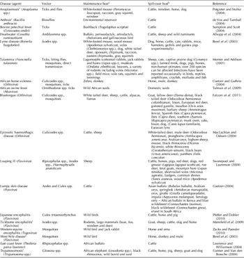
Probably the most ancient viral disease is rabies that is found worldwide. The primary hosts include carnivores, bats and cattle (Swanepoel, Reference Swanepoel, Coetzer and Tustin2004). Species-to-species transmission occurs when there is contact with infectious saliva through a bite, mucous membranes or breaks in the skin. All mammals are susceptible to rabies, but reservoir hosts are geographically dependent (Table 1). There are many wildlife vectors, and the disease normally is spread from bites; however, horizontal transmission has been reported in kudu antelope in southern Africa (Mansfield et al., Reference Mansfield, McElhinney, Hubschle, Mettler, Sabeta, Nel and Fooks2006). Respiratory transmission has been documented from bats to other species in enclosed areas, such as caves and houses. In South America, there are significant losses of cattle due to vampire bat bites. To prevent transmission in domestic animals, vaccination, nocturnal housing and the reduction of roaming are useful. Wild European red fox (Vulpes vulpes) populations have been successfully immunized with oral bait vaccines. There is some progress in wildlife vaccine use in the United States, but complex carnivore communities prevent its use in some continents like Africa due to difficulties in achieving adequate coverage of target species. Control programs are best targeted at domestic species (Rupprecht et al., Reference Rupprecht, Stohr, Meredith, Williams and Barker2001).
Infectious vector-borne diseases that are important at the wildlife–livestock interface

a Usually asymptomatic, reservoir and naturally infected; bsymptomatic, mortality and possible vectors, cZoonotic disease.
Bacterial diseases
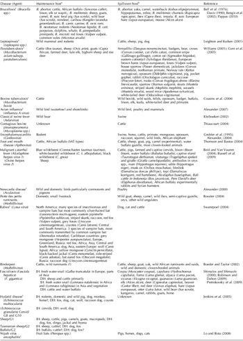
Brucellosis is spread worldwide and consists of six species, Brucella abortus, Brucella melitensis, Brucella suis, Brucella ovis, Brucella canis and Brucella neotomae. The most common species is B. abortus that primarily infects bovines causing abortion leading to economic loss and barriers to international trade. The infection is transmitted through contact with fetal membranes, lochia, post-parturient discharges and milk. However, in wildlife little is known about the occurrence of abortions due to brucellosis. B. abortus and B. suis have been isolated worldwide from a range of wildlife species (Table 2) (Davis, Reference Davis, Nielsen and Duncan1990; Pappas, Reference Pappas2010). Only a few true reservoirs can sustain infection with B. abortus, bison (Bison bison) and elk or wapiti (Cervis canadensis) in North America and African buffalo in southern Africa (Godfroid, Reference Godfroid2002). In South Africa, hippopotamus (Hippopotamus amphibious), zebra (Equus burchellii), eland (Taurotragus oryx), waterbuck (Kobus elipsiprymnus) and impala have tested positive serologically for brucellosis. Apart from B. abortus and B. suis, the other brucella strains are seldom reported in wildlife. Prevention of wildlife contacting livestock has proven to be effective in controlling the spread of brucellosis in South Africa (Godfroid, Reference Godfroid2002). Vaccines are still a work in progress as they do not have the same protection in wildlife but have shown promise in red deer (Cervus elaphus) (Godfroid, Reference Godfroid2002).
Infectious non-vector-borne diseases that are important at the wildlife–livestock interface

a Asymptomatic and reservoir; bsymptomatic, mortality and possible vectors, cZoonotic disease; DH, definitive host, IH, intermediate host.
Great economic loss due to chronic disease and death and trade restrictions in cattle can occur with bovine tuberculosis caused by Mycobacterium bovis (Pollock and Neill, Reference Pollock and Neill2002). Cattle are the primary host but many domestic and wild animals can also vector the disease. Brush-tailed opossums (Trichosurus vulpecula), badgers (Melus melus), bison, elk, kudu, African buffalo and white-tailed deer (Odocoileus virginianus) can all act as reservoir hosts There are numerous spill-over hosts, and species with large populations can become reservoir hosts, e.g. wild boar (Table 2) (Cousins et al., Reference Cousins, Huchzermeyer, Griffin, Van Rensburg, Kriek, Coetzer and Tustin2004; Corner, Reference Corner2006). Wild birds appear to be susceptible to M. bovis when challenged experimentally (Butler et al., Reference Butler, Fitzgerald, Berry, Church, Reed and Kaneene2001). Currently, there are active elimination programs in the Americas, Japan and New Zealand. The disease is widespread in Africa, Asia and the Middle East (de Lisle et al., Reference de Lisle, Bengis, Schmitt and O'Brien2002) and persists in parts of Europe e.g. United Kingdom, Spain and Portugal. Transmission between animals is by inhalation, ingestion or contamination of an open wound by infected material (respiratory secretions, feces, milk, urine, vaginal secretions or semen) (Cousins et al., Reference Cousins, Huchzermeyer, Griffin, Van Rensburg, Kriek, Coetzer and Tustin2004; Wilkinson et al., Reference Wilkinson, Bennett, McFarlane, Rushton, Shirley and Smith2009).
Test and slaughtering or segregation can control tuberculosis outbreaks. Affected herds should be re-tested periodically to eliminate shedding in cattle. In the final stages of elimination, surveillance by slaughter and disinfection may reduce the spread of the agent within the herd. The occurrence of M. bovis in wildlife reservoirs greatly complicates these efforts. Culling may decrease transmission by decreasing the population density of the reservoir; however, recent findings demonstrated the incidence of cattle tuberculosis was not sustainable once badger culling stopped suggesting that badger culling is not an effective method to control the disease in cattle (Jenkins et al., Reference Jenkins, Woodroffe and Donnelly2010). Implementation of animal-proof barriers around feeding and storage areas and decreasing interactions between wildlife and domestic animals can decrease transmission. Vaccination of animals against M. bovis is key for control where test and slaughter campaigns are not feasible, but its effectiveness in free-ranging wildlife populations is yet to be proven although studies are ongoing (Buddle et al., Reference Buddle, Wedlock and Denis2006).
Paratuberculosis, caused by Mycobacterium avium paratuberculosis (MAP), causes muscle wasting, decreased milk yield, diarrhea and severe malnutrition leading to death in ruminants which are the primary host. Many infected animals become chronic carriers with only a few developing disease, most commonly presenting as a decrease in milk production. It is spread worldwide except for Sweden and some regions of Australia. In some cases, wildlife can maintain the organism, such as the tule elk (Cervus elaphus nannodes) (Manning et al., Reference Manning, Kucera, Gates, Woods and Fallon-McKnight2003) and bighorn sheep (Ovis canadensis) in the United States (Jessup and Williams, Reference Jessup, Williams, Fowler and Miller1999). In The Netherlands and Italy, it is likely that roe deer (Capreolus capreolus) play a role in infecting livestock. Similarly, in Scotland, rabbits are thought to have a role in the maintenance and spread of the disease (Daniels et al., Reference Daniels, Henderson, Greig, Stevenson, Sharp and Hutchings2003a, Reference Daniels, Lees, Hutchings and Greigb). Wild animals found to be infected with the bacterium include a wide variety of small mammals, lagomorphs, bears, primates and pigs (Table 2). MAP bacteria have been detected in birds (Corvidae), but their role in transmission is unknown.
The fecal–oral route, fomites and insects are the primary routes of transmission between animals (Buergelt et al., Reference Buergelt, Bastianello, Michel, Coetzer and Tustin2004). Wildlife may be able to spread the disease between farms; however, little is known about the transmission in wildlife. Experimental studies and surveys suggest that transmission between ruminants and wildlife can occur (Beard et al., Reference Beard, Stevenson, Pirie, Rudge, Buxton, Rhind, Sinclair, Wildblood, Jones and Sharp2001; Corn et al., Reference Corn, Manning, Sreevatsan and Fischer2005). Potential routes of transmission from wildlife to livestock could be contamination of pastures, water supplies or feed. There is no treatment so control and/or eradication of the organism are important. For control, wildlife should be kept away from livestock and a test and slaughter program should be implemented for control in cattle (Williams, Reference Williams, Williams and Barker2001).
One of the oldest documented bacterial diseases is anthrax, a highly infectious disease of domestic and wild mammals. The disease is common in Eastern Europe, Asia, central and south America and Africa (Beyer and Turnbull, Reference Beyer and Turnbull2009). The disease is caused by Bacillis anthracis that has a biotic (amplification phase in mammalian host) and abiotic (dormant spore phase) cycle. When an infected animal dies, sporulation takes place once the vegetative form of the bacterium is exposed to air and spores can remain infectious for decades in the soil. Spores can be disseminated by insects that feed on a carcass and then contaminate browse, by scavengers that feed off the carcass and then contaminate water supplies, by water run-off contaminating pastures and directly by ingesting the carcass.
Herbivores and suids are the primary hosts. Death due to anthrax has been recorded in at least 52 species (Hugh-Jones and de Vos, Reference Hugh-Jones and de Vos2002) including many antelope hunted for human meat consumption. Oftentimes, wildlife epizootics follow severe outbreaks in livestock; they also occur when there is a high infection load in the environment and at the wildlife–livestock interface when land and resources are being shared by both. However, outbreaks in cattle have occurred in the absence of any wildlife and vice versa (Beyer and Turnbull, Reference Beyer and Turnbull2009). Anthrax once introduced into wildlife populations remains one of the most devastating diseases shown by recently reported outbreaks affecting wildlife in the Americas, Africa and Asia (Mlengeya et al., Reference Mlengeya, Mbise, Kilewo, Mlengeya, Gereta and Moshy1998; Manyibe et al., Reference Manyibe, Low and Chege2006; Clegg et al., Reference Clegg, Turnbull, Foggin and Lindeque2007; ProMed-mail, 2010a).
Cattle and wildlife can be protected by vaccination, but in wildlife there are few examples where it has been applied early in an epidemic. One apparently successful example of vaccine use was during a recent outbreak among endangered Grevy's zebra (Equus grevyi) in northern Kenya (Muoria et al., Reference Muoria, Muruthi, Kariuki, Hassan, Mijele and Oguge2009), where both cattle and zebra were vaccinated but it is extremely difficult to scientifically assess the efficacy and impact on the epidemic. Other means of control are burning, burying or plastic covering of infected carcasses, preventing scavenging, disinfecting contaminated sites and controlling watering sites at which scavengers bathe by replacing them with concrete water troughs (Hugh-Jones and de Vos, Reference Hugh-Jones and de Vos2002). Development of an oral vaccine for wildlife is warranted.
Leptospira has over 200 serovars that belong to 10 species (Murray, Reference Murray2003). Leptospirosis is becoming more common in tropical areas but is not exclusive to hot climates. Increased prevalence of this zoonosis has been associated with rising human and livestock populations. The rising populations lead to increases in contamination and deterioration of water sources as well as adaptation of certain wildlife species to degraded human landscapes, thriving on human waste and detritus (Vinetz et al. Reference Vinetz, Wilcox, Aguirre, Gollin, Katz, Fujioka, Maly, Horwitz and Chang2005). The organism is excreted in the urine and through secretions associated with calving, and it can be transmitted directly or indirectly between hosts (Hunter, Reference Hunter, Coetzer and Tustin2004). All mammals are susceptible to at least one species of Leptospira, and this pathogen has been identified in over 180 species of animals.
The primary reservoir hosts for most species of Leptospira are wild mammals, particularly rodents, and domestic hosts are cattle, pigs, sheep and dogs (Hunter, Reference Hunter, Coetzer and Tustin2004). Rats are a reservoir for Leptospira icterohaemorrhagiae that affects cows, pigs and mice and are also a reservoir for Leptospira ballum which causes disease in sheep (Leighton and Kuiken, Reference Leighton, Kuiken, Williams and Barker2001). The most prominent signs in affected sheep and cattle are abortions, decreased fertility or decreased milk yield and in swine, stillbirths and increased neonatal mortality (Hunter, Reference Hunter, Coetzer and Tustin2004). Infection in reservoirs is most commonly asymptomatic (Leighton and Kuiken, Reference Leighton, Kuiken, Williams and Barker2001) but wildlife can be adversely affected, with certain marine mammal species being highly susceptible and therefore good indicators of estuary pollution (Vinetz et al. Reference Vinetz, Wilcox, Aguirre, Gollin, Katz, Fujioka, Maly, Horwitz and Chang2005). For control in pigs, cattle and dogs, there are vaccines; however, vaccines prevent symptoms, not necessarily infection or shedding of organisms (Hunter, Reference Hunter, Coetzer and Tustin2004). Immunity is serovar specific and vaccines are not cross-protective. Preventing contact with infected wildlife, livestock or contaminated environments and controlling rodent populations and good sanitation in kennels and areas where livestock are bred or give birth will reduce occurrence of the disease. Longer-term solutions to reduce the risk of exposure require restoration of ecological services and improving agricultural systems or practices that reduce contaminated run-off entering waterways (Vinetz et al. Reference Vinetz, Wilcox, Aguirre, Gollin, Katz, Fujioka, Maly, Horwitz and Chang2005).
Porcine viral diseases
African swine fever virus (ASFV) is highly contagious and spreads rapidly in susceptible pig populations (Fauquet et al., Reference Fauquet, Mayo, Maniloff, Desselberger and Ball2005). Isolates of ASFV vary in terms of virulence from being highly pathogenic, causing nearly 100% mortality, to low-virulence isolates that can be difficult to diagnose. The disease is endemic in most of sub-Saharan Africa, the Caucasus and the island of Sardinia, Italy. The primary hosts are domestic and wild boar, which are equally susceptible to the disease. Warthog (Phacochoerus africanus) are the natural host in sub-Saharan Africa, and they tolerate infection without clinical disease (Tulman et al., Reference Tulman, Delhon, Ku and Rock2009). In the presence of Ornithodoros spp. ticks, the cycle is maintained through the infection of piglets in the burrow. Maintenance in domestic pigs in the absence of Ornithodoros spp. ticks or reservoirs is dependent on susceptible hosts (pigs and wild boar) in high density and with high reproductive or turnover rates to ensure the presence of naïve hosts. Free-ranging village pigs and feral pigs are important reservoirs of infection in many parts of western Africa. However, ASFV-infected products often act as a reservoir, causing the disease to reappear when infected products are fed to pigs. For example, an outbreak of ASFV was recorded in pigs after being fed guinea fowl that had eaten ASFV-infected ticks (Kleiboeker, Reference Kleiboeker2002).
Although not fully understood, the role wild boar may play in the introduction, persistence and spread of the virus in Eurasia must be further investigated. Whether wild boar are spill-over hosts or whether they can maintain the disease is still in debate. Transmission occurs by direct contact with infected animals through the oro-nasal route, indirect contact on fomites (vehicles, feed and equipments), aerosol transmission (only over very short distances) and by the bite of infected Ornithodoros spp. ticks. For control measures in endemic areas, it is difficult to eliminate the natural reservoir; however, control of the soft tick vectors, regulation and/or banning swill feeding and adoption of high biosecurity when infection occurs are important in preventing and controlling the disease (Tulman et al., Reference Tulman, Delhon, Ku and Rock2009).
Classical swine fever (CSF) is another highly contagious and economically significant viral disease of pigs (Fauquet et al., Reference Fauquet, Mayo, Maniloff, Desselberger and Ball2005). There have been outbreaks of CSF in the United Kingdom, Netherlands, Romania, Slovakia, Spain and Germany; although, many countries have eliminated this disease from domestic swine (Stegeman et al., Reference Stegeman, Elbers, de Smit, Moser, Smak and Pluimers2000; Artois et al., Reference Artois, Depner, Guberti, Hars, Rossi and Rutili2002). The primary hosts are the same as for ASFV, domestic pigs; however, feral pigs are thought to be susceptible to infection and are therefore potential reservoirs. Although it is still debated, some believe that wild pigs cannot sustain an infection without the presence of CSF virus (CSFV)-infected domestic pigs (Kleiboeker, Reference Kleiboeker2002). In Sardinia, however, it appears that wild boar-to-wild boar transmission played an important role in virus persistence; in contrast, ASFV was unable to persist in the absence of free-roaming domestic swine (Artois et al., Reference Artois, Depner, Guberti, Hars, Rossi and Rutili2002). Transmission of CSFV occurs by direct contact or ingesting infected food through unregulated swill feeding. For controlling the disease in affected areas, the following are necessary: quarantine, banning wild boar hunting, oral vaccination of wild boars, culling and testing of wild boars and vaccination of domestic pigs in the region affected (Artois et al., Reference Artois, Depner, Guberti, Hars, Rossi and Rutili2002; Kleiboeker, Reference Kleiboeker2002).
Nipah virus infection is emerging in Southeast Asia. The virus is carried in fruit bats (reservoir) and affects humans and pigs (hosts). The virus began circulating in pigs in Malaysia in 1996 and then emerged in humans in 1998 when over 260 people became ill and 1 million pigs had to be culled. Nipah virus is now endemic in Southeast Asia. Swine are extremely susceptible and can act as an amplifying host for the virus. The primary reservoirs are fruit bats of the genus Pteropus. Transmission is unknown but is considered to be from eating fruit that has been contaminated with bat saliva or urine, by drinking contaminated water or by eating aborted bat fetuses. Where close contact with infected pigs has occurred, infections have been reported in dogs, horses and cats (Daniels et al., Reference Daniels, Shahirudin, Aziz, Ong, Coetzer and Tustin2004). For control, contact between pigs and fruit bats needs to be decreased. Fruit tree plantations should be removed from areas where pigs are kept, wire screens should be placed on open-sided pig sheds and run-off from the roof should be prevented from entering pens where pigs reside. Animal models used for vaccine development appear promising (Lo and Rota, Reference Lo and Rota2008).
Avian viral diseases
Newcastle disease virus (NDV) is caused by avian paramyxovirus type 1 and is an important viral disease of domestic birds causing morbidity and mortality rates up to 100% in poultry (Alexander, Reference Alexander2000). Poultry are the primary host, but over 250 species of birds in 27 orders have been reported infected (Alexander, Reference Alexander2000). Wild birds can act as a reservoir and large die-offs have occurred as a result of ND virus (NDV). For example, there was 92% mortality in juvenile cormorant populations in Canada and the United States (Glaser et al., Reference Glaser, Barker, Weseloh, Ludwig, Windingstad, Key and Bollinger1999). Columbiformes are also susceptible to NDV (ProMed-mail, 2009a), and NDV is suspected of causing large mortalities among pigeons and doves in East Africa over the last decade (R.A. Kock, personal observation Reference Kock, Wamwayi, Rossiter, Libeau, Wambwa, Okori, Shiferaw and Mlengeya2006). Wild birds can carry the virus that becomes more virulent once established in poultry.
A strain of virus found in cormorants has been detected in domestic turkeys, although the route of transmission between the wild and domestic birds is unclear (Kuiken, Reference Kuiken1999). Recently a strain of NDV has been confirmed in another large-scale die-off of double-crested cormorants in the United States (ProMed-mail, 2010b). Transmission is through inhalation or ingestion of the virus in feces and respiratory secretions or by fomites (Kuiken, Reference Kuiken1999). To reduce transmission of NDV, good poultry biosecurity is necessary, which includes bird and pest proofing of housing, clean feed and water supplies, minimizing travel on and off the facility and disinfecting vehicles and equipment before and after they enter the farm (Alexander, Reference Alexander2000).
Avian influenza viruses are contagious and extremely variable viruses that are widespread in birds, particularly waterfowl and shorebirds (Webster et al., Reference Webster, Bean, Gorman, Chambers and Kawaoka1992). Epidemics of highly pathogenic avian influenza can spread rapidly, and devastate the poultry industry, resulting in high mortality and severe trade restrictions (Alexander, Reference Alexander2007). Avian influenza is caused by viruses in the influenza virus A genus of the family Orthomyxoviridae that are closely related to human, equine, swine and canine influenza viruses. There are two surface antigens, the hemagglutininin (HA) and neuraminidase (NA) proteins, which are used to name the type of influenza A, and the combination of 16 HA and 9 NA proteins determines the subtype (Webster et al., Reference Webster, Bean, Gorman, Chambers and Kawaoka1992).
Low-pathogenicity avian influenza (LPAI) and highly pathogenic avian influenza (HPAI) viruses are categorized according to the severity of disease in chickens. The vast majority of viruses found in birds are LPAI; however, HPAI viruses are predominately a cause of disease of poultry where intensive rearing conditions appear to encourage some strains of virus to evolve into a pathogenic form (Alexander, Reference Alexander2007). These viruses rarely spill over into wild birds but recent wild bird outbreaks indicate a possibly changing epidemiology with H5N1 having been reported causing wild bird epidemics in China (Liu et al., Reference Liu, Xiao, Lei, Zhu, Qin, Zhang, Zhang, Zhao, Wang, Feng, Ma, Liu, Wang and Gao2005), Mongolia and Russia in 2005, 2006, 2009 and 2010; but these appear to be self-limiting and persistence has not been demonstrated. These epidemics are likely dependent on continued spill-over from a high infection load in the environment, associated with domestic poultry production systems and especially, domestic and semi-domestic waterfowl (Newman et al., Reference Newman, Siembieda, Kock, McCracken, Khomenko and Mundkur2010).
Waterfowl and shorebirds appear to be the natural reservoirs of LPAI and carry all the known avian influenza subtypes. Transmission of the virus is by contact with infected feces, saliva and nasal secretions (Alexander, Reference Alexander2007). Fomites are important in the transmission of both LPAI and HPAI and flies may be mechanical vectors. In countries where HPAI has been eliminated from domesticated poultry, potential routes of introduction into flocks are infected migratory waterfowl, poultry, pet birds or fomites (Alexander, Reference Alexander2007), but the wild bird route of infection remains largely unconfirmed and anecdotal (Newman et al., Reference Newman, Siembieda, Kock, McCracken, Khomenko and Mundkur2010). A wild bird reservoir for H5N1 is yet to be established and possibly never will be, given the widespread infection of wild birds with mild strains of viruses and considerable resilience to H5N1 infection through cross protection. The quite remarkable expression of H5N1 pathogenicity in some wild bird populations is cause for alarm, however, and shows a considerable shift in character of these viruses through co-evolution with poultry.
Control of the disease in poultry is based on preventing the introduction of infected birds, contact with contaminated fomites and contact with wild birds and their water sources. Birds should not be returned to the farm from live bird markets and strict hygiene and biosecurity measures are necessary to prevent virus transmission on fomites. Outbreaks can be controlled by rapid depopulation of infected and exposed flocks, proper disposal of carcasses and contaminated materials and strict biosecurity measures. Vaccines are not often used; however, countries or regions may consider vaccination as a preventive or adjunct control measure during an outbreak (Capua and Marangon, Reference Capua and Marangon2006, Reference Capua and Marangon2007). Ultimately, control of this disease will most likely require a re-examination of poultry production systems that appear to be a driver for the evolution of highly pathogenic influenza viruses (Wallace et al., Reference Wallace, Bergmann, Hogerwerf, Gilbert, Giles-Vernick, Craddock and Gun2010).
Future research needs
There are some diseases where the role of wildlife is still open to question. Contagious bovine pleuropneumonia, a very important disease of cattle in Africa, can cause 80% mortality in naïve herds, with the majority of the surviving cattle remaining chronic carriers of the causative bacterium. The last large outbreak was in Zambia in 2006 and approximately 15,000 cattle were culled (ProMed-mail, 2009b). To date, there is no evidence indicating that wildlife is a reservoir for this disease; however, few investigations have been completed to disprove the potential (Barber et al., Reference Barber, Stone and Delay1970; Paling et al., Reference Paling, Waghela, Macowan and Heath1988). Lumpy skin disease is another example where there are too few investigations to delineate the role of wildlife in the epidemiology of this disease. There have been only a few documented cases in Asian water buffalo (Bubalus bubalis) (Ali et al., Reference Ali, Esmat, Attia, Selim and Abdel-Hamid1990) and Arabian oryx (Oryx leucoryx) (Greth et al., Reference Greth, Gourreau, Vassart, Nguyen Ba, Wyers and Lefevre1992). Rift valley fever (RVF), a vector-borne viral disease that causes high mortality rates in young domestic ruminants and abortion in adults, is another example where the role of wildlife needs be determined. Increased surveillance and research on the disease ecology of pathogens will help to define and clarify the role of wildlife in these diseases.
Vector-borne diseases
Vector-borne diseases have the potential to become more prevalent as they are affected by climate change. Although there are many vector-borne diseases that have a wildlife reservoir and are economically important in livestock such as RVF, Japanese encephalitis in swine and trypanosomiasis in cattle, these diseases are not considered contagious because spread of the disease is via insect vectors (Culex spp. and tsetse fly, respectively). Therefore, rapid transboundary spread does not usually occur. Another vector species for which climate patterns impact populations and disease prevalence are rodents, which are a reservoir for the agent of encephalomyocarditis. This disease of rodents in which there are spill-over infections to pigs and free-ranging elephants occurs when there are peaks in the rodent population which are driven by rainfall (Grobler et al., Reference Grobler, Raath, Braack, Keet, Gerdes, Barnard, Kriek, Jardine and Swanepoel1995; Bengis et al., Reference Bengis, Kock and Fischer2002). For prevention and control of insect vector-borne diseases, combinations of aerial spraying, adhesive pyrethroid insecticides, impregnated screens and traps, vaccines if available, movement of animals to higher altitudes and confining animals to insect-proof stables hold the greatest promise of recovering infested areas for animal production. Rodent vectors can be controlled through trapping, poisoning and rodent-proofing livestock housing and feed stores.
Parasitic diseases such as hydatid disease and fascioliasis, vector-borne and parasitic diseases are also not covered in this paper but are included in the tables that follow (Tables 1 and 2). From a production viewpoint, parasites and tick-borne diseases are particularly important because, while rarely fatal, they can seriously affect productivity and profitability. Although parasites can be effectively controlled, parasite resistance to drugs, arising from inappropriate use, is a growing problem. For ticks, the cost of traditional dipping with acaracides for control is becoming prohibitive, and raises environmental concerns about disposal of the waste chemical (Williams et al., Reference Williams, Yuill, Artois, Fischer and Haigh2002).
Conclusion
Although this paper only summarizes the important TADs at the wildlife–livestock interface, it emphasizes that there is an extensive list of diseases with the potential to be exchanged at this interface and many of the known diseases have unidentified reservoirs and hosts (Tables 1 and 2). Globally, novel diseases are being discovered and many pathogens are re-emerging or moving into new hosts and environments. Human population growth is predicted to reach 9 billion within a few decades, leading to increased encroachment into wildlife-occupied regions for settlement and food production. Furthermore, as urban areas expand and income levels rise in emerging economies, the demand for livestock-based protein will result in increases in the number of domestic animals raised in these areas. This will cause increased contact between the sectors and therefore create more opportunities for pathogen migration and mutation. It is crucial to learn more about the complex ecology of these diseases, identify the reservoirs and vectors for pathogens and identify new and changing epidemiological patterns to reduce future disease transmission.
There is also a need to initiate regional training for capacity building to improve detection of disease in wildlife and domestic livestock, and enhance surveillance to facilitate early detection of disease. It is critical that veterinary and wildlife health professionals work across disciplines to share information about outbreaks for both disease monitoring and control. Specifically, this approach needs to be focused in regions where disease is more likely to emerge, such as tropical countries with growing populations where encroachment into previously ‘wild’ areas is creating closer contact between domestic and wild animals. These countries include India, China, the Democratic Republic of Congo, Bangladesh, Indonesia, Pakistan and Ethiopia where, according to a 2008 report by FAO, 65% of the world's hungry live (FAO, 2010). When considering the effects of disease on livestock and livelihoods, future studies and surveillance efforts need to be focused on the diseases that have the greatest impact on agriculture, trade and food security in these countries.
Understanding wildlife and their role is a vital part of understanding the epidemiology and ecology of transborder infectious diseases and is necessary for effective control of these diseases. The role of agroecological systems providing conducive environments for pathogen evolution needs to be examined, in light of increasing emergence of diseases in livestock, some with possible origins in wildlife species, based on phylogenetic associations of strains and types of agent. Subsequent spill-back into the environment and to humans and other animals from livestock is also of considerable concern. In order to improve this understanding, the following components are necessary: capacity building and training, cross-sectoral collaboration and information sharing between health professionals, increased wildlife disease surveillance and monitoring, wildlife–livestock interface and disease ecology studies that include habitat use, migration ecology, population monitoring methods and a collaborative approach outbreak investigation. Cross-sectoral cooperation and a multi-faceted One Health approach are crucial to clarify disease dynamics at the wildlife and livestock interface and to address important pathogens impacting agricultural livelihoods and conservation of valuable wildlife resources globally.