Partial anomalous pulmonary venous connection is a rare condition that is often clinically silent and may go unrecognised into adulthood. Anomalous right pulmonary vein connection to the inferior vena cava can be associated with dextrocardia and other cardiopulmonary anomalies, such as right pulmonary hypoplasia and abnormal pulmonary arterial supply in the well-known scimitar syndrome. Reference Schramel, Westermann, Knaepen and van den Bosch1
We report a unique case of a scimitar syndrome variant in a young adult, who was successfully treated percutaneously, after the first misdiagnosis of arrhythmogenic ventricular cardiomyopathy.
Case report
A 19-year-old male patient experienced syncope after a heavy physical effort. Because of dilation of right cardiac chambers apparently without any cardiovascular defects, arrhythmogenic ventricular cardiomyopathy was suspected. The cardiac MRI detected neither right ventricle wall motion abnormalities nor fibrosis nor fatty infiltration. Anyway, a loop recorder was implanted and the patient was followed up with arrhythmogenic ventricular cardiomyopathy, but no tachyarrhythmias or other episodes of syncope were reported afterwards.
Two years later, the second outpatient cardiologist addressed the patient to our Pediatric Cardiology Unit, suspecting a partial anomalous pulmonary venous connection to the inferior vena cava.
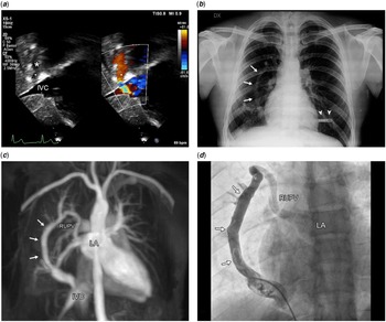
Physical examination revealed a fixed S2 split; the electrocardiogram showed a sinus rhythm and incomplete right bundle branch block. Echocardiography confirmed right chambers dilation and volume overload and an abnormal vessel draining into a dilated inferior vena cava through a narrow connection (Fig 1a). The chest X-ray displayed a scimitar sign along the right heart border (Fig 1b).
( a ) Echocardiography: an abnormal vessel (asterisk) draining into the IVC through a narrow connection. ( b ) Chest X-ray: the scimitar sign (arrows) along the right heart border. Arrowheads: implantable loop recorder. ( c ) MRA and ( d ) angiography. The scimitar vein (arrows) has a double connection with both IVC and LA, via the RUPV. IVC = inferior vena cava; LA = left atrium; MRA = magnetic resonance angiography; RUPV = right upper pulmonary vein.

A repeat cardiac MRI confirmed the presence of a right-to-left shunt lesion, as the Qp:Qs was 1.8:1. Gadolinium-enhanced 3D magnetic resonance angiography showed a vertical vein draining the right upper and middle pulmonary lobes with a double connection both with the inferior vena cava and the left atrium via the right upper pulmonary vein (Fig 1c). The inferior vena cava connection was preceded by a slightly wide ampulla. Hypoplasia of the right lung, pulmonary sequestration, and anomalous systemic arterial supply were excluded.
Repair of the cardiovascular anomaly was indicated. Thanks to its peculiar anatomy, a percutaneous treatment was amenable with a device occluding the anomalous connection to the inferior vena cava connection, redirecting the right pulmonary venous return completely to the left atrium. An alternative would have been the occlusion from the left atrium side. While this would have had a reduced risk of systemic embolism, the left-to-right shunt would have been markedly increased. According to our plans, the device landing zone should have included the wide ampulla at the inferior vena cava connection, a likely highly compliant venous structure requiring a very large vascular plug device. We hypothesised that a double retention disc device might have had greater stability. The distance between the inferior vena cava connection and the nearest hepatic vein was measured by cardiac MRI, excluding potential hepatic vein obstruction caused by the retention disc.
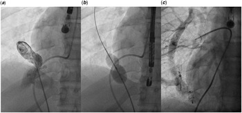
A static sizing of the inferior vena cava connection (Fig 2b) was performed using an 18-mm Amplatzer® sizing balloon (Abbott, Plymouth, MN). The inferior vena cava connection and the ampulla were easily stretched during the test, confirming the pre-procedural hypothesis. A 12-mm Amplatzer® Septal Occluder (Abbott, Plymouth, MN) was chosen. The device was placed with good stability. Before the delivery, the trans-oesophagal echocardiography excluded a residual shunt through the device and hepatic vein obstruction. Pulmonary artery pressure and pulmonary capillary wedge pressure remained unchanged. The levophase of a selective right pulmonary artery angiography confirmed the complete redirection of the right pulmonary vein return to the left atrium (Fig 2c). The procedure had no complications. Daily low-dose aspirin 100 mg was prescribed for 6 months. The patient is now doing well at a 1-year follow-up. An echocardiogram showed no obstruction of the inferior vena cava or hepatic veins, no residual shunt, and a right chambers volume reduction.
( a ) Selective contrast injection in the caudal portion of the scimitar vein confirmed the narrowing close to the IVC connection. ( b ) Balloon sizing of the scimitar vein across IVC connection. ( c ) The levophase of a selective right pulmonary artery angiography confirmed the complete right pulmonary venous redirection to the LA and the absence of residual shunt.

Discussion
Although rare, partial anomalous pulmonary venous connection should always be suspected in the presence of right chambers dilation, with signs of volume overload. In most cases, it coexists with intra-cardiac defects, including sinus venous or secundum atrial septal defects. The partial anomalous pulmonary venous connection needs surgical repair, but some uncommon variants might be treated percutaneously. Reference Luciano, Laux and Boudjemline2-Reference Forbess, O’Laughlin and Harrison4 Partial anomalous pulmonary venous connection diagnosis can be challenging, especially in adult patients. Scimitar syndrome may be even more challenging since it is associated with more cardiopulmonary anomalies, for which echocardiography may be not sensitive enough. Cross-sectional imaging such as CT or cardiac MRI may be useful both in diagnosis and treatment planning. In cardiac MRI, functional parameters such as cardiac output or pulmonary blood flow or ventricular stroke volumes are sometimes even more informative than images. In this case, the difference in the stroke volumes of the two ventricles would have been a strong hint for a shunt lesion in the first MRI, where the Qp:Qs ratio was not calculated. When dealing with a right ventricular enlargement, the Qp:Qs ratio should be always calculated and included in the analysis, to exclude a pre-tricuspid shunt lesion. In this report, we described a rare variant of scimitar syndrome with only a few cases reported in the literature. Reference Luciano, Laux and Boudjemline2-Reference Forbess, O’Laughlin and Harrison4 In our patient, there was normal cardiac position, normal right pulmonary artery, and no lung malformation (e.g. pulmonary sequestration, horseshoe lung). The scimitar vein had double drainage with both the inferior vena cava and the left atrium, without any abnormal arterial supply to the right lung, a peculiar anatomy suitable for complete percutaneous repair.
Conclusion
Partial anomalous pulmonary venous connection is a rare condition that may go unrecognised into adulthood and should be suspected in presence of right chambers dilation. Cross-sectional imaging is extremely useful in the diagnosis of CHD in adults. In our case, the cardiac MRI was essential in unveiling the uncommon anatomical pattern, avoiding surgery and choosing the right device for percutaneous treatment.
Financial support
This work received no specific grant from any funding agency, commercial or not-for-profit sectors.
Conflicts of interest
None.