Introduction
In general, neuronal types are unevenly distributed within the plane of the vertebrate retina. The best-known instances of this are found in those retinas with a specialized high acuity region, concentrically organized to form an area centralis, or as a horizontal stripe called a visual streak (Hughes, Reference Hughes and Crescitelli1977). The chicken retina, the subject of this study, possesses an area centralis (Bruhn & Cepko, Reference Bruhn and Cepko1996; Weller et al., Reference Weller, Lindstrom, De Grip and Wilson2009) characterized by a high density of ganglion cells that falls off radially from this central region (Ehrlich, Reference Ehrlich1981; Straznicky & Chehade, Reference Straznicky and Chehade1987). In addition to this familiar radial decline in density, there are reports of particular neuronal types, in various retinas, whose distribution follows some other pattern. Common patterns include a dorsal distribution, a ventral distribution, and a peripheral distribution. The significance of these distributions is unknown; moreover, basic data such as the fraction of neurons corresponding to particular patterns, and whether nonuniform distribution is associated with particular transmitters or neuromodulators, are unreported. An underlying question concerns the extent to which the retina uses “generic” machinery to process all parts of the visual world or alternatively has specialized neurons found only in particular retinal regions.
In this study, we examine the distribution of putative nitrergic neurons within the inner retina of the chicken. Nitrergic neurons are characterized by their ability to generate nitric oxide (NO), a short-lived modulator of cell function, made on demand by the enzyme nitric oxide synthase (NOS), and shown in imaging studies of live retina to be produced by a variety of retinal neurons (Eldred & Blute, Reference Eldred and Blute2005). Three putative nitrergic neuronal types with highly nonuniform distribution have already been reported in the chicken retina and a more systematic study seemed warranted. These three types are (1) the “restricted” efferent fibers, (2) their postsynaptic target amacrine cells whose distributions are exclusively ventral (Lindstrom et al., Reference Lindstrom, Nacsa, Blankenship, Fitzgerald, Weller, Vaney and Wilson2009), and (3) a ganglion cell whose reported distribution is entirely dorsal (Cellerino et al., Reference Cellerino, Novelli and Galli-Resta2000).
Many studies have described nitrergic neurons in the retina (Sandell, Reference Sandell1985; Sagar, Reference Sagar1986; Vaney & Young, Reference Vaney and Young1988; Weiler & Kewitz, Reference Weiler and Kewitz1993; Yamamoto et al., Reference Yamamoto, Bredt, Snyder and Stone1993; Morgan et al., Reference Morgan, Miethke and Li1994; Huxlin, Reference Huxlin1995; Kurenni et al., Reference Kurenni, Thurlow, Turner, Moroz, Sharkey and Barnes1995; Perez et al., Reference Perez, Larsson, Alm, Andersson and Ehinger1995; Blute et al., Reference Blute, Mayer and Eldred1997; Haberecht et al., Reference Haberecht, Schmidt, Mills, Massey, Nakane and Redburn-Johnson1998; Fischer & Stell, Reference Fischer and Stell1999; Cellerino et al., Reference Cellerino, Novelli and Galli-Resta2000), either through the use of antibodies to the neuronal isoform of NOS, nNOS, or through the use of an indirect method, NADPH diaphorase (NADPHd). This method (Thomas & Pearse, Reference Thomas and Pearse1961), which antedates the discovery of NO as a signaling molecule (Dawson et al., Reference Dawson, Bredt, Fotuhi, Hwang and Snyder1991), works by allowing NOS to catalyze electron transfer from NADPH to a soluble tetrazolium salt, that is, thereby reduced to a colored and insoluble formazan. In favorable preparations, this method can reveal the detailed structure of individual cells with almost Golgi-like clarity and has the advantage that the synthetic enzyme survives aldehyde fixation (Matsumoto et al., Reference Matsumoto, Nakane, Pollock, Kuk and Forstermann1993; Huxlin, Reference Huxlin1995). A drawback of this method is its inability to distinguish the neuronal isoform of NOS from other isoforms, or indeed other reductases, though the degree to which this is problematic varies between preparations and probably is reduced by fixation, to which nNOS is relatively insensitive (Matsumoto et al., Reference Matsumoto, Nakane, Pollock, Kuk and Forstermann1993).
In this study, we have used both the NADPHd method and antibodies to nNOS, accompanied by neurobiotin injection of some neuronal types. Some of the neurons we identify in the inner retina of the chicken clearly correspond to neuronal types described in previous studies (Morgan et al., Reference Morgan, Miethke and Li1994; Fischer & Stell, Reference Fischer and Stell1999; Cellerino et al., Reference Cellerino, Novelli and Galli-Resta2000), but we also describe putative nitrergic cells not previously reported. We find that nitrergic neuronal types peculiar to a region of the retina outnumber those that are found throughout the retina.
Materials and methods
Animal procedures
Forty-eight 3-week-old White Leghorn chickens (Gallus gallus), obtained from the University of Queensland Central Animal house, were used in this study, in accord with the Australian code of practice for the care and use of animals for scientific purposes. A further 23 White Leghorn chickens were hatched from eggs obtained from the Avian Sciences Facility of the University of California, Davis, CA. All methods were reviewed and approved by the Institutional Animal Care and Use Committee. Birds were killed with an intraperitoneally injected overdose of sodium pentobarbitone and the eyes quickly enucleated. Vitreous was removed from the eyecup, and the eye hemisected along the line of the pecten, which was subsequently excised. Retina from the larger nasal and smaller temporal halves of the eyecup was gently removed from the pigment epithelium and mounted photoreceptor-side down, on black filter paper (Millipore, Billerica, MA, AABG02500). Retinal pieces were maintained, until processing, in carbogenated Ames medium (Sigma, St. Louis, MO) containing 2.0 g/l of NaHCO3 (pH 7.4 at room temperature) and adjusted to 320 mOsmol with NaCl.
Histochemistry
Some initial experiments employed an NADPHd method in which NADPH itself was supplied (Fischer & Stell, Reference Fischer and Stell1999). However, we found cleaner results with a two-step procedure in which NADPH was synthesized in situ by reduction of NADP, using malic acid as the substrate for endogenous malic enzyme (Sagar, Reference Sagar1986; Vaney & Young, Reference Vaney and Young1988; Vaney, Reference Vaney2004). Retinas were fixed in 4% paraformaldehyde (PFA; Sigma, P6148) in 0.1 m phosphate-buffered saline (PBS) for 20 min at room temperature, followed by several washes and overnight in 0.1 m Tris buffer at pH 8.2 to which 0.2% Triton X100 had been added. Solutions used for the NADPHd reaction were as described in Vaney and Young (Reference Vaney and Young1988). The reaction was allowed to proceed for 90 min at 37°C and was then stopped by extensive washing in Tris-buffered saline at pH 7.6. After retinas were removed from their Millipore filters, they were coverslipped as flat-mount preparations with 50% glycerol in PBS. Cells of interest were examined on a Zeiss Axioskop microscope and photographed with an Insight monochrome digital camera (1,600 × 1,200 pixel resolution; Diagnostic Instruments). Images were adjusted for brightness, contrast, and sharpness in Adobe Photoshop. Measurements of cell density were obtained from tracings of identified cell bodies made with a drawing tube. The drawing tube was also used to record the structure of multistratified cells that were not easily photographed.
Antibodies and immunohistochemistry
Details of the antibodies used in this study are listed in Table 1. For anti-nNOS staining, we employed a polyclonal antiserum raised against pig brain nNOS (Mayer et al., Reference Mayer, John and Bohme1990) previously used in pigeon visual system (Cuthbertson et al., Reference Cuthbertson, Zagvazdin, Kimble, Lamoreaux, Jackson, Fitzgerald and Reiner1999) and whose specificity (in human) has been shown by preadsorption assay and correlation with in situ hybridization with NOS mRNA (Yew et al., Reference Yew, Wong, Li, Lai and Yu1999). As we describe below, we used Western blotting to show that this antiserum identifies a single band from chicken retina corresponding to nNOS. For most immunohistochemistry, retinas were fixed for 20–45 min in 4% PFA and then washed in 0.1 m PBS and 0.5% Triton overnight. One percent donkey serum or 1% bovine serum albumin was coapplied with the primary antibody (rabbit polyclonal anti-nNOS; Alexis Biochemicals, Enzo, Plymouth Meeting, PA, ALX-210-501-R025) used at the dilution of 1:5000. Incubations of 4–6 days were followed by a wash overnight in 0.1 m PBS. Secondary antibodies were usually Cy5-conjugated AffiniPure F(ab’)2 fragment donkey antirabbit IgG (H + L) (Jackson ImmunoResearch, West Grove, PA, 711-176-152) diluted to 1:500, again with 0.5% Triton and 1% donkey serum or 1% bovine serum albumin as blocker. Following 1–2 days in secondary antibody, retinas were washed overnight in 0.1 m PBS and then mounted in Prolong Gold antifade solution (Invitrogen, Carlsbad, CA, P36934). Retinas were subsequently examined on a Zeiss LSM510 laser scanning confocal microscope equipped with the META fluorescence discrimination capacity. Additional antibodies were used in conjunction with anti-nNOS in some experiments.
Primary antibodies

DSHB, Developmental Studies Hybridoma Bank, GST, Glutathione S-transferase.
An affinity purified goat anti-Choline Acetyltransferase (ChAT), previously described by Kihara et al. (Reference Kihara, Paschon, Cardoso, Higa, Castro, Hamassaki and Britto2009), was used to identify strata 2 and 4 of the inner plexiform layer (IPL) (Millar et al., Reference Millar, Ishimoto, Johnson, Epstein, Chubb and Morgan1985; Drenhaus et al., Reference Drenhaus, Morino and Veh2003). The secondary antibody was Cy3-conjugated AffiniPure donkey antigoat IgG (Jackson ImmunoResearch, 705-165-147) used at 1:500. We found the staining pattern to be identical to that shown by Drenhaus et al. (Reference Drenhaus, Morino and Veh2003) and Kihara et al. (Reference Kihara, Paschon, Cardoso, Higa, Castro, Hamassaki and Britto2009).
Affinity purified goat polyclonal anti-Brn3 raised against the C-terminus of human Brn3b was used to identify ganglion cells (Fischer et al., Reference Fischer, Dierks and Reh2002). This antibody recognizes all three Brn3 family members in rat, mouse, and human (manufacturer’s technical information), which have high homology with the C-termini of the chicken orthologs of Brn3. The most commonly used secondary antibody was Cy3-conjugated donkey antigoat, at the same dilution used in the anti-ChAT protocol. Mouse antichick Nkx2.2 (74.5A5, Developmental Studies Hybridoma Bank (DSHB), Iowa City, IA) was coapplied with anti-nNOS to identify oligodendrocytes and Nonastrocytic Inner Retinal Glial (NIRG) cells (Fischer et al., Reference Fischer, Scott, Zelinka and Sherwood2010). This antibody was used at a dilution of 1:50 and incubated for 7 days at 4°C. Secondary antibodies in these experiments were goat antirabbit conjugated to Alexa Fluor 488 (A11008; Invitrogen) and goat antimouse conjugated to Alexa Fluor 568 (A11004; Invitrogen).
Protein isolation and Western blotting
In order to confirm that the anti-nNOS polyclonal antibody was recognizing only nNOS, we performed Western blotting on proteins isolated from homogenized retinas.
The procedure for protein isolation was adapted from that of Weller et al. (Reference Weller, Lindstrom, De Grip and Wilson2009). Retinas were homogenized on ice in 20 mM Tris (Sigma; T3253 and T1503, pH 7.4), 150 mM NaCl, 10 μg/ml aprotinin (Sigma, A6279), 4 μg/ml leupeptin (Roche, Pleasanton, CA, 1017101), and 4 μl/ml 0.25 m phenylmethylsulphonyl fluoride (Roche, 236608) and then centrifuged for 5 min at 4000 × g at 4°C. The supernatant was ultracentrifuged for 15 min at 45,000 × g at 4°C, and the resulting pellet resuspended in the homogenization solution. Dynabeads Protein A were used to remove any endogenous antibodies. Electrophoresis was carried out as previously described, with the exception of the use of a 3–8% Tris-Acetate gradient gel, Tris-Acetate sodium dodecyl sulfate running buffer, and the standard HiMark Protein Ladder. All supplies were obtained from Invitrogen, unless otherwise noted. Proteins were transferred as described in Weller et al. (Reference Weller, Lindstrom, De Grip and Wilson2009). The membrane was blocked in 1% nonfat dry milk (w/v) for 45 min, followed by 1 h of incubation with the primary antibody, rabbit anti-nNOS, diluted to a concentration of 1:4000 in Tris-buffered saline with 0.05% Tween-20 (TBST). Three 5-min washes in TBST both preceded and followed 1 h incubation with peroxidase-conjugated goat antirabbit secondary antibody (Jackson Immunoresearch, 111-035-008) diluted to a concentration of 1:80,000 in TBST. The resulting protein bands were visualized via Enhanced Chemiluminescence (ECL) detection with ECL Western Blotting Detection Reagents (Amersham, GE LifeSciences, Piscataway, NJ, RPN 2106). On visualization, a single band was apparent at ~160 kDa, corresponding to the molecular mass of nNOS (Goureau et al., Reference Goureau, Regnier-Ricard, Jonet, Jeanny, Courtois and Chany-Fournier1997).
Cell filling
In order to visualize the complete structures of axon-bearing amacrine cells, these cells were injected with neurobiotin in live whole-mount retinas. For these experiments, both nasal and temporal pieces of retina were cut equatorially to yield the dorsal and ventral pieces that were subsequently attached to Millipore filters. These pieces were exposed to 35 μl of filtered 0.1% acridine orange in 20 ml superfusate for 20 s, a procedure that initially labeled the perinuclear cytoplasm of the cells in the ganglion cell layer (GCL). Retinal pieces were then maintained in normal superfusate for 1–4 h, during which labeling of the GCL faded relative to the labeling seen in the inner nuclear layer (INL). The tips of injection micropipettes were filled with 0.4% Lucifer yellow (Sigma) and 4% N-(2-aminoethyl)-biotinamide hydrochloride (neurobiotin; Vector Laboratories, Burlingame, CA) in 0.1 m Tris buffer, pH 8.2. Neurobiotin was iontophoresed with +1 nA current for 60–90 s, and the cells were then left to equilibrate in normal superfusate for 30–120 min before fixation in 4% PFA in 0.1 m phosphate buffer for 20–30 min. Following overnight wash in 0.1 m PBS with 0.2% Triton, retinas were incubated in anti-nNOS antibodies, and any other antibodies, as already described. After the final PBS wash, retinas were incubated for 1 day in 1:500 Cy2-conjugated Streptavidin (Jackson ImmunoResearch, 016-220-084) to label the neurobiotin-injected cells. Another 0.1 m PBS wash and mounting followed, as described previously. Z stacks from the confocal microscope were examined in Zeiss LSM image browser and, where necessary, volume rendered in imageJ.
In some experiments, NADPHd, instead of anti-nNOS, was used in conjunction with cell filling. When this was done, the procedure was modified so that, after filling, the retina was incubated in 1% donkey serum blocker with 1:500 Cy2-conjugated Streptavidin for 1 day, following an overnight wash in 0.1 m PBS prior to the NADPHd reaction.
Nomenclature
The position of somata within the INL and the depth of dendritic branching within the IPL were calculated by focusing through the thickness of the retina and measuring depth relative to the inner and outer margins of the IPL. Following convention, we have considered the IPL to comprise five strata of equal thickness, although it is clear that many neurons restrict stratification more finely. Some neuronal types in our study, with varying degrees of certainty, correspond to types described in other studies, notably that of Fischer and Stell (Reference Fischer and Stell1999). For those neurons whose identity is incontrovertible (e.g., target cells and bullwhip cells), we have used established names but, where correspondence is not completely certain, we have used our own names and pointed out similarities and differences with neurons described by other authors.
Results
Retinas from 62 chickens were stained and viewed in whole mount, using either NADPHd or antibody staining with a polyclonal anti-nNOS. In favorable preparations, the NADPHd method revealed neuronal anatomy with excellent clarity, so that very fine dendritic branching of individual cells was visible. Staining with anti-nNOS gave strong staining of cell bodies but seldom allowed dendrites to be seen, except when these were very thick (e.g., Fig. 5b). Like Fischer and Stell (Reference Fischer and Stell1999), we found that anti-nNOS and NADPHd staining techniques interfered with each other, regardless of which procedure was applied first, and we were therefore unable to achieve satisfactory double labeling of neurons. Nevertheless, comparison of material treated with the two methods gave strong support to the idea that the same neurons were identified by the two methods and, in particular, the NADPHd method was not giving false positives. Three pieces of evidence lead to this conclusion. First, most cell types seen with NADPHd could be identified with anti-nNOS, the only exceptions being the diffusely branching amacrine cells, whose somata were not sufficiently distinctive to allow positive identification with anti-nNOS. Second, the characteristic staining of individual cells was very similar using the two methods. For example, the amacrine cells that we have called light cells had weak staining at the very margin of the soma with both methods. Third, those cells that were weakly stained with NADPHd were also positive when examined with anti-nNOS, although weakly so. Bullwhip cells and displaced ganglion cells were examples of this.
In conjunction with NADPHd and anti-nNOS, we used microelectrode filling of cells with neurobiotin, later processed with a fluorophore-conjugated streptavidin. Filling of cells was carried out in live, isolated retinas in which the cell bodies of the INL were first lightly labeled with acridine orange (Sandell & Masland, Reference Sandell and Masland1988; Dacey, Reference Dacey1989). By targeting somata of the appropriate size, we were able to visualize more complete structures for some larger cells and provide corroboration that some neuronal types were indeed absent from some regions of retina.
General
A surprising feature of the chicken retina is both the density and diversity of nitrergic neurons. Furthermore, as described by Cellerino et al. (Reference Cellerino, Novelli and Galli-Resta2000), there is an immediately recognizable difference between dorsal and ventral retina. In the dorsal retina, the heaviest staining, whether from anti-nNOS or NADPHd, is found in the GCL, whereas, in the ventral retina, structures in the INL and IPL have the strongest signal. In all regions of the retina, the nitrergic neurons form a dense plexus in stratum 1 of the IPL, adjacent to the INL. With either method, this stratum, unlike the other strata of the IPL, appears densely sprinkled with positive dots that at first sight appear to be nonspecific background staining. The axons of target amacrine cells are a major contributor to this plexus and provide a majority of the strongly positive puncta.
GCL
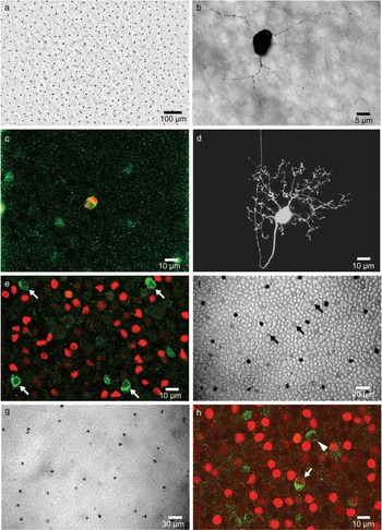
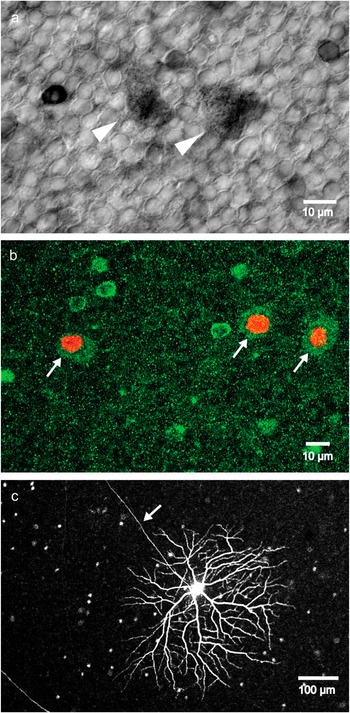
The strongly positive cells found in the GCL of the dorsal retina are characterized by cell bodies in which the cytoplasm is typically asymmetrically distributed, having a long axis of ~9.7 μm. These cells are spaced with very high regularity (Fig. 1a) and, over much of the dorsal retina, have a density of ~240 mm−2, though density declines several fold at the periphery. In some NADPHd preparations, it was possible to see two or three very thin dendrites leaving the cell body and traveling up to 50 μm with occasional branching (Fig. 1b). These dendrites typically ran at the boundary of the GCL and the IPL but in no cells were we able to see an axon. In some preparations, we used anti-nNOS or NADPHd in conjunction with an antibody to Brn3, a subfamily of transcription factors unique to ganglion cells (Gan et al., Reference Gan, Xiang, Zhou, Wagner, Klein and Nathans1996, Reference Gan, Wang, Huang and Klein1999; Liu et al., Reference Liu, Khare, Liang, Peters, Liu, Cepko and Xiang2000) and expressed by >93% of ganglion cells (Fischer et al., Reference Fischer, Dierks and Reh2002). Surprisingly, the NADPHd-positive cells in the GCL always appeared Brn3-negative (Fig. 1e), suggesting that they might not be ganglion cells but might instead be displaced amacrine cells, as in the cat (Wassle et al., Reference Wassle, Chun and Muller1987), or conceivably glial cells of which two types have cell bodies in the GCL (Fischer et al., Reference Fischer, Scott, Zelinka and Sherwood2010; Rompani & Cepko, Reference Rompani and Cepko2010). Strongly nNOS-positive cells, like the weakly nNOS-positive cells described below, were found to be negative for Nkx2.2, a glial marker found in both oligodendrocytes and NIRG cells (Fischer et al., Reference Fischer, Scott, Zelinka and Sherwood2010). Furthermore, one good fill with neurobiotin revealed that a strong nNOS-positive cell in the dorsal retina had an axon in the fiber layer and a small dendritic field of ~70 μm diameter (Fig. 1c and 1d), comparable to the somatic spacing of the strongly positive cells in the GCL. Based on its small soma and highly branched but small dendritic tree, this ganglion cell would be included in Group 1c by Naito and Chen (Reference Naito and Chen2004).
The GCL in the dorsal and ventral retina. (a) The very regular distribution of the intensely NADPHd-positive cells in the dorsal retina is apparent at low magnification. (b) These strongly staining cells typically show two or three very fine dendrites emerging from the cell body and branching occasionally; these dendrites run in the GCL and ascend to the inner margin of the IPL. (c) Anti-nNOS (green) and injected neurobiotin (red) are colocalized in this cell body in dorsal retina. (d) Neurobiotin injection reveals that the nNOS-positive cell is a ganglion cell with a small dendritic tree. (e) In the dorsal retina, the nNOS-positive cells (green) never colocalize with anti-Brn3 staining (red). (f) A separate population of lightly stained and irregularly distributed NADPHd-positive cells is also visible in the dorsal retina (three examples shown with arrows). (g) In the ventral retina, there are NADPHd-positive cells ranging from moderate to weakly stained, but no neurons are as strongly stained or as regular as those in the dorsal GCL. (h) Anti-nNOS staining reveals a pattern similar to that seen by NADPHd in the ventral retina but, unlike in the dorsal retina, some nNOS-positive cells are also Brn3-positive (arrow), whereas others are negative (arrowhead).

In addition to the regularly spaced and strongly nNOS or NADPHd-positive cells, a much fainter population of cells in the GCL of the dorsal retina could be seen with NADPHd (Fig. 1f) and was also discernable with anti-nNOS. Although these somata were Brn3 negative, like the strongly staining somata in the dorsal GCL, we count them as a different cell type, on the basis that they are not part of the regular array and are distinctly fainter. Like the strongly nNOS-positive cells, these weakly staining cells were found to be negative for Nkx2.2 but since we were unable to visualize any part other than the soma, we cannot rule out the possibility that they are displaced amacrine cells.
Although Cellerino et al. (Reference Cellerino, Novelli and Galli-Resta2000) report that NADPHd-positive cells in the ventral GCL are rare, we found them to be common and clearly visible, perhaps due to the greater sensitivity of our two-step NADPHd method. The clear regularity and intense staining of the dorsal retina was absent in the ventral retina. Instead, both NADPHd and anti-nNOS showed a distribution of staining from moderate to very faint (Fig. 1g). We measured the density of the darker cells of the GCL in the median ventral retina as 124 mm−2, with lighter cells about twice that density. While the variation in staining might imply the presence of more than one type of cell, we were unable to make categorical distinctions based on this. Unlike the situation in the dorsal retina, we found some nNOS-positive cells in the ventral GCL to be Brn3-positive, although others were Brn3 negative (Fig. 1h). The Brn3-positive cell has no counterpart in the dorsal retina and must be counted as a ganglion cell confined to the ventral retina. The Brn3-negative cell in the ventral retina, on the other hand, cannot be distinguished from the lightly staining Brn3-negative cell in the dorsal GCL and we count it as the same cell type.
INL and IPL
Ubiquitous cells
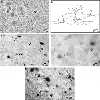
Several types of nitrergic neuron were found throughout the retina, regardless of region. The commonest nitrergic amacrine cell seen with either anti-nNOS or NADPHd has a soma of ~7.2 μm diameter lying at the border of the IPL and INL. Typically these cells, which we call light cells, are very lightly stained except just beneath the somatic plasmalemma, where staining typically has a beaded appearance (Fig. 2a–2c). A single process descends vertically through the IPL and gives rise to thin, minutely punctate, symmetrical branches at an IPL depth of ~80%. Dendritic branches from adjacent cells appear to be nonoverlapping and give rise to a narrowly defined stratum. The density of these cells is highest around the horizontal meridian (1040 mm−2), high in the dorsal retina (800 mm−2), and about half that in the ventral retina. These neurons are almost certainly those classified as type 1 by Fischer and Stell (Reference Fischer and Stell1999).
Common ubiquitous cells seen with NADPHd staining. (a) The delicate and minutely punctate dendrites of light cells seen at a depth of 80% in the IPL. (b) Camera lucida drawings of four adjacent light cells showing that their nonoverlapping dendrites tile the retina. (c) At the lowest level of the INL, light cells (white arrowheads) and knob cells (arrows) have somata of the same size but are distinguishable by their different degrees of staining. Four medium-dark knob cells and three light cells lie in this plane; two bistratified cells (dark arrowheads) sit higher in the INL and are out of focus in this image. (d) The same field as in (c) but at a depth of 40% in the IPL; at this level, the characteristic swellings of the four knob cells are apparent (arrows). (e) At a depth of 20% in the IPL, the main dendrites of bistratified cells (arrowheads) are visible; the cell bodies are out of focus, but the stout primary dendrite from the cell body can be seen (arrow).

A second type of amacrine cell, the bistratified cell, has a cell body of ~7.5 μm diameter and sits a little higher in the INL than the light cells (Fig. 2c). A single thick process, often running obliquely, enters the IPL and continues to a depth of ~20%, just below the main nitrergic plexus in stratum 1, at which point it forms branches that run horizontally. Branches are typically varicose and asymmetrical, extending predominantly on one side (Fig. 2e). Although neighboring cells typically branched on different sides, adjacent cells generally did not have overlapping dendrites. From these main dendrites, it was often possible to see fine secondary dendrites descend to ~50% depth in the IPL, where they branched again and ran a short distance horizontally. Very likely, these cells correspond to the type 2 cells of Fischer and Stell (Reference Fischer and Stell1999), although the fine processes that descend to the deeper layer of the IPL are not mentioned by them and could easily have been missed.
A third ubiquitous type of amacrine cell, the knob cell, resembles the light cell in position and size of the cell body (~7.4 μm diameter) but differs in having a somewhat darker stain (Fig. 2c). A single process, so thin as to be usually invisible, descends to an IPL depth of 55% where it swells into a knob about 1–2 μm diameter (Fig. 2d). From this swelling, a pair of dendritic branches emerge opposite each other, and branch and descend further to terminate at an IPL depth of ~70%.
Regional cells
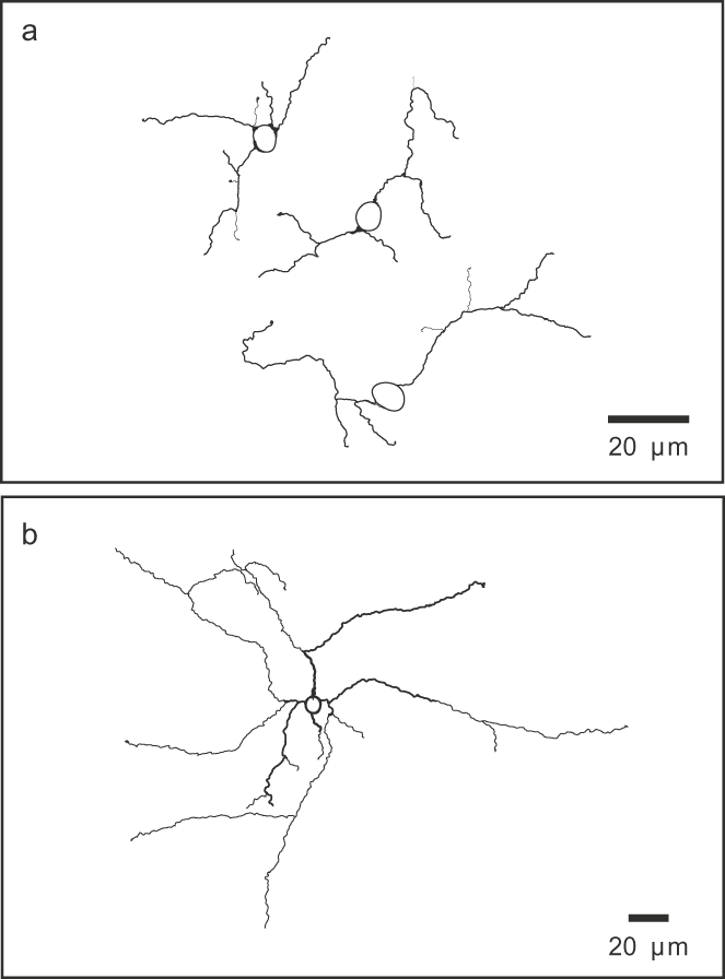
Diffuse amacrine cells of two types are found in an equatorial band. The cell bodies of the smaller and more abundant of these lie at the border of the INL and are typically ~7.3 μm in diameter and moderately stained. At their densest, these cells are found at ~490 mm−2. Typically one or two branches emerge from the cell body and descend into the IPL, branch irregularly and extend down almost to the GCL (Fig. 3a). The second type of diffuse cell is less than a tenth as common and is characterized by a densely staining cell body of ~9 μm diameter and widely branching dendrites, giving a total dendritic spread of ~200 μm (Fig. 3b). Like the smaller diffuse cell, the larger one extends dendrites throughout the IPL.
Camera lucida drawings of NADPHd staining of diffuse amacrine cells found in an equatorial band. (a) Small diffuse amacrine cells, seen here in a single field, generally give rise to two or three dendrites that descend through the depth of IPL; the dendrites branch infrequently to produce a sparse dendritic tree of <100 μm diameter. (b) The less common large diffuse amacrine cell is characterized by a darkly staining cell body that typically gives rise to four dendrites, which also descend through the depth of the IPL.

In the ventral retina, the “restricted” efferent fibers previously described in chicken and also in pigeon and quail (Maturana and Frenk, Reference Maturana and Frenk1965; Uchiyama and Ito, Reference Uchiyama and Ito1993; Morgan et al., Reference Morgan, Miethke and Li1994; Nickla et al., Reference Nickla, Gottlieb, Marin, Rojas, Britto and Wallman1994; Fischer and Stell, Reference Fischer and Stell1999; Uchiyama et al., Reference Uchiyama, Aoki, Yonezawa, Arimura and Ohno2004; Lindstrom et al., Reference Lindstrom, Nacsa, Blankenship, Fitzgerald, Weller, Vaney and Wilson2009) ascend through the IPL and swell to form large synapses with the cell bodies of target amacrine cells, also known as association amacrine cells (Fig. 4a) (Ramón y Cajal, Reference Ramón y Cajal1893; Uchiyama and Stell, Reference Uchiyama and Stell2005; Lindstrom et al., Reference Lindstrom, Nacsa, Blankenship, Fitzgerald, Weller, Vaney and Wilson2009). Although ~15% of efferent fibers branch at least once in the outer sublamina of the IPL, only a single target cell was ever seen to be contacted by each efferent fiber. Target amacrine cells are themselves positive for both NADPHd and anti-nNOS, as are their axons (Fig. 4b). As previously mapped (Lindstrom et al., Reference Lindstrom, Nacsa, Blankenship, Fitzgerald, Weller, Vaney and Wilson2009), target cells as well as efferent fibers were seen throughout the ventral retina but were densest in the horizontal meridian where they occurred at ~290 mm−2. In rare instances, it was possible to follow a target cell axon to its termination. Axons always ran in stratum 1 of the IPL and, in those we were able to follow, headed dorsally. As shown in Fig. 4c, and similar to the finding in quail (Uchiyama et al., Reference Uchiyama, Aoki, Yonezawa, Arimura and Ohno2004), axons characteristically made a few 90 deg turns before terminating millimeters away in a field of little branches covering several 100 μm2, still in stratum 1 (Fig. 4d).
Efferent fibers and their target cells seen with NADPHd. (a) In the inferior temporal retina, close to the top of the pecten, efferent fibers in the outer IPL are seen ascending to their target cells within the INL; both the target cells and the synapses formed by efferent fibers are strongly NADPHd positive. (b) In stratum 1 of the IPL, which is the main nitrergic plexus, an efferent synapse and target cell soma are out of focus, but the efferent fiber itself may be seen (arrow), as well as the much finer target cell axon (arrowhead), which travels a great distance in this stratum. (c) Target cell axons running in stratum 1 typically execute abrupt turns and sometimes loops (arrow). (d) A target cell axon (arrow) makes numerous branches at its termination in stratum 1.

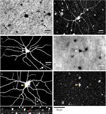
Efferent fibers and their target cells were very noticeable because of their large size and heavy staining. No less striking were the large axon-bearing amacrine cells characteristic of the ventral retina. These cells resemble, and are probably a subset of, the type I “proprioretinal neurons” described by Catsicas et al. (Reference Catsicas, Catsicas and Clarke1987) who used diI labeling to identify neurons with very long intraretinal processes. At their densest, these cells number ~75 mm−2. The somata of these neurons are often irregularly shaped with a long axis of up to 17 μm lying at the border of the INL, where they give rise to several stout dendrites running in stratum 1 of the IPL (Fig. 5a and 5b). Dendrites from these neurons overlapped considerably and were frequently observed to run together in antiparallel beside those of neighboring axon-bearing cells. Dendritic spread in these cells often exceeded 500 μm, although not all were this extensive, and there was considerable variation not only in the size of the dendritic field but also in the thickness of dendrites and the degree of staining. In some NADPHd preparations, it was possible to see a fine axon emerging from the cell body and running laterally in stratum 1. Although these axon-bearing amacrine cells were seen by both anti-nNOS and NADPHd to be exclusive to the ventral retina, their distribution was not identical to that of efferent fibers and target amacrine cells. Axon-bearing amacrine cells were seen in the extreme ventral retina, where efferents and target amacrine cells are absent (Lindstrom et al., Reference Lindstrom, Nacsa, Blankenship, Fitzgerald, Weller, Vaney and Wilson2009); moreover, the distribution of these axon-bearing amacrine cells did not extend as far dorsally as that of the efferent fibers.
Axon-bearing amacrine cells from the ventral retina. (a) A small axon-bearing amacrine cell stained with NADPHd visible at the margin of the IPL; also visible here are two light cells (arrows) and the synaptic terminal of an efferent fiber (arrowhead). (b) Anti-nNOS identifies an amacrine cell seen together with numerous other nNOS-positive somata. (c) A neurobiotin-filled axon-bearing amacrine cell that gives rise to a single intraretinal axon (arrow). (d) The same neuron as in (c), with the position of the soma marked with an arrowhead; NADPHd staining identifies this as a nitrergic cell. (e) In this neurobiotin-filled cell, an axon (arrow) exits the soma (yellow arrow) and gives rise to a thin collateral (white arrowhead). The lower panel shows a transverse section through the confocal z-stack. This section cuts through the dendrites and axon, shown in green, together with anti-ChAT staining shown in white. The two horizontal white bands represent the stratification of starburst amacrine cells and define strata 2 and 4 of the IPL. White arrowheads indicate the dendrites of the injected amacrine cell running in stratum 1 and the red arrowhead indicates the axon, lying deeper at the margin of strata 1 and 2. (f) The cell body (yellow arrowhead) of this same amacrine cell is anti-nNOS positive; the apparent difference in soma shape is due to leaked neurobiotin in (e). Fifty micrometers the 50 μm scale bar applies to (f) and the small lower panel.

To examine the structure of these neurons in greater detail, we injected them with neurobiotin, targeting large cell bodies at the border of the INL as revealed by acridine orange staining and using either anti-nNOS or NADPHd to confirm the presence of nNOS (Fig. 5c–5f). Several general results are significant. First, of the 62 axon-bearing amacrine cells in the ventral retina identified by intracellular filling, 37 (~60%) were negative for either NADPHd or anti-nNOS. As shown in Fig. 6, positive and negative neurons were otherwise indistinguishable. Second, although it was possible to see and fill large cell bodies in the dorsal INL, none of the resulting fills were of axon-bearing amacrine cells. This supports our finding that nitrergic axon-bearing amacrines are exclusively ventral and goes further to argue that even those axon-bearing amacrine cells that are nNOS-negative are absent from the dorsal retina. Third, even though the dendrites of these nNOS-positive axon-bearing amacrine cells were often seen running together, in no instance did we see the tracer spread beyond the injected cell. Among the nNOS-negative axon-bearing amacrine cells, only a single fill resulted in the spread of tracer. In this instance, the tracer was seen in dozens of small (~6.5 μm diameter) amacrine cell somata at the inner margin of the INL, having a density of ~1500 mm−2.
Neurobiotin-filled axon-bearing amacrine cells from the ventral retina. (a) An nNOS-positive axon-bearing amacrine cell of the smaller type; characteristically, these cells have a single axon and dendrites that are fairly symmetrically distributed. (b) An nNOS-positive axon-bearing amacrine cell of the larger type; this example has two axons (arrows) and relatively thin tapering dendrites that are asymmetrically distributed. (c) A small nNOS-negative amacrine cell morphologically similar to the small nNOS-positive type shown in (a). (d) The larger of the two cells shown here bears two axons (arrows) but is nNOS negative.

Among the 25 axon-bearing amacrine cells that were injected, there were at least two types of cells that were nNOS or NADPHd positive. Seven cells possessed an approximately round soma and roughly symmetrical dendrites with an average dendritic field diameter of 320 μm (250–404 μm) (Fig. 6a). The other 18 cells characteristically had irregular or spindle-shaped somata and asymmetrical dendritic fields with an average diameter of 596 μm (450–736 μm) (Fig. 6b). These differences in dendritic spread were not attributable to differences in eccentricity since the two types of cell could be seen adjacent to each other. Both types of cell bore axons originating from the cell body or the base of a primary dendrite, and the larger type of neuron frequently had two equivalent axons (Fig. 6b). In both types of neuron, axons were sometimes seen to produce thin collaterals (Fig. 5e). The filling of axons was never sufficient to allow them to be traced to their terminations, although it was often possible to see that they extended for at least several millimeters. We observed that occasionally the axon, soon after leaving the cell body, would dive down into the fiber layer before turning back and reentering stratum 1 of the IPL. In some preparations, we used an antibody to ChAT that labeled strata 2 and 4 of the IPL. From z sections of this material, it was clear that the axon, though slightly deeper than the dendrites, nevertheless also traveled within stratum 1 (Fig. 5e).
As expected, injection of cells based on soma size yielded filled cells other than axon-bearing amacrine cells. Three other types of nNOS-positive cells were seen in the INL. The first of these was the distinctive bullwhip cell found only in the ventral retina, originally described as substance P-like immunoreactive cells (Ehrlich et al., Reference Ehrlich, Keyser and Karten1987) and subsequently shown to contain glucagon and regulate the growth of the eye (Fischer et al., Reference Fischer, Omar, Walton, Verrill and Unson2005, Reference Fischer, Skorupa, Schonberg and Walton2006, Reference Fischer, Stanke, Ghai, Scott and Omar2007, Reference Fischer, Ritchey, Scott and Wynne2008). The very large and oblate cell bodies of these neurons give rise to a single stout process ~7 μm in diameter extending in the outer sublamina of the IPL. From this continues a long and apparently unbranched axon and, laterally, dendrites that descend to the top of stratum 3 and a few to the bottom of stratum 4 (Fig. 7a). The somata of these cells were weakly, but unambiguously, positive with both NADPHd and anti-nNOS (Fig. 7b).
Bullwhip cell somata found in the ventral retina are weakly nNOS-positive. (a) The extensive dendritic branching of this unusual cell type is seen from an x–y projection of confocal images (upper panel) of a neurobiotin fill; fine dendrites are varicose, but the thin extensive axon (arrow) is smooth. Lower panel, the same cell projected in the z–y plane shows the main dendritic branching in the top of stratum 3, a small number of centrally placed dendritic branches in the bottom of stratum 4, and the axon (arrow) in stratum 1. (b) Upper panel, a confocal section through a neurobiotin-filled bullwhip cell showing the large cell body (arrow) and stout horizontal process. Lower panel shows weak anti-nNOS staining of the same soma.

The other neurons inadvertently filled, and found in both dorsal and ventral retina, were displaced ganglion cells. Like Fischer and Stell (Reference Fischer and Stell1999), we saw weak NADPHd staining of these neurons (Fig. 8a) but, unlike them, we found that anti-nNOS also gave a weak but definite signal (Fig. 8b). From our sample of the 19 displaced ganglion cells filled with neurobiotin, it was clear that there was more than one cell type. About two-thirds of the filled cells were negative for nNOS, and those that were positive constituted more than one anatomical class. We encountered cells whose branching was confined to stratum 2 (Fig. 8c) as reported by Yang et al. (Reference Yang, Millar and Morgan1989) and whose projection is to the nBOR nucleus (Karten et al., Reference Karten, Fite and Brecha1977) but also some with branching throughout the IPL. All nNOS-positive cells had cell bodies >20 μm in diameter and obvious axons that ran into the fiber layer. We found that all nNOS-positive displaced ganglion cells were Brn3-positive (Fig. 8b) in apparent disagreement with Fischer et al. (Reference Fischer, Dierks and Reh2002), who report displaced ganglion cells as Brn3-negative. A probable resolution of this discrepancy lies in the different selectivities of the antibodies employed. Fischer et al. (Reference Fischer, Dierks and Reh2002) used antibodies to Brn3.0, also called Brn3a, whereas the affinity purified polyclonal antibody we used in this study recognizes Brn3a, Brn3b, and Brn3c, any of which can be expressed in subsets of ganglion cells (Xiang et al., Reference Xiang, Zhou, Macke, Yoshioka, Hendry, Eddy, Shows and Nathans1995). In support of this interpretation, we note that Liu et al. (Reference Liu, Khare, Liang, Peters, Liu, Cepko and Xiang2000) show a displaced ganglion cell that is strongly positive for Brn3b in the INL of chick.
Displaced ganglion cells found in the INL. (a) With NADPHd staining, the large cell bodies of displaced ganglion cells (arrows) had a characteristically fuzzy appearance. (b) With anti-nNOS, a similar weak cytoplasmic staining (green) is seen and the cells are unambiguously identified as displaced ganglion cells because they are the only Brn3-positive cells (red) found in the INL. (c) Neurobiotin fill of an nNOS-positive displaced ganglion cell from the ventral retina; a single axon (arrow) descends to the fiber layer and the dendritic branching is confined to stratum 2 of the IPL.

Discussion
Comparison with previous studies on chicken retina
A main finding of this study is that a surprising number of cell types in the inner retina of the chicken are likely to be nitrergic. Conservatively, we estimate 15 neuronal types: at least 9 types of amacrine cell, 5 types of ganglion cell, and 1 centrifugal midbrain neuron. The mechanism of action of NO via the modulation of soluble guanylyl cyclase is well established in the retina. Acting through cGMP, NO closes gap junctions between horizontal cells (DeVries & Schwartz, Reference DeVries and Schwartz1989, Reference DeVries and Schwartz1992; Pottek et al., Reference Pottek, Schultz and Weiler1997) and between AII amacrine cells (Mills & Massey, Reference Mills and Massey1995). It opens ion channels in both cones and ganglion cells (Ahmad et al., Reference Ahmad, Leinders-Zufall, Kocsis, Shepherd, Zufall and Barnstable1994; Savchenko et al., Reference Savchenko, Barnes and Kramer1997) and stimulates the release of GABA, probably through multiple mechanisms (Yu & Eldred, Reference Yu and Eldred2005). Our finding that a large number of neuronal types in the inner retina likely synthesize NO suggests that there are functions for NO that are yet to be discovered.
Some of the 15 neuronal types we found can clearly be identified with cell types described in previous studies of this retina, some neurons are probably those described elsewhere, and other neurons are described for the first time. The restricted efferent fibers and their postsynaptic partners, the target amacrine cells, with whom they have a one-to-one synaptic relationship, have been described as strongly positive for both nNOS and NADPHd in several studies (Morgan et al., Reference Morgan, Miethke and Li1994; Goureau et al., Reference Goureau, Regnier-Ricard, Jonet, Jeanny, Courtois and Chany-Fournier1997; Fischer & Stell, Reference Fischer and Stell1999; Posada & Clarke, Reference Posada and Clarke1999; Cellerino et al., Reference Cellerino, Novelli and Galli-Resta2000; Rios et al., Reference Rios, Lopez-Costa, Fosser, Brusco and Saavedra2000; Lindstrom et al., Reference Lindstrom, Nacsa, Blankenship, Fitzgerald, Weller, Vaney and Wilson2009). The unique anatomy of this pair of cells has allowed their unambiguous identification in this study.
We have been able to confirm a population of small, darkly staining cells in the GCL, as described by Cellerino et al. (Reference Cellerino, Novelli and Galli-Resta2000). Although the soma shape, asymmetrical and teardrop-like, is unusual for ganglion cells (Ehrlich, Reference Ehrlich1981), Cellerino et al. (Reference Cellerino, Novelli and Galli-Resta2000) identified these as ganglion cells because they degenerated following cuts through the fiber layer made near the optic nerve head. We found these cells to be uniformly negative for Brn3, a transcription factor expressed in almost all ganglion cells (Fischer et al., Reference Fischer, Dierks and Reh2002) and, although dendrites were often visible with NADPHd, we were never able to see an axon with this method. The suggestion that these cells might in fact be displaced amacrine cells was refuted by their complete anatomy as revealed by neurobiotin filling. Although we were only successful in filling one nNOS-positive cell out of ~60 injections in the GCL, the axon of this ganglion cell was clearly visible.
Unlike Cellerino et al. (Reference Cellerino, Novelli and Galli-Resta2000), we identified other asymmetrical somata in the dorsal GCL. These were consistently shown with either NADPHd staining or with anti-nNOS but were much more lightly stained. These cells were also Brn3 -negative but, because of their much lighter staining and also because they clearly were not part of the very regular array of darkly staining cells, we presume them to be a different cell type. While Cellerino et al. (Reference Cellerino, Novelli and Galli-Resta2000) described the ventral retina as almost devoid of nitrergic cells in the GCL, we were able to see a variety of cells ranging from moderately to very faintly stained. Some cells were Brn3-negative and might belong to the same population as the lightly staining cells of the dorsal retina, but others were Brn3-positive, and therefore represent a third cell type in the GCL. The true number of types, however, could be larger.
In a careful study, Fischer and Stell (Reference Fischer and Stell1999) used both NADPHd and anti-nNOS to identify nitrergic cells in transverse sections of chicken retina taken from the medial retina. The cells we designate as light cells are almost certainly their type 1 amacrine cells. The amacrine cells we have called bistratified cells are almost certainly their type 2 amacrine cells, although they describe dendritic branching only in stratum 1 and not extending down into stratum 3 as we observed. However, the processes that extend down this far are extremely fine and relatively sparse, so they might easily have been overlooked. The amacrine cells we have called knob cells possess a process that descends to a depth of ~60% in the IPL but was so narrow as to be invisible in all but a few NADPHd preparations. In whole mount, it was possible to focus through the retina and roughly align the swelling at 60% depth with the position of the cell body at the margin of the INL. In transverse sections, it would be much harder to make this association. The type 3 amacrine cells of Fischer and Stell might conceivably correspond to our knob cells because they have cell bodies at the margin of the INL and are described as having very thin dendrites. Fischer and Stell, however, show dendritic stratification at ~40% depth, whereas for knob cells we find that branching extends down to ~70%. The Fischer and Stell type 4 amacrine cell seems to have no equivalent in our study. This cell is described as having a cell body high in the INL and multiple levels of branching 40–70% deep. We describe two diffusely branching amacrine cells, a larger and a smaller type, both of which have dendrites that branch throughout the whole depth of the IPL; however, we observed the somata of both these cell types to lie at the margin of the IPL and INL.
Fischer and Stell report that NADPHd stains displaced ganglion cells weakly, but they suggest that since, in their hands, anti-nNOS failed to recognize displaced ganglion cells, this is likely to be an artifact. Our results confirm that some displaced ganglion cells are positive for NADPHd, but we also show that some, but not all, displaced ganglion cells are weakly positive for anti-nNOS. Intracellular filling of these cells with neurobiotin has allowed us to see that at least two types of displaced ganglion cells are positive for nNOS. One type has dendritic branching confined to stratum 2 and the other type has dendrites throughout the IPL.
In this study, we find that the unusual amacrine cells originally described by Ehrlich et al. (Reference Ehrlich, Keyser and Karten1987) and named bullwhip cells are weakly positive for both NADPHd and nNOS. These cells contain glucagon and project an axon to the circumferential proliferative zone of the retina where they are thought to regulate retina and eye growth (Fischer et al., Reference Fischer, Omar, Walton, Verrill and Unson2005, Reference Fischer, Skorupa, Schonberg and Walton2006, Reference Fischer, Stanke, Ghai, Scott and Omar2007, Reference Fischer, Ritchey, Scott and Wynne2008). Since these cells are found only in the ventral retina, it is understandable they were not described by Fischer and Stell.
Also found in the ventral retina are the axon-bearing amacrine cells that contribute to the dense weave of processes that constitutes the nitrergic plexus in stratum 1. Many of these neurons are strikingly large and stain intensely with NADPHd or anti-nNOS. Neurobiotin filling of these cells has allowed us to resolve several issues concerning their anatomy. It is clear from our fills that not all axon-bearing amacrine cells are nitrergic, in fact only between one-third and one half are nNOS-positive, although these are morphologically indistinguishable from those that are nNOS-negative. We can discriminate two types of nNOS-positive axon-bearing amacrine cells, differing not only in their size but also in the appearance of their somata and dendritic trees. Very likely, these two types correspond to the small and large semilunar amacrine cells described by Cajal in the bird retina, both of which he reports as having extraordinarily long processes running in stratum 1 (Ramón y Cajal, Reference Ramón y Cajal1972).
Comparison with other species
We confirmed the finding of Fischer and Stell (Reference Fischer and Stell1999) that stratum 1 of the IPL represents the major nitrergic plexus in the chicken retina, containing not only the axons of target amacrine cells but also the axons and dendrites of the two types of large axon-bearing amacrine cells. This is not a conserved feature of the vertebrate retina and is remarkably variable in reality. In the rabbit retina, stratum 3 represents the main nitrergic plexus (Haberecht et al., Reference Haberecht, Schmidt, Mills, Massey, Nakane and Redburn-Johnson1998; Vaney, Reference Vaney2004) and in rat retina, two plexuses of NADPHd-positive fibers are found, one in stratum 2 and one at the border between strata 3 and 4 (Palanza et al., Reference Palanza, Jhaveri, Donati, Nuzzi and Vercelli2005).
The total of 15 neuronal types we infer to be nitrergic in the inner retina of the chicken is considerably more than the number reported for other species. For example, the turtle reportedly has five nitrergic amacrine cells and possibly one displaced amacrine cell (Haverkamp et al., Reference Haverkamp, Kolb and Cuenca2000). In the rabbit, only two wide-field amacrine cells are reported (Sagar, Reference Sagar1986; Vaney & Young, Reference Vaney and Young1988), and only two types of amacrine cell are reported in the frog retina (Sato, Reference Sato1990). For some retinas, it is probable that the number of nitrergic types is underestimated. This is likely because most studies examine only a single region of the retina and would be expected to miss neurons particular to other regions. It is also likely that in some instances relatively insensitive methods underestimate the true number of nitrergic cells. In the pigeon retina, for example, only three types of amacrine cells have been reported as NADPHd positive (Sato, Reference Sato1990), though the phylogenetic proximity of pigeon and chicken suggests that most, if not all, neuronal types are probably common to both. However, in other instances, such as the rabbit retina, it seems likely that only two amacrine cell types are nitrergic. In this retina, staining quality is excellent and the result agreed upon by numerous authors (Sandell, Reference Sandell1985; Sagar, Reference Sagar1986; Vaney & Young, Reference Vaney and Young1988; Koistinaho et al., Reference Koistinaho, Swanson, De Vente and Sagar1993; Perez et al., Reference Perez, Larsson, Alm, Andersson and Ehinger1995; Haberecht et al., Reference Haberecht, Schmidt, Mills, Massey, Nakane and Redburn-Johnson1998; Volgyi et al., Reference Volgyi, Xin, Amarillo and Bloomfield2001; Vaney, Reference Vaney2004; Wright & Vaney, Reference Wright and Vaney2004). The significance of this difference between rabbit and chicken is obscure but might simply reflect the greater thickness and complexity of the avian retina. However, it is notable that the intensely NADPHd-positive ND1 cells in the rabbit retina are axon-bearing amacrine cells that have a similar morphology to the large, intensely stained cells in the INL of the chicken retina, although the two types of axon-bearing amacrine cells branch in different strata of the IPL.
Regionality
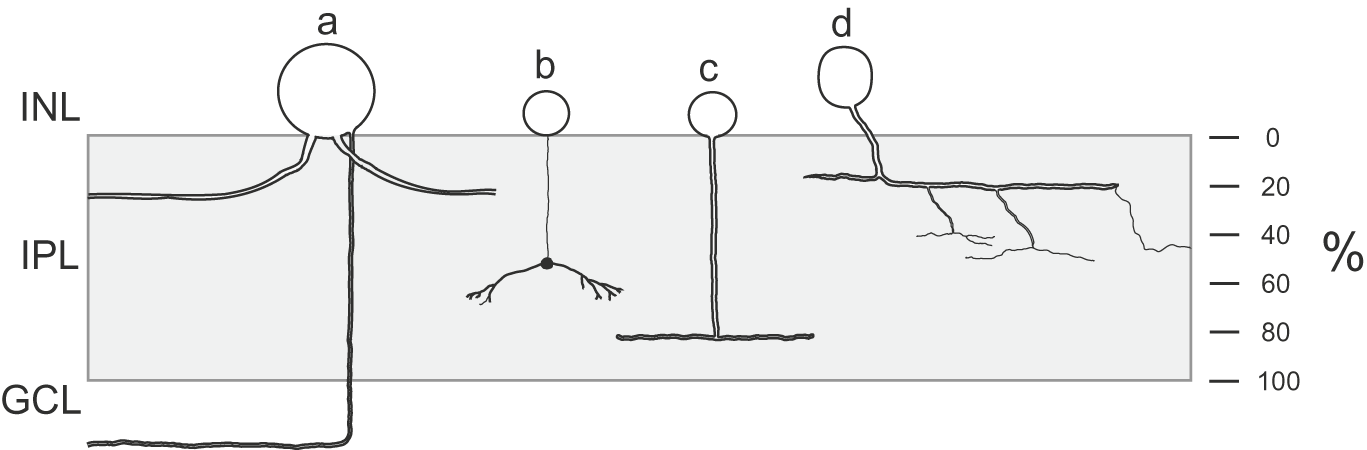
At least six putative nitrergic neuronal types are found ubiquitously throughout the retina (Fig. 9). These include three amacrine cells, the light cell, the knob cell, and the bistratified cell; two types of displaced ganglion cell, one that stratifies exclusively in stratum 2 and one that branches diffusely throughout the IPL. As we discuss below, we infer that a presumed ganglion cell is also ubiquitously distributed.
Summary of the nonregional nitrergic cells of the inner retina showing their branching within the IPL. (a) Displaced ganglion cell; in reality, there are two types, one as shown here with dendritic branching within stratum 2 and another with branching throughout the depth of the IPL. Three amacrine cell types are (b) knob cell, (c) light cell, (d) the bistratified cell. Not shown here is a lightly staining soma in the GCL, that is Brn3-negative; whether this is a ganglion cell or displaced amacrine cell is unclear.

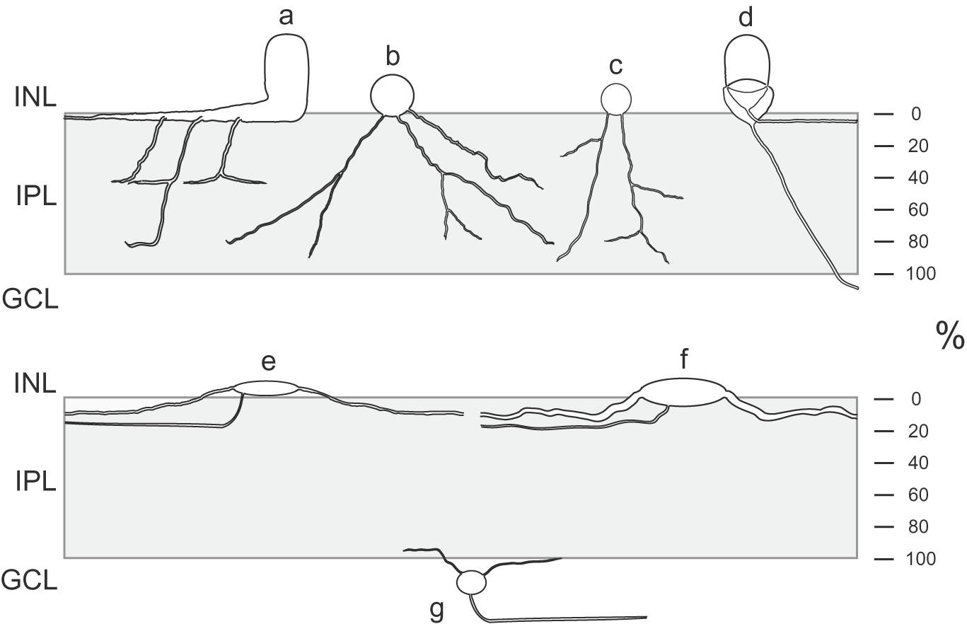
A greater number of nitrergic neuronal types, however, have a spatially restricted distribution (Fig. 10). These include efferent axons and their postsynaptic partners, the target amacrine cells, both of which have been shown to be NADPHd and nNOS-positive and are found exclusively in the ventral retina (Lindstrom et al., Reference Lindstrom, Nacsa, Blankenship, Fitzgerald, Weller, Vaney and Wilson2009). Also in the ventral retina, we identify bullwhip amacrine cells as weakly nNOS-positive and two types of large axon-bearing amacrine cell confined to the ventral retina. Two diffuse amacrine cells are confined to an equatorial band of the retina. Within the GCL, the heavily staining Brn3-negative ganglion cell is confined to the dorsal retina, and a lightly staining Brn3-positive cell is confined to the ventral retina. Lightly staining Brn3-negative cells are found throughout the retina, and we parsimoniously count this as a single population with ubiquitous distribution. Altogether, we estimate conservatively that there are nine neuronal types that have regional restriction.
Summary of the regional nitrergic cells of the inner retina. (a) Bullwhip cell confined to the ventral retina. (b) Large diffuse cell and (c) small diffuse cell, both of which are restricted to an equatorial band. (d) Efferent fiber and target amacrine cell entirely ventral. (e) Small axon-bearing amacrine and (f) large axon-bearing amacrine, both of which are exclusive to the low ventral retina. (g) A Brn3-negative ganglion cell branching in the extreme lower IPL is found in the dorsal retina. In addition, there is a Brn3-positive ganglion cell of unknown stratification found in the ventral retina.

Several neuronal types are described as regionally restricted in the turtle retina. Two types of amacrine cell that are immunoreactive for corticotropin releasing factor have been described, one of which is restricted to the visual streak and the other is exclusively ventral (Williamson & Eldred, Reference Williamson and Eldred1989; Zhang & Eldred, Reference Zhang and Eldred1992). In contrast to this, the mammalian retina has fewer reported examples of regionally restricted neurons. In the rat, amacrine cells staining with anti-phenylethanolamine-N-methyltransferase antiserum are confined to the dorsal retina (Versaux-Botteri et al., Reference Versaux-Botteri, Martin-Martinelli, Nguyen-Legros, Geffard, Vigny and Denoroy1986). Somatostatin-immunoreactive amacrine cell somata are confined to the ventral retina in both rabbit (Sagar, Reference Sagar1987) and cat (White et al., Reference White, Chalupa, Johnson and Brecha1990), whereas, in guinea pig, somatostatin-positive cells are confined to the retinal periphery (Spira et al., Reference Spira, Shimizu and Rorstad1984). There can be little doubt, however, that these somatostatin-positive amacrine cells are homologs in the three species since they all show peculiar branching at the retinal margin. Like the glucagon-immunoreactive bullwhip cells described in bird retina and its homolog in turtle (Wetzel & Eldred, Reference Wetzel and Eldred1997), these unusual neurons may all play a role in regulating retinal growth.
Conclusions
In this study, we show both that a surprisingly large number of neuronal types in the inner retina of chicken are likely to be nitrergic and that a majority of these are regionally specific. The paucity of similar studies focusing on other neuromodulators leaves unresolved the question of whether regional restriction is greater in nitrergic neurons than other retinal neurons. Leaving this question aside, the presence of regionally specific neuronal types implies that different processing strategies are appropriate to different regions of the retina. Since our understanding of the working of the inner retina is only rudimentary, the nature of these regional strategies is presently obscure. To sharpen an experimental attack on this problem, it might first be productive to pose the general question: what unique information, useful to a chicken, might be gleaned from the different regions of the visual image falling upon its retina? An answer to this question would have to begin with a more complete understanding of the visual world of the chicken.
Acknowledgments
Grant sponsor: National Eye Institute (EY04112 & EY12576 to M.W.); Australian Research Council, Queen Elizabeth II Fellowship to N.H.; National Health & Medical Research Council (Australia) Principal Research Fellowship to D.V. We thank Sal Borges for help in preparing the figures.