Twin–twin transfusion syndrome complicates 10–30% of monochorionic twin pregnancies, and results in increased incidence of cardiac malformations to more than 10 times as that of general population.Reference Karatza, Wolfenden and Taylor 1 , Reference Bahtiyar, Dulay and Weeks 2 Up to 70% of recipient twins experience some cardiac compromise, either anatomically or functionally, often pulmonary stenosis or endocardial fibroelastosis, with the ultimate possibility of foetal hydrops.Reference Karatza, Wolfenden and Taylor 1 – Reference Van Den Boom, Battin and Hornung 3 Donor twins rarely suffer from left ventricular outflow obstruction, and the possibility of coarctation of the aorta.Reference Van Den Boom, Battin and Hornung 3
Surgery has long been the superior method of treatment of neonatal coarctation.Reference Fiore, Fischer and Schwartz 4 , Reference Rao, Chopra and Koscik 5 Although successful repair in premature babies of weight <2 kg have also been reported,Reference Sudarshan, Cochrane and Jun 6 there is a relatively higher mortality and morbidity rates compared with mature newborns. Balloon angioplasty is generally safe; however, with significant restenosis and aneurysm formation rates,Reference Rothman, Galindo and Evans 7 its use as a palliative procedure in cases of high-risk surgery may be acceptable.Reference Bouzguenda, Marini and Ou 8
Case report
Male monozygotic twins were born prematurely at 30 weeks of gestation. The pregnancy was complicated by twin–twin transfusion. Birth weight of the donor was 900 g, and the recipient was 1550 g. On the 5th day of life, the donor was noted to have symptoms of heart failure; blood pressure in the right arm was 75/35, and unrecordable in the legs with diminished femoral pulses. Echocardiographic evaluation confirmed the presence of severe coarctation with depressed ventricular function (ejection fraction 45%). The Ductus arteriosus was closed. The recipient, the larger twin, had normal cardiac evaluation.
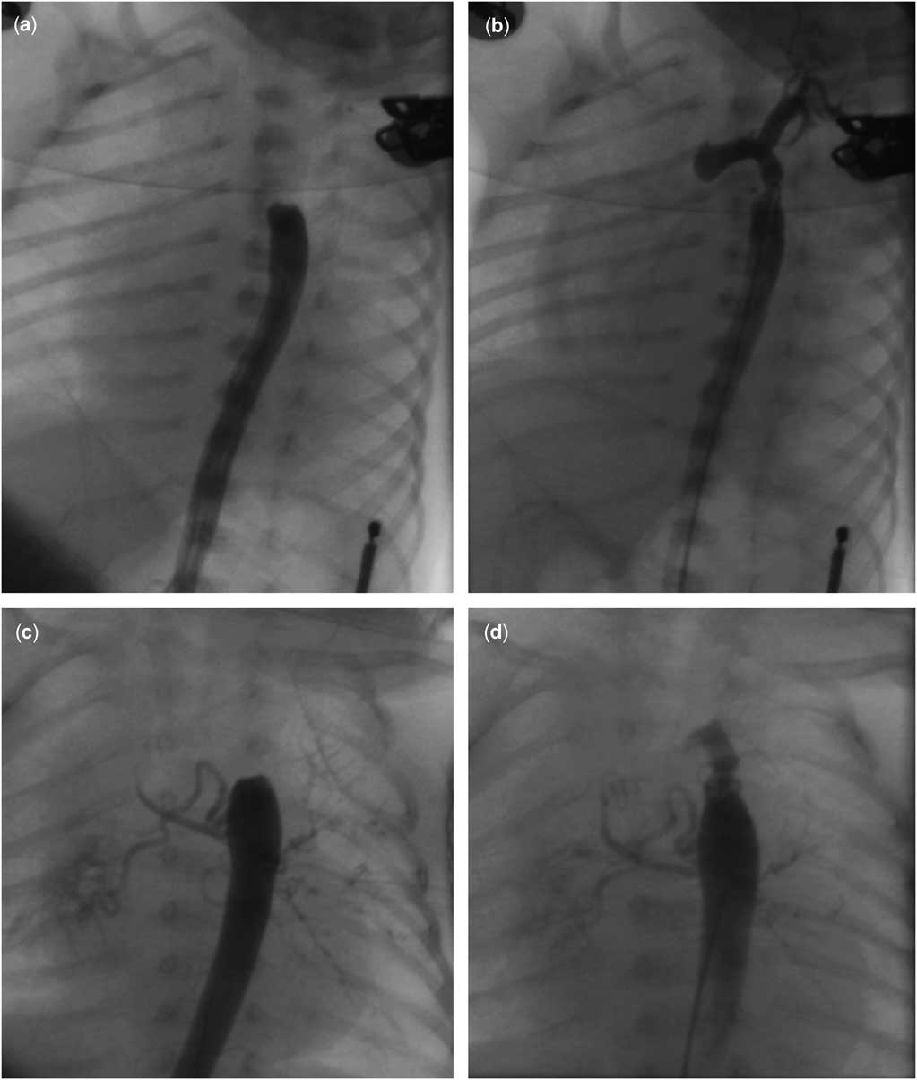
Owing to high-risk surgical intervention in the small and sick infant, decision was made to perform angioplasty. First, cardiac catheterisation was performed on the 7th day of life via umbilical arterial approach under sedation with midazolam. After hand injection aortography through a 3.5 Fr umbilical arterial catheter, a 0.014-inch floppy-tip guidewire was manipulated across the coarctation site. The aortic diameter measured 2.5 mm at the level of the diaphragm, which was used to select the balloon size. The narrowest diameter could not be assessed angiographically, but estimated to be <1 mm, which is the diameter of the umbilical catheter that could not be passed over a wire through the coarctation segment. A 2.5 mm coronary angioplasty balloon was inserted over the wire without a sheath or guiding catheter. The 10 mm long balloon was centred across the coarctation site and inflated to 10 atm. Direct pressure measurements showed residual gradient of 14 mmHg. Procedure was completed with no complications and with satisfactory angiographic results (Fig 1a and b; Supplementary Movies 1 and 2).
( a ) Descending aortogram obtained by hand injection through an end-hole umbilical catheter in antero-posterior projection in a 900 g infant. There is severe coarctation of the aorta with no retrograde filling of the aortic arch. ( b ) Aortogram obtained by hand injection following balloon angioplasty using 2.5 mm coronary balloon, which is equal to the aortic diameter at the level of the diaphragm. There is improvement in the flow across the coarcation, with intimal tear and mild residual obstruction. ( c ) Descending aortogram in the same patient 40 days later at a weight of 1500 g done by hand injection through a dilator of micropuncture kit (external diameter of catheter 0.035 inch), which reached only to the mid-thoracic aorta showing recurrence of severe coarctation with no retrograde filling of the aortic arch. ( d ) Aortogram with the same dilator placed to the midthoracic aorta following angioplasty using a 4 mm coronary balloon (same as aortic diameter at diaphragm) showing retrograde filling of the aortic arch with intimal tear and residual obstruction.

Over the next few days, heart failure improved clinically. Follow-up echocardiography showed the normalisation of cardiac function (ejection fraction of 73%) with moderate residual coarctation (peak gradient of 28 mmHg).
Over the next few weeks, the residual gradient across coarctation continued to rise gradually, reaching 45 mm Hg. The patient’s weight was 1500 g. Owing to high surgical risks, and after consultation with colleagues from cardiothoracic surgery at our and other institutions in the country, decision was made for percutaneous reintervention after the patient was declined surgery because of his low weight. A second catheterisation was performed with sedation. The right femoral artery was accessed with a 21-gauge needle and a 0.014-inch wire. The inner dilator of 15 cm-long micropuncture set (0.035 inch external diameter) was placed in the femoral artery and used for angiography, because of the unavailability of a 3 Fr sheath in the country, and because a 4 Fr sheath was believed to be too large for the patient’s femoral artery. Aortography was performed by hand in the thoracic aorta and showed filling of the aorta to the site of recoarctation, without retrograde flow into the arch (Fig 1c), and with no evidence of aneurysm. The descending aorta measured 4 mm at the level of the diaphragm, which was used to select the balloon size for the reintervention. A 0.014-inch wire was manipulated across the coarctation, the dilator was exchanged for a 4 mm coronary balloon, again without a sheath, and fully inflated to 12 atm. The balloon was then exchanged for the same dilator that was placed to the mid-thoracic aorta for angiography, which showed retrograde filling of the aortic arch (Fig 1d). During exchange procedures, haemostasis was maintained as much as possible by finger pressure. Estimated blood loss was 20 ml. Gradient was <10 mmHg measured by non-invasive pressure measurement.
The patient was observed in the hospital until his weight reached 1700 g, and then discharged home. Follow-up in the cardiology clinic 1 month after the procedure showed normal pulses; blood pressure in the right arm was 76/35 and in the leg 60/32. The last follow-up was at 13 months of age; echocardiography showed no aortic obstruction with peak gradient of 12 mm across the coarctation site, and with no evidence of aneurysm. Blood pressure was normal.
Discussion
Although cardiac disease in donors of twin–twin transfusion syndrome is generally less common than in recipients, coarctation appears to be more associated with donors. Mechanism of the development of coarctation is hypothesised to be due to decreased blood volume and placental return in the donor foetus that results in decreased blood flow through aortic isthmus, resulting in its underdevelopment.Reference Bahtiyar, Dulay and Weeks 2 , Reference Van Den Boom, Battin and Hornung 3
Low birth weight premature infants are considered high-risk group for both surgical and percutaneous interventions. Surgical complications include residual gradient, lung injury, bleeding, phrenic, and vagal nerve injury. Pecutaneous complications include bleeding, thrombosis, and vessel avulsion, as well as residual gradient and aneurysm formation. Risk increases exponentially for smaller patients.Reference Rothman, Galindo and Evans 7
In our patient, both the umbilical and femoral approaches were used during the repeated angioplasty to address recoarctation. Ideally, the use of a 3-Fr sheath would have been preferred because of the very small size of the infant.Reference Rothman, Galindo and Evans 7 Unfortunately, this sheath size was not available at our institution, or in the country. A 4-Fr sheath was too large for the patient’s arteries, and therefore both the catheterisations were performed with no sheath. This increases the risk for bleeding and vessel injury by the bare wire.
Although the gold standard for treatment of neonatal coarctation is surgery, percutaneous intervention was performed for the purpose of palliation because of the the small size of the patient. The decision in the first intervention at a weight of 900 g was justifiable for the significant surgical risk; however, the authors considered the second intervention at a weight of 1500 g only after the patient was declined for surgery at both the home institution and at another institution in the country. The authors do not advocate palliation for neonatal coarctation to be done routinely, and the decision should be considered individually for every patient. The angiographic results of these two interventions demonstrated residual obstruction. However, follow-up echocardiography imaging at 13 months showed no obstruction. This is believed to be secondary to favourable remodelling of the arterial wall, which has been described.Reference Rothman, Galindo and Evans 7 , Reference Fletcher, Nihill and Grifka 9 Further interventions in our patient were not needed; however, continuous follow-up is advised to evaluate for recurrence of coarctation and aneurysm formation.
Conclusion
Primary balloon angioplasty for small babies with severe aortic coarctation can be performed as a palliative procedure with the success in improving heart failure and relieving obstruction. Repetition of balloon angioplasty may be considered for individual patients if surgery is not possible.
Acknowledgment
None.
Financial Support
This research received no grant from any funding agency, commercial, or not-for-profit.
Conflicts of Interest
None.
Supplementary material
To view supplementary material for this article, please visit http://dx.doi.org/10.1017/S1047951114000365.