During normal pregnancy, there is a bidirectional passage of cells across the placenta, both from the fetus to the mother and vice versa (Ref. Reference Lo1). It has been appreciated for many years that fetal cells such as trophoblasts and fragments of syncytiotrophoblast circulate in the maternal circulation during pregnancy (Ref. Reference Douglas2). However, it has only become accepted more recently, that cells of fetal origin can be detected within the mother's circulation (Ref. Reference Bianchi3) and tissues (Ref. Reference O'Donoghue4), even decades after pregnancy. This persistence of relatively low numbers of fetal cells in the mother's circulation is termed ‘fetal microchimerism’ (Ref. Reference Liegeois5). When the traffic of cells is in the opposite direction and maternal cells reside in the child it is termed ‘maternal microchimerism’. This review will focus specifically on the phenomenon of fetal microchimerism.
A chimera is an organism in which there are two or more different populations of genetically distinct cells that are derived from separate zygotes. The term originates from the mythological chimera, which was a monstrous creature made up of the body of a lion, the head of a goat and the tail of a serpent (Ref. Reference Bazopoulou-Kyrkanidou6).
When a mother carries a population of cells derived from her children within her own tissues it indicates that she has herself become a chimera. This observation provokes many important questions. Cells of fetal origin will be semiallogenic, with half their genes originating from the father, and as such would be expected to elicit an immune response. It has indeed been shown that such a maternal cellular immune response directed against antigens expressed on the fetus can be detected in humans (Refs Reference Piper7, Reference Verdijk8, Reference James9). The persistence of these fetal cells, and the mother's immune response to them, are also thought to have important effects on the mother's health, which can be potentially either beneficial or detrimental. These clinical consequences, and the mechanisms behind them, will be the subject of this review.
Detection of fetal cells in the maternal circulation
Fetal cells are found at very low frequency in the maternal circulation and their identification requires a sensitive methodology. One strategy commonly adopted to facilitate the identification of fetal cells is the utilisation of the Y chromosome as a surrogate to identify cells of male origin in a female host. It is then presumed that male cells are derived from previous male children and are fetal in origin (Refs Reference Bianchi3, Reference Lo10). Initially, the impetus behind research in this field was the desire to isolate fetal cells from the maternal circulation during pregnancy as a source of fetal chromosomes or DNA to enable prenatal diagnosis of genetic disease, without the need for invasive tests (Ref. Reference Bianchi11).
Fluorescent in-situ hybridisation (FISH) with chromosome-specific probes for markers such as the Y chromosome can be used to microscopically identify fetal cells. In addition, extra probes can be used to recognise clinically important aneuploidies in fetal cells. Using this technique, fetal cells can be seen in samples of maternal blood at a frequency of less than 20 fetal cells per ml of maternal blood. This frequency was found to be 2-5 times higher in pregnancies with an aneuploid fetus (Ref. Reference Krabchi12). However, the practicality of using FISH directly on maternal blood is limited by the difficulty of screening the very large number of maternal cells necessary to detect rare fetal cells. Therefore several strategies have been used to enrich the fetal cells before FISH analysis. These include sorting cells by flow cytometry or by using antibody-labelled beads and magnetic columns to enrich populations of fetal nucleated erythrocytes, trophoblast cells and fetal leukocytes (Refs Reference Bianchi3, Reference Guetta13, Reference Lim, Tan and Goh14).
DNA amplification using the polymerase chain reaction (PCR) provides a sensitive technique to identify and quantify the presence of fetal cells in the maternal circulation during pregnancy. Quantitative PCR has also shown that the number of fetal cells in the maternal circulation is small during normal pregnancies, with a mean of one fetal nucleated cell per ml of maternal venous blood (Ref. Reference Bianchi3). Subsequent studies identified that following elective first trimester termination of pregnancy, approximately 500,000 nucleated fetal cells might transfer into the maternal circulation (Ref. Reference Bianchi15). It is therefore clear that a pregnancy does not need to continue beyond the first trimester for there to be a significant transfer of fetal cells to the mother. It is presumed that this fetal–maternal transfer would also occur in early miscarriage, but this has not yet been well studied. However, the particularly large fetal–maternal transfusion that is seen after a surgical termination of pregnancy might be partly due to mechanical disruption of the fetal–maternal barrier during the procedure.
The use of quantitative real-time PCR enables precise quantification of the transfer of fetal cells into the maternal circulation and minimises the risks of contamination because it is carried out in a closed system. A fluorescent probe is used, which elicits signal during the process of DNA amplification. Continuous optical monitoring of fluorescence shows the progress of the amplification reaction and enables accurate target quantification over a wide concentration range. A majority of studies now use this technology to quantify the presence of Y chromosome DNA as a measure of male fetal cell exchange. The single-copy gene encoding sex-determining region Y protein (SRY) (Refs Reference Lo10, Reference Zhong, Holzgreve and Hahn16, Reference Zimmermann17, Reference Lo18) or the multi-copy gene DYS14 (Refs Reference Zhong, Holzgreve and Hahn16, Reference Zimmermann17, Reference Lambert19, Reference Adams20) are both commonly detected as markers for the Y chromosome. Direct comparisons have shown that PCR for the multicopy DSY14 is more sensitive at low copy numbers than the single-copy SRY gene (Ref. Reference Zhong, Holzgreve and Hahn16).
Free fetal DNA is present in the maternal plasma during pregnancy and this increases with advancing gestation and is easier to detect than nucleated fetal cells. A direct comparison was performed using a sensitive quantitative real-time PCR assay. Although male fetal DNA could be detected free in the maternal plasma of all mothers with a male pregnancy, it could be detected in only 26% of mothers when the cellular fraction alone was examined (Ref. Reference Lo10). In those mothers in whom fetal cellular material was detected, the magnitude of DNA was in the range of 0.5–2.3 genome equivalents per ml (Ref. Reference Zhong, Holzgreve and Hahn16). However, several studies have shown the range to be much higher when plasma is examined for free fetal DNA, where it is in the range of 292–443 genome equivalents per ml (Refs Reference Lo10, Reference Lo21).
Longitudinal studies have been carried out during pregnancy that clearly show increases in the level of free fetal DNA throughout pregnancy, with a particularly marked increase over the last 8 weeks of pregnancy (Ref. Reference Lo18). However, following delivery, free fetal DNA is cleared very rapidly from maternal plasma, becoming undetectable after just 1 day. This rapid clearance implies that to maintain the high levels of fetal DNA found in maternal plasma during pregnancy, there must be a large liberation of free fetal DNA into the maternal circulation. In fact, it has been estimated that over 2000 copies of fetal DNA must be liberated into the maternal circulation every minute to maintain this steady state.
It is also interesting that there appears to be little correlation between the levels of fetal plasma DNA and fetal cellular material detected in the maternal circulation. This might reflect the differing cellular origins of the material, with free fetal DNA possibly being derived mainly from fetal trophoblast. This lack of correlation is an important consideration, because the easily detectable free fetal DNA within the plasma might also not correlate well with persistent fetal microchimerism, which would originate from intact fetal cells (Ref. Reference Lo10).
Persistence of fetal cells
That fetal cells can persist in the maternal circulation and tissues for extended periods after pregnancy was identified first in a murine model (Ref. Reference Liegeois5). This was extended to humans in 1996 when it was demonstrated using PCR for sequences on the Y chromosome that male haematopoietic stem cells, presumed to be of fetal origin, could be detected in healthy women up to 27 years after the birth of a male child (Ref. Reference Bianchi22). Subsequent research with fluorescent probes specific for the Y chromosome, has demonstrated persistent fetal cells in a wide range of maternal tissues. These include skin (Ref. Reference Ohtsuka23), liver (Ref. Reference Stevens24), thyroid (Refs Reference Srivatsa25, Reference Klintschar26, Reference Klintschar27), intestine (Ref. Reference Johnson28), lymph node (Ref. Reference Khosrotehrani29) and bone (Ref. Reference O'Donoghue4). It therefore appears that fetal cells are widely distributed within the body and become incorporated into a range of differentiated cell types. The traffic of fetal cells across the placenta and the establishment of fetal microchimerism in a range of maternal tissues is summarised in Figure 1.
Fetal microchimerism. (a) During normal pregnancy, fetal cells and free fetal DNA are transferred across the placenta into the mother. (b) Fetal cells can be detected in the maternal circulation during pregnancy. These cells are rare (~1 cell per ml) and increase in frequency during the third trimester. After delivery, small numbers of these fetal cells persist and engraft into the mother. Free fetal DNA can also be detected in the maternal plasma or serum during pregnancy. This is at a 3–10 times higher concentration than fetal cellular DNA. Following pregnancy, fetal free DNA appears to be rapidly cleared from the maternal circulation. However, fetal cells can persist in the mother's blood and tissues for decades. This is termed fetal microchimerism. (c) Techniques such as fluorescent in situ hybridisation (FISH) and polymerase chain reaction (PCR) have enabled the detection of fetal cells in a wide range of maternal tissues, including those indicated.

Murine studies have demonstrated that fetal cells are present in all previously pregnant mice and that these cells are detectable in a range of tissues (Ref. Reference Khosrotehrani30). In humans, fetal microchimerism has also been consistently shown to be common, even after normal pregnancy (Refs Reference Adams20, Reference Loubiere31) or after pregnancy loss (Ref. Reference Khosrotehrani32), but the exact proportion of women in whom this occurs, or the factors that are responsible are difficult to determine. This might be due in part to the diverse range of methodologies utilised and inconsistency in factors such as the quantity of DNA examined or the nature of the target gene. In addition, a number of diverse cohorts have been studied to date.
Some of the findings of these studies can appear difficult to explain. For example, 21% of women who did not report any previous male pregnancies were found to harbour low levels of male, presumed fetal, cells in their circulation. When the pregnancy history of these women was examined, both the prevalence and level of male microchimerism were greater in women with a history of induced abortion (Ref. Reference Yan33). However, some women in whom persistent male cells were found reported no previous known pregnancies. A possible explanation may be that such women have suffered a subclinical pregnancy loss. It has been shown from longitudinal studies that around 24% of pregnancies detectable by sensitive HCG assays result in early pregnancy losses before the pregnancy is clinically detected (Ref. Reference Wang34). However, microchimerism might also result from nonpregnancy-related sources such as blood transfusion or transplantation. The mother might also have been a twin herself, either from a recognised twin pregnancy or as a result of the ‘vanishing twin’ phenomenon might have been part of a twin pregnancy that was never recognised. Finally, it has also been suggested that microchimeric cells can originate from older siblings, having been transferred to the mother during pregnancy and then in a future pregnancy transferred from the mother to a subsequent child (Ref. Reference Yan33).
Fetal microchimerism and disorders of pregnancy
The passage of fetal cells across the placental barrier into the mother's circulation is dependent on the anatomical integrity of the placental ‘barrier’. It has been shown consistently that conditions that are associated with placental dysfunction can result in increased passage of fetal cells into the mother. These conditions can also include an abnormal fetal karyotype, such as trisomy 21, in which the frequency of fetal cells was reported to be six times higher in maternal blood than in control pregnancies (Ref. Reference Bianchi22). Increased frequencies of fetal DNA in the maternal circulation have also been reported in pre-eclampsia (Ref. Reference Holzgreve35) and preterm labour (Ref. Reference Leung36). Therefore elevated levels of fetal cells or free fetal DNA are a possible marker of disease activity in disorders associated with placental dysfunction.
Polymorphic eruption of pregnancy is a benign rash that can occur towards the end of pregnancy and resolves after delivery. Interestingly, fetal cells have been found in the skin lesions of this disorder and are not present in unaffected control skin. It is therefore hypothesised that this disorder may be associated with the migration of fetal cells to the skin (Ref. Reference Aractingi37).
The identity of fetal microchimeric cells
The term ‘pregnancy-associated progenitor cells’ (PAPCs) was proposed to describe the fetal stem cell population transferred during pregnancy. PAPCs have the capacity for engraftment in the maternal host and lead to multilineage differentiation and generation of a persistent fetal cell population. It has been suggested that an embryonic-like stem cell transferred during pregnancy could then differentiate into circulating, lineage-specific stem cells. It is, however, also possible that the established plasticity of bone-marrow-derived stem cells could allow circulating haematopoietic stem cells to transdifferentiate into another lineage (Ref. Reference Rossi38).
In the mouse, fetal stem cells with lymphoid progenitor capacity have been shown to transfer into the maternal circulation. They developed into functional T and B cells in the maternal host. T cells of fetal origin even underwent selection in the maternal thymus (Ref. Reference Khosrotehrani39). In humans, fetal haematopoetic and mesenchymal stem cells have been identified in maternal blood during and after pregnancy (Refs Reference O'Donoghue4, Reference Bianchi22, Reference O'donoghue40). It has also been possible to culture pure fetal mesenchymal stem cells from maternal blood after termination of pregnancy and differentiate these cells into bone and fat. In a striking piece of work, the bone marrow of women was examined many years after their last pregnancy and mesenchymal stem cells of male, presumed fetal, origin could be found in all the women examined who had previously given birth to sons (Ref. Reference O'Donoghue4).
The immune response to persistent fetal cells
The transfer of fetal cells into the maternal circulation has been shown to be a natural consequence of normal pregnancy. The question arises as to whether this can elicit a fetus-specific immune response either during or after pregnancy. The maternal immune system is modulated during pregnancy to promote acceptance of the fetal allograft, with a transient state of tolerance induced, despite maternal immune ‘awareness’ of fetal antigen (Ref. Reference Tafuri41). However, the maternal immune system can still mount an effective cellular immune response to viruses during pregnancy and generate virus-specific memory T cells (Ref. Reference Constantin42).
If a fetal-specific immune response does indeed exist, then any resultant immunopathology is likely to be modulated by the small cellular burden of fetal microchimeric tissue. In a close parallel with transplantation biology, immune recognition of fetal tissue by maternal T cells may be direct, by engagement with fetal antigen-presenting cells, or indirect, through interaction with fetal-derived peptides presented on the surface of maternal cells (Ref. Reference Nelson43). It has also been suggested that fetal T cells that have crossed into the maternal circulation, even if present in only relatively small numbers, may be directly involved in recognition of maternal antigen. Despite the small numbers of fetal T cells, this could have more wide-ranging effects on the mother's immune system through cytokine production and alteration of the immunological milieu (Ref. Reference Nelson43). Fetal cells might also be actively recruited to sites of tissue damage and disease. This may mean that immunopathological effects become concentrated in particular tissues and that small cell numbers could thereby have a significant local effect. The mechanisms by which fetal microchimeric cells could be directly involved in generating an immune response, or maternal T cells could be primed against fetal antigens and then generate a response against fetal microchimeric cells or subsequent pregnancies is summarised in Figure 2.
Potential mechanisms of immune responses to fetal microchimerism. Persistent fetal cells include immunologically active cells, such as cytotoxic T cells and T-helper cells. These may respond directly to maternal cells owing to variation in major or minor histocompatibility antigens. This can be considered a ‘fetal-versus-maternal’ reaction, analogous to ‘graft-versus-host’ disease. An alternative mechanism is that the maternal lymphocytes will be primed against fetal antigens. These can be presented directly by persistent fetal antigen-presenting cells or by an indirect pathway, with fetal minor histocompatibility antigens processed by maternal antigen-presenting cells. The development of primed maternal T cells directed against fetal antigens has been suggested to have a role in conditions such as autoimmune disease and recurrent miscarriage, and in antitumour effects. CTL, cytotoxic T lymphocyte; Th, T-helper cell; APC, antigen-presenting cell; TCR, T cell receptor; MHC, major histocompatibility complex.

Study of the maternal responses to antigen derived from the Y chromosome offers a convenient surrogate for maternal responses against the fetus. It has been demonstrated clearly in humans that CD8+ maternal T cells specific for fetally derived antigens can be isolated from peripheral blood of women post partum (Refs Reference Piper7, Reference Verdijk8, Reference James9). In particular, human leukocyte antigen (HLA) peptide tetramers containing immunogenic epitopes derived from SMCY on the Y chromosome have been used to isolate maternal T cells that are specific for fetal antigens. Responses were detected in 37% of women with a history of previous pregnancy and women who had had two or more sons demonstrated an HY-specific response in 50% of cases. T cells specific for fetal antigen are rare and their detection is facilitated by short-term T cell culture to allow cell expansion in vitro. These HY-specific CD8+ T cells are functional and produce interferon-γ (IFNG) following stimulation with either peptide or whole male cells in vitro. HY-specific T cell clones are able to kill male cells expressing the appropriate HLA allele, whereas female cells are not recognised (Ref. Reference Piper7).
Peripheral tolerance to fetal-specific T cells
Despite the documented evidence of fetus-specific immunity, it is clear that normal pregnancy is not followed by overt autoimmunity or evidence of responses similar to those observed in graft-versus-host disease. This has led to suggestions that there must be mechanisms of peripheral tolerance that can counteract the maternal immune response against persistent fetal antigens. These mechanisms are likely to be closely related to those by which the mother tolerates the fetal allograft during pregnancy, but there will be important physiological differences, such as the absence of any placental or decidual effects and a return of the hormonal environment to the nonpregnant state. If there are instances when tolerance is not complete following pregnancy it would be expected that maternal immune responses to persistent fetal cells could have important effects on maternal health. Several mechanisms exist by which peripheral T cell tolerance can occur, and these can be classified as T cell intrinsic mechanisms, such as ignorance, anergy and deletion, or T cell extrinsic mechanisms, which include regulatory T cells and antigen presentation by immature dendritic cells (reviewed in Refs Reference Walker and Abbas44, Reference Wood and Sakaguchi45).
There is a substantial literature examining animal models in which donor cell chimerism is associated with T cell unresponsiveness and graft acceptance (Refs Reference Lubaroff and Silvers46, Reference Cosimi and Sachs47, Reference Ehl48). What is more difficult to determine is whether the chimerism directly causes T cell unresponsiveness or is a consequence of it (Refs Reference Claas49, Reference Monaco50). The effect of antigen exposure during pregnancy on subsequent immunological tolerance has been investigated. Mouse models have demonstrated that exposure to male antigen through pregnancy makes these mice subsequently tolerant of syngenic male skin grafts and unable to generate cytotoxic responses to male antigen (Ref. Reference Simpson, Benjamin and Chandler51). The critical determinant of this effect in murine models seems to be the induction of regulatory T cells during antigen exposure during pregnancy. These regulatory T cells have been shown to have a potent effect on preventing rejection of the fetal allograft and subsequently inducing tolerance to fetal antigens (Ref. Reference Aluvihare, Kallikourdis and Betz52).
An increase in T regulatory cells during human pregnancy has also been seen and they are thought to have a role in preventing rejection of the fetal allograft (Ref. Reference Somerset53). It has recently been demonstrated in women after pregnancy, that not only can cytotoxic T lymphocytes specific for fetal cells be isolated, but a population of fetal-specific CD8+ T cells, which are regulatory in function can also be detected. This demonstrates how exposure to fetal microchimeric cells can trigger both tolerance and immunity simultaneously, and how the net result on maternal health and disease will be determined by the delicate balance of these effects (Ref. Reference van Halteren54).
Murine models have also demonstrated that clonal deletion within the thymus could be operating as a mechanism to maintain peripheral tolerance. Bonilla and co-workers showed that the presence of persistent microchimerism actively maintains cytotoxic T cell unresponsiveness by deletion of the self-reactive T cell repertoire (Ref. Reference Bonilla55). However, as described earlier, the detection of functional cytotoxic T cell responses in humans following pregnancy makes it less likely that this is the dominant effect providing tolerance in human females who display microchimerism following pregnancy (Refs Reference Piper7, Reference Verdijk8). With the increasing knowledge of mechanisms governing peripheral tolerance following transplantation and during pregnancy, there is now the opportunity to clarify in humans the mechanisms by which tolerance to microchimeric fetal cells is maintained after pregnancy.
Fetal microchimerism in transplantation biology
Fetal microchimerism has important implications for both the clinical practice of transplantation and for our understanding of transplantation biology. It is recognised that female donors are associated with a higher risk of graft-versus-host disease in the setting of stem cell transplantation and that this risk is particularly high when the donor has a history of pregnancy (Refs Reference Kollman56, Reference Flowers57). Graft-versus-host disease that occurs in the setting of HLA-matched bone marrow transplantation results from the activity of donor T cells that recognise minor histocompatibility antigens presented by the host tissues, thereby causing an immune response to these tissues (Ref. Reference Mutis58). In both murine models and human pregnancy, it has been shown that pregnancy can prime the maternal T cell immune response to minor histocompatibility antigens from paternal genes (Refs Reference Piper7, Reference Verdijk8, Reference James9). This observation thus provides a biological explanation for the effect of parity on increasing the incidence of graft-versus-host disease.
A crucial corollary of this effect is related to the observation that in bone marrow transplantation for malignant disease, donor T cell responses are also directed against the tumour itself. This is termed the ‘graft-versus-tumour’ effect, and is clearly demonstrated by the effectiveness of donor lymphocyte infusion. In this technique, patients who have relapsed after allografting are given an infusion of lymphocytes from the original donor to induce a potent immune-mediated ‘graft-versus-tumour’ effect (Ref. Reference Dazzi and Goldman59). This understanding of graft-versus-leukaemia explains why multiparous female donors are associated not only with an increased risk of graft-versus-host disease, but also with a reduced risk of leukaemia relapse (Refs Reference Gratwohl60, Reference Randolph61, Reference Gahrton62). It is therefore possible that a greater understanding of fetal microchimerism will assist in optimal selection of donors for transplantation, with the aim of optimising graft-versus-tumour responses while minimising the graft-versus-host effect.
The potential for these effects to be translated to solid organ transplantation is now being actively investigated. In renal transplantation, it has been suggested there may be responses by the female host against minor histocompatibility antigens if male kidneys are transplanted into females. These could potentially worsen graft survival through chronic rejection (Ref. Reference Gratwohl63). The persistence of fetal cells is effectively a naturally occurring haploidentical transplant. Understanding how this chimeric population is able to establish and persist in a fully functioning immune system is likely to reveal valuable lessons applicable to transplantation. Understanding and harnessing these processes is an exciting frontier in this field.
Fetal microchimerism and recurrent miscarriage
Recurrent miscarriage, which is defined as three or more consecutive miscarriages, is a common problem. There are many causes of recurrent miscarriage (reviewed in Ref. Reference Rai and Regan64), but in many couples no cause is identified, even after detailed investigation. In a proportion of these, immune-mediated mechanisms might play a role. The association between certain HLA types and recurrent miscarriage seems particularly strong in those women who have an initial successful pregnancy but then go on to suffer recurrent miscarriages. This is termed secondary recurrent miscarriage, and it might be this group for whom immunological causes are most important (Ref. Reference Kruse65). It has been established that T cell immunity might develop during pregnancy against minor histocompatibility antigens from the Y chromosome (Ref. Reference Piper7). Therefore, it has been suggested that the birth of a boy in the first pregnancy predisposes to subsequent recurrent miscarriage because of immunisation against Y chromosome antigens. Indeed, epidemiological evidence from Dutch cohorts with secondary recurrent miscarriage have found that a male first child is associated with a reduced chance of having another successful pregnancy (Refs Reference Nielsen66, Reference Christiansen67). The suggested mechanism for this is that the initial male pregnancy primes the maternal immune system and initiates a response to antigens, including those encoded by the Y chromosome. In subsequent pregnancies, T-cell-mediated responses to these antigens are therefore suggested to cause immune recognition of the developing fetus, resulting in miscarriage. This hypothesis is further supported by a recent finding in women with secondary recurrent miscarriage. If these women have a class II HLA type known to be able to present epitopes from the Y chromosome, this is associated with a reduction in chances of a subsequent live birth, but this effect is not seen in women with these HLA types who had a daughter (Ref. Reference Nielsen68). This hypothesis has not yet been supported by in vitro evidence of alterations in the immune response to fetal antigens in recurrent miscarriage patients. This is an important area for further research, because if such an effect was demonstrated, it would enable novel therapeutic strategies to be pursued.
Fetal microchimerism in autoimmune disease
The striking clinical similarities of autoimmune diseases such as systemic sclerosis with chronic graft-versus-host disease (Refs Reference Chosidow69, Reference Jaffee and Claman70) led to the suggestion that an association may exist between fetal microchimerism and autoimmune disease. Other circumstantial factors that support this concept are the female predominance of many autoimmune diseases and the peak incidence in women of childbearing age. If this theory is true, then diseases that have been traditionally considered as autoimmune processes might in fact have an alloimmune component (Ref. Reference Nelson71).
Initial work examined the association between fetal microchimerism and the autoimmune disease systemic sclerosis. A prospective, blinded study of microchimerism in women with systemic sclerosis identified fetal cells in peripheral blood using a quantitative PCR method. Seventeen women with systemic sclerosis were compared with 16 healthy women. Only nonpregnant women with previous sons were recruited. The results demonstrated greater quantities of male, presumed fetal, DNA in those women who had systemic sclerosis. However, fetal DNA was also detected in healthy controls (Ref. Reference Nelson72). Further studies have reported similar findings that fetal microchimerism is more prevalent in the peripheral blood mononuclear cells, skin lesions and other organs of women with the disease compared with controls (Refs Reference Lambert19, Reference Johnson28, Reference Artlett, Smith and Jimenez73, Reference Ichikawa74). Furthermore, the chimeric cells detected were more likely to be dendritic cells or B cells, which are cell types with an important role in disease pathogenesis (Ref. Reference McNallan75). T cells of fetal origin have also been isolated in women with systemic sclerosis, and these appear to have a Th-2-oriented profile, which is again consistent with the nature of this disease (Ref. Reference Scaletti76). However, even in healthy controls, it was clear that fetal microchimerism was present, and other studies have not definitively identified a correlation between systemic sclerosis and the presence or magnitude of fetal microchimerism (Refs Reference Selva-O'Callaghan77, Reference Murata, Nakauchi and Sumida78, Reference Gannage79). An additional finding was of increased HLA class II compatibility between patients with scleroderma and their children, compared with controls (Ref. Reference Nelson72). It has been suggested that HLA compatibility is associated with increased microchimerism by allowing fetal cells to cross the placenta undetected. An alternative hypothesis is that it might allow increased engraftment of fetal material by minimising the magnitude of the alloreactive immune response. Previous studies that have examined the prevalence of fetal microchimerism in the peripheral blood of women with systemic sclerosis using PCR are summarised in Table 1. Despite the variation observed between these studies, a majority show increased levels of fetal microchimerism in women with systemic sclerosis. Insight into the observed variation in the degree of association is provided by a recent study that examined clinical subtypes of systemic sclerosis. Fetal microchimerism was found in different cellular compartments in the distinct subtypes of the disease. This highlights the importance of the nature of the cell or tissue type examined and of having complete clinical information (Ref. Reference Rak80).
Prevalence of fetal microchimerism in the peripheral blood of women with systemic sclerosis

Despite the wide variations in patient cohorts studied and exact PCR methodology used, the majority of studies have shown an association between systemic sclerosis and the presence of fetal microchimerism.
aQuantitative PCR showed statistically significant increase in quantity of fetal DNA in women with systemic sclerosis despite no change in the percentage of cases in which microchimerism detected.
bQuantitative PCR showed circulating fetal microchimerism was quantitatively greater in systemic sclerosis patients compared with healthy women.
cDetection in whole blood.
dDetection in peripheral blood mononuclear cells.
Abbreviations: dcSSC, diffuse cutaneous systemic sclerosis; lcSSC, limited cutaneous systemic sclerosis.
A range of other autoimmune diseases has also been examined for their association with fetal microchimerism. Primary biliary cirrhosis shares pathological similarities with hepatic graft-versus-host disease and affects women in their childbearing years. Fanning and colleagues demonstrated by in situ hybridisation of liver biopsies that that cells containing a Y chromosome of presumed fetal origin could be identified in 8 out of 19 patients with primary biliary cirrhosis. They did not identify any cells of fetal origin in their control group, which consisted of women with chronic hepatitis C or alcoholic liver disease, and therefore concluded that there may be a role for microchimerism in the pathogenesis of the disease (Ref. Reference Fanning81). However, other studies have not found any such clear association (Refs Reference Tanaka82, Reference Rubbia-Brandt83, Reference Corpechot84, Reference Invernizzi85, Reference Schoniger-Hekele86). These studies also demonstrated that fetal cells can be detected in the liver of control women.
Sjogrens syndrome, an autoimmune disease of the exocrine glands, has been similarly scrutinised, and fetal microchimeric cells have been detected in inflammatory lesions of these patients (Ref. Reference Kuroki87); however, fetal cells were not detected in peripheral blood (Ref. Reference Toda88). Similarly, studies using nested PCR to examine samples of salivary gland tissue found male DNA of presumed fetal origin more frequently in patients with the disease. This finding was confirmed by FISH examination. In peripheral blood, fetal DNA was equally common in patients with Sjogrens syndrome and controls. This demonstrates the crucial importance of the nature of the tissues studied and reveals the tissue-specific distribution of fetal microchimeric cells. Variation in distribution of the microchimeric cells means that the nature of the tissue examined has a crucial influence on the findings (Ref. Reference Endo, Negishi and Ishikawa89).
The study of autoimmune thyroid disease is of particular interest because of its high postpartum incidence and the propensity for it to affect females. A murine model of experimental autoimmune thyroiditis showed the accumulation of fetal cells during pregnancy and the early post partum period (Ref. Reference Imaizumi90). In Hashimoto's thyroiditis an increase in fetal microchimerism in affected thyroid tissue of women with previous pregnancies was detected using PCR (Ref. Reference Klintschar26) and quantitative PCR (Ref. Reference Klintschar27). However, using FISH, others have found similar frequencies of fetal microchimeric cells in both Hashimoto's thyroiditis specimens and thyroid neoplasms (83% versus 75%, respectively) (Ref. Reference Srivatsa25). Fetal microchimerism was also detected more commonly in thyroid tissue of patients with Graves disease compared with those with thyroid adenomas. Although again, there was no difference in the frequency at which fetal cells were detected in peripheral blood (Ref. Reference Ando91). It still remains unclear whether fetal cells have a direct involvement in the aetiology of thyroid disease or whether its increased prevalence is a reflection of inflammation causing increased recruitment of fetal cells to sites of disease.
Systemic lupus erythematosis (SLE), a chronic multisystem autoimmune disease, has also been the subject of investigation to determine whether there is an association with fetal microchimerism. Initial findings of microchimeric cells detected by FISH in all histologically abnormal tissue types examined from a mother with SLE, but not in her histologically normal tissues, raised the prospects of a role for fetal microchimerism in the disease, and again demonstrated the diversity of tissues in which fetal microchimeric cells can be found (Ref. Reference Johnson92). However, subsequent studies have been much less clear. PCR of peripheral blood has demonstrated an increased frequency of microchimerism in women with SLE (Ref. Reference Abbud93), but other studies have demonstrated no association with the presence of disease, or any variation with disease activity (Ref. Reference Mosca94).
Making sense of these contradictory findings is made difficult because of variations in study design. The methodologies for detecting fetal cells vary widely. The nature of the tissue examined is also crucial. Studies examining peripheral blood often have different findings from those examining tissues that are the site of disease activity or inflammation. Furthermore, these studies have often suffered from inadequate controls and a paucity of clinical information. Clinical details, such as the extent of organ involvement, disease severity, subtype and disease activity might be crucial factors that are often omitted. Complete pregnancy histories are also not always included. This is vital information to establish the presence of alternative causes of fetal microchimerism other than live children. These include early pregnancy loss, termination of pregnancy, blood transfusion and the mother having been a twin herself.
Overall, the association of fetal microchimerism with systemic sclerosis seems to be the most clearly established. Whether the role of fetal cells is an integral component of the pathogenesis of the disease or whether the finding of fetal cells in affected tissues, or at increased frequency in blood, is a reflection of the recruitment or mobilisation of fetal cells as part of the inflammatory process, remains controversial.
Fetal microchimerism, immunity to fetal antigens and malignancy
It has been suggested that fetal microchimism or the immune response to persistent fetal cells could protect a mother against future malignancy. Conversely, persistent fetal cells recruited to sites of tumour development may in fact have a role in tumour pathogenesis. Maternal T cells exposed to fetal antigen during pregnancy can be primed during this exposure and have been shown to be functional and able to kill cells expressing these antigens (Ref. Reference Piper7). It is well recognised from experience with bone marrow transplantation, that maternal T cells specific for tumour-related minor histocompatibility antigens play an important role in eliminating tumour cells in what is termed the ‘graft-versus-tumour’ effect. Some minor histocompatibility antigens are only expressed on haematopoietic or tumour cells, and these are termed tumour-specific minor histocompatibility antigens. One such antigen, HA-1, has been shown to induce a maternal response during pregnancy (Ref. Reference Verdijk8). Priming of maternal T cells against these tumour-related antigens could provide a mechanism by which pregnancy could enhance immune tumour surveillance and protect a mother from future cancer.
In this context, it is fascinating that there is epidemiological evidence that fetal microchimerism may indeed be associated with a protective effect against malignant disease. A previous history of pregnancy is, in the long term, a protective factor against the future development of breast cancer and this effect is generally presumed to operate through hormonal mechanisms. However, Gadi and co-workers compared levels of fetal microchimerism in a cohort of women with breast cancer and a well-matched control cohort. They demonstrated that the cohort with breast cancer had a significant reduction in both the presence and quantity of fetal microchimeric cells (Ref. Reference Gadi95). It is possible that this potentially protective mechanism could either be due to the fetal cells themselves being immunologically active and eliminating tumour cells, or because, as suggested above, an increased level of fetal microchimerism is associated with an increased maternal cellular immune response against fetal and tumour antigens.
The presence of persistent fetal cells has also been investigated in a range of tumours that have arisen during pregnancy or in tumours from women who had previous pregnancies (Refs Reference Cha96, Reference Cirello97, Reference O'donoghue98, Reference Dubernard99, Reference Dubernard100, Reference Nguyen Huu101, Reference Nguyen Huu102). Dubernard demonstrated the frequent presence of fetal-derived cells in tumour stroma of breast carcinoma associated with pregnancy. They suggest that the unusual properties of fetal cells, such as the presence of HLA-G, which assists in immune evasion, could influence breast tumour pathogenesis (Ref. Reference Dubernard100). Further work with a murine model of pregnancy-associated breast tumours demonstrated that high-grade tumours contained significantly more fetal cells (Ref. Reference Dubernard99).
Fetal progenitor cells can also form endothelial cells that can be recruited to maternal inflammatory skin and form functional blood vessels (Ref. Reference Nguyen Huu102). Since angiogenesis is crucial in tumour pathology, it is interesting that fetal cells have been shown to home to maternal melanoma and give rise to blood vessels and lymphatics. It is hypothesised that recruitment of fetal progenitor cells to tumour sites could facilitate tumour growth or spread through this mechanism (Ref. Reference Nguyen Huu101). This is an area that deserves further investigation.
Involvement of fetal cells in tissue repair
As a result of investigations into the association of fetal microchimerism with autoimmune disease, it has become clear that fetal cells are present in a wide range of tissues, not only those affected by autoimmune processes. An alternative hypothesis to explain the presence of fetal cells in these tissues is that, rather than these cells having a detrimental effect, they could be homing to areas of damaged tissue and participating in the repair process.
Well-differentiated tissues of male fetal origin have been detected by FISH in both the thyroid (Ref. Reference Bianchi103) and liver (Refs Reference Stevens24, Reference Johnson104). It has been suggested that the progeny of fetal stem cells that crossed into the maternal circulation migrated to these diseased or damaged areas and have differentiated so they are morphologically indistinguishable from the maternal cells. This implies that fetal stem cells might be an alternative source of cells for tissue repair.
Murine models utilising green fluorescent protein labelling of fetal cells have enabled this concept to be examined further. Models of injury to the maternal liver and kidney following delivery of a fetus labelled with green fluorescent protein showed that fetal cells were detectable in maternal liver, spleen and kidney after injury, suggesting that they are playing a role in the repair process, or are at least migrating to areas of tissue damage (Refs Reference Khosrotehrani105, Reference Wang106). Fetal cell recruitment has also been shown to occur across the blood–brain barrier to sites of injury in the mouse brain (Ref. Reference Tan107).
Appendix specimens obtained from women with appendicitis during pregnancy were examined by FISH for male cells of presumed fetal origin. Male cell microchimerism was found in all women with a current male pregnancy or previous sons. These cells were more common in women with a current male pregnancy and at sites of increased tissue inflammation. This again suggests that fetal cells are present more generally at sites of maternal tissue injury during pregnancy and may have an active role in the repair of maternal tissues (Ref. Reference Santos108).
Conclusion
For many years, the conventional view of pregnancy has been that the fetal and maternal vascular systems are kept separate. More detailed observation has shown that transplacental passage of cells is in fact the norm. It is a startling discovery that fetal cells can persist in a wide range of a woman's tissues following pregnancy and she often becomes a chimera. Furthermore, it has been shown that maternal CD8+ T cell responses directed against fetal antigens can also be detected following pregnancy. However, the impact that the persistence of allogenic cells of fetal origin and the maternal immunological response to them has on the mother's health and whether it is detrimental or beneficial to the mother is still not clear.
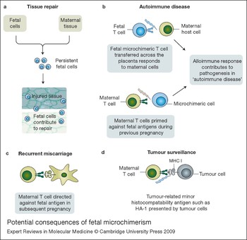
It has been suggested that autoimmune diseases, in particular systemic sclerosis, are associated with an increase in fetal microchimerism. However, whether fetal microchimeric cells play an important direct role in the pathogenesis of autoimmune disease, or whether this association is related to the recruitment of fetal cells to sites of tissue damage and inflammation is unclear. Indeed, it could be that the fetal cells are actually beneficial rather than detrimental and are playing a role in tissue repair. The possible implications of fetal microchimerism on maternal health and disease are summarised in Figure 3.
Potential consequences of fetal microchimerism. Fetal microchimerism and its associated immune responses have been suggested to have several effects on maternal health. (a) Microchimeric cells may actually be beneficial for maternal health owing to the recruitment of fetal cells to sites of tissue damage and their contribution to repair. (b) An association is suggested between fetal microchimerism and some autoimmune diseases. The microchimeric cells could be in themselves immune mediators, or the maternal immune response to the fetal cells might, through an alloimmune effect, contribute to diseases traditionally considered to have a solely ‘autoimmune’ pathogenesis. (c) The priming of maternal T cell responses to fetal antigens during pregnancy might also have a role in recurrent miscarriage. (d) Immune responses against tumour-related minor histocompatibility antigens are increasingly being found to have a role in immune surveillance against tumours. Some tumour-related antigens are also present on fetal cells, therefore pregnancy might have a role in priming the immune system against such tumour-related antigens.

This exciting field offers the promise of enhancing our understanding, not only of the immunology of pregnancy, but also of transplant immunology, autoimmunity and peripheral tolerance. The potential influence of fetal–maternal cellular trafficking is only just starting to be elucidated. There are many questions which remain unanswered, and important areas for future research include the possible role of microchimerism in recurrent miscarriage, the role of fetal microchimerism in cancer and the influence of microchimerism on outcomes following solid organ transplantation.
Acknowledgements
D.M.L. is supported by a Clinical Research Training Fellowship funded by Wellbeing Of Women. M.D.K. is funded by MRC grant RRAK11071 to investigate T-regulatory cells in pregnancy and K.P.P./P.A.H.M. are funded by LRF grant 9904. The authors thank the peer reviewers for their insightful advice and helpful comments.