Introduction
Acute kidney injury (AKI) is defined by an acute rise in serum creatinine levels or decrease in urine output (Ref. Reference Moe1). AKI is associated with increased short-term and long-term mortality and with progression of chronic kidney disease (CKD). However, there is no satisfactory therapy (Ref. Reference Bellomo, Kellum and Ronco2). Recent consensus definitions allow recognition of AKI at earlier stages and the even earlier concept of incipient AKI has been advanced (Refs Reference Perazella and Coca3, 4). This will hopefully facilitate clinical research examining early management strategies. A correct understanding of the pathophysiology of AKI will allow the identification of the specific therapeutic agents and earlier biomarkers for clinical practice. The bulk of renal cell mass is composed of tubular cells. Tubular cell death or injury is the main morphological feature of AKI. In addition, certain nephrotoxic drugs promote specifically proximal tubular cell injury or death. A key role of mitochondria in AKI was long suspected based on electron microscopy morphological assessments and ex-vivo and cell culture studies. Mitochondrial dysfunction might result in limited ATP availability and increased oxidative stress. Mitochondria are also responsive to cell stress and their role in apoptosis was recently reviewed (Refs Reference Sanz5, Reference Martinou and Youle6, Reference Vaux7, Reference Wang and Youle8). More recent evidence indicates that the specific therapeutic targeting of mitochondria beyond targeting apoptosis-specific regulatory factors may be beneficial in AKI. Clinical trials are addressing the potential of mitochondria-targeted approaches in AKI. This review is focused on in vivo evidence of mitochondrial injury or dysfunction during AKI and on how a better understanding of these processes may lead to novel diagnostic and therapeutic approaches that prevent AKI or accelerate recovery. Mitochondria-targeted therapies are discussed in-depth.
Mitochondria in tubular cells
Cells from tubular segments mostly involved in molecular transport are rich in mitochondria. The proximal convoluted tubule reabsorbs the bulk of the glomerular ultrafiltrate. Energy is provided by numerous elongated mitochondria (Fig. 1). Thick ascending cuboidal cells and distal convoluted tubule cells are also rich in mitochondria although not to the same extent as the proximal tubules.
Mitochondria in proximal tubular cells. Proximal tubular cells perform the bulk of energy-driven reabsorption of glomerular ultrafiltrate molecules (>1 kg of solutes daily). This is achieved by an increased apical cell membrane surface in the form of microvilli and a highly invaginated basolateral membrane containing transporters involved in solute reabsorption and in proximity to abundant mitochondria. The key ATP-consuming step is creation of a transmembrane ion gradient by the Na+/K+ ATPase in the basolateral membrane.

Mitochondrial structure
Mitochondria have a double membrane (Ref. Reference Eaton, Bartlett and Pourfarzam9) (Fig. 2). The outer mitochondrial membrane (OMM) contains enzymes as well as porins and translocases that allow the exchange of metabolites and cations with the cytosol. An excessive permeabilisation of the OMM allows the release of pro-apoptotic factors from the intermembrane space. The inner mitochondrial membrane (IMM) is highly impermeable and has a characteristic lipid pattern, which includes the presence of the phospholipid cardiolipin. The IMM contains transporters and the electron transport chain. Cardiolipin is required for the assembly, proper structure and activity of several mitochondrial respiratory chain complexes also electrostatically anchors cytochrome c to the IMM and may play an important regulatory role in cytochrome c release, which triggers the downstream events in apoptosis. Cardiolipin also plays an essential role in mitochondrial biogenesis (Ref. Reference Chicco and Sparagna10). Opening of the IMM mitochondrial permeability transition pore (MPTP) triggers cell death leads to mitochondrial swelling and cell death. The MPTP is formed by dimers of ATP synthase (Ref. Reference Giorgio11). Cyclophylin D (CypD) is thought to be an additional component while adenine nucleotide translocator (ANT) and voltage-dependent anion channel (VDAC) are now thought not to be required, since the pore opens in both ANT−/− and in VADC−/− animals (Refs Reference Kokoszka12, Reference Baines13). The mitochondrial matrix within the IMM contains the mitochondrial DNA (mtDNA) and tricarboxylic acid (TCA) cycle enzymes. The human mitochondrial genome encodes for 13 protein subunits of the oxidative phosphorylation chain, while most of the 1098 mitochondrial proteins in Mitocarta are encoded by nuclear DNA (Refs Reference Anderson14, Reference Pagliarini15).
Mitochondrial structure and site of action of mitochondria-targeted therapeutic strategies. OMM separates the intermembrane space from the cytosol. The intermembrane space contains molecules involved in the initiation of apoptotic cascades and signalling pathways that regulate respiration and metabolic functions. ROS and high mitochondrial Ca2+ may trigger MPTP opening. Persistent MPTP opening leads to the loss of mitochondrial membrane potential, mitochondrial swelling and outer membrane rupture. The exact composition of the MPTP remains debated. Cyclophilin D (CypD) and ATP synthase may be components. CsA, an inhibitor of CypD, may prevent MPTP opening. ROS are generated physiologically during oxidative phosphorylation. However, excess intramitochondrial calcium may cause uncoupling of oxidative phosphorylation and excess production of ROS. Excess ROS-induced oxidative stress may damage DNA, proteins and lipids, in addition to opening the MPTP. MitoQ, Mito-CP and SkQ1 are positively charged antioxidant agents that selectively accumulate in the mitochondrial matrix by virtue of the mitochondrial membrane potential. Bendavia is a SS peptide that accumulates in the IMM where it binds cardiolipin, protecting cardiolipin from peroxidation and thus stabilizing mitochondrial cristae and preventing cytochrome c (CytC) release. Bax and Bak recruit the mitochondrial apoptosis pathway and permeabilise the OMM to release cytochrome c and other proapoptotic mediators. Cristae remodelling favours the release of apoptogenic factors from mitochondria since 85% of cytochrome c is sequestered within the mitochondrial cristae by cardiolipin. EndG: endonuclease G; AIF: apoptosis inducing factor.
Mitochondrial turnover and dynamics
Mitochondrial biogenesis is the process of increasing the total mitochondrial mass and functional capacity or compensating the loss of damaged mitochondria by mitophagy (Ref. Reference Weinberg16). PPAR gamma coactivator-1 (PGC-1α) regulates mitochondrial biogenesis and associates with nuclear respiratory factors (NRFs) and mitochondrial transcription factor A (Tfam) to promote transcription of nuclear genes encoding mitochondrial proteins (Ref. Reference Wu17). In cultured proximal tubular cells enforced overexpression of PGC-1α increased mitochondrial number, respiratory capacity and mitochondrial proteins (Ref. Reference Rasbach and Schnellmann18). Mitophagy is the selective engulfment of mitochondria by autophagosomes and their subsequent catabolism by lysosomes that allows the selective degradation of defective mitochondria (Ref. Reference Jin and Youle19).
The mitochondrial dynamics concept includes changes in mitochondrial morphology, distribution and connectivity (tethering and fusion/fission) (Refs Reference Liesa, Palacin and Zorzano20) (Fig. 3). Under physiological conditions, mitochondria are elongated and filamentous. Mitochondrial fission and fusion are tightly regulated (Ref. Reference Bereiter-Hahn and Voth21, Reference Kageyama, Zhang and Sesaki22). The key regulators of mitochondrial fission are dynamin-related protein (Drp1) and mitochondrial fission-1 (Fis-1) (Refs Reference Labrousse23, Reference Smirnova24). Mitofusins 1 and 2 (Mfn1 and Mfn2) are required for fusion of the OMM and optic atrophy 1 (OPA1) for fusion of the IMM, cristae remodelling and protection from cytochrome c release (Reference Iglewski25). Drp1, Mfn1, Mfn2 and OPA1 are GTPases of the dynamin family.
Mitochondrial fusion and fission. Mitochondria move, fuse and divide shuttling between an elongated interconnected mitochondrial network or a fragmented discrete phenotype. Fission (Fis1 and Drp1) and fusion proteins (Mfn1, Mfn2 and OPA1). Drp1 cycles between cytosol and the OMM. High cytosolic Ca2+-activated calcineurin dephosphorylates and activates Drp1, leading to Drp1 translocation to the mitochondria to induce mitochondrial fission and fragmentation that under pathological conditions favour mitochondria-initiated cell death. Fis1 on the OMM may function as a receptor for Drp1. mdivi-1 is a pharmacological inhibitor of Drp1 and of mitochondrial fission that prevents experimental kidney injury.
Mitochondrial functions in health and disease
The main physiological mitochondrial functions are generation of energy in the form of ATP, regulation of the redox state, intracellular calcium and iron homoeostasis, trafficking of phospholipids, regulation of danger signalling and synthesis of 1,25-(OH)2-vitamin D (Ref. Reference Nunnari and Suomalainen26). Under stress conditions, mitochondria also contribute to the regulation of apoptosis, necrosis and autophagy.
Mitochondria generate ATP to provide energy for the cell through a process called aerobic respiration or oxidative phosphorylation, which takes place at the electron transport chain (respiratory chain) in the IMM (Ref. Reference Lanza and Nair27). Maintenance of the mitochondrial membrane potential by electron transport is critical for ATP generation.
Reactive oxygen species (ROS) are generated as by-products of oxidative phosphorylation. Mitochondria are both key generators and primary targets of oxidative stress. Up to 2% of the oxygen consumed by mitochondrial oxidative phosphorylation undergoes one electron reduction to generate the superoxide radical, and subsequently other ROS such as hydrogen peroxide and the hydroxyl radical (Ref. Reference Kowaltowski and Vercesi28). ROS have key roles in cell signalling (Ref. Reference Thannickal and Fanburg29). However, uncoupling of oxidative phosphorylation secondary to excess Ca2+ may cause excess mitochondrial ROS generation which may lead to irreversible oxidative damage of mtDNA, membrane lipids and proteins, resulting in mitochondrial dysfunction and ultimately cell death.
Mitochondria maintain the homoeostasis of cytosolic calcium by taking up and buffering Ca2+ in the matrix during physiological Ca2+ signalling. However, excess mitochondrial calcium may uncouple oxidative phosphorylation, generate excess ROS and trigger cell death by opening the MPTP in the IMM. The recent availability of mice lacking the mitochondrial calcium uniporter (MCU−/− mice) confirmed the existence of additional calcium channels, the role of MCU in rapid mitochondrial uptake calcium and the role of rapid matrix calcium changes in regulating mitochondrial respiration, activity of TCA enzymes such as pyruvate dehydrogenase and MPTP opening (Ref. Reference Pan30). However, deficiency in MCU did not prevent eventual cell death during ischaemia–reperfusion.
Mitochondria are key regulators of iron metabolism. Iron is a cofactor for enzymes involved in many metabolic processes, but may be harmful by enhancing the production of ROS and contributes to ferroptosis. Iron is transformed into its biologically available form in the mitochondrion by the iron–sulphur (Fe/S) cluster and haeme synthesis pathways (Ref. Reference Levi and Rovida31).
Mitochondria are key regulators of apoptotic, necrotic and autophagy cell death. OMM and IMM permeabilisation play key roles in cell death. OMM permeabilisation may activate apoptosis by releasing into the cytosol proteins from the intermembrane space that activate caspases (cytochrome c, SMAC/DIABLO) or promote caspase-independent cell death (AIF, endonuclease G) (Ref. Reference Tait and Green32). Integrity of the OMM is regulated by interactions between pro- and anti-apoptotic members of the Bcl-2 protein family (Ref. Reference Chipuk33). Necrosis following ischaemia/reperfusion results from the opening of the MPTP in the IMM, although additional forms of cell death are activated (Ref. Reference Linkermann34). Apoptosis and MPTP opening may interact, depending on cell type and death stimulus, to amplify death signals (Ref. Reference Kinnally35). MPTP opening results in mitochondrial swelling and cristae remodelling, thus facilitating a more complete release of mitochondrial pro-apoptotic factors (Ref. Reference De36).
Mitochondria regulate autophagy through generation of ATP and regulation of ADP and intracellular calcium levels (Ref. Reference Cardenas37). Mitochondrial Ca2+ inhibits autophagy through repression of AMP-activated protein kinase (AMPK) activity (Ref. Reference Eng38). Nutrient starvation reduces mitochondrial ATP generation, activation of AMPK and promotes autophagy (Refs Reference Egan39, Reference Inoki, Zhu and Guan40, Reference Kim41).
Mitochondria injury promotes inflammation by releasing ROS (Refs Reference West, Shadel and Ghosh42, Reference Bulua43), regulating phospholipid biosynthesis, ceramide and cholesterol metabolism, and glycosphingolipid anabolism (Refs Reference Vance and Shiao44, Reference Lebiedzinska45) and decreasing mitochondria-generated anti-inflammatory molecules such as 1α,25-(OH)2-vitamin D (Refs Reference Sanchez-Nino46, Reference Rojas-Rivera47).
Mitochondria in AKI
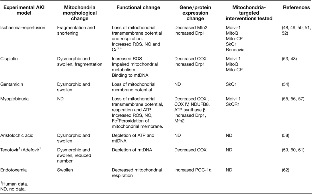
AKI has been associated with altered morphology and function of mitochondria as well as with altered expression of mitochondria-related genes and proteins indicating mitochondrial injury (Table 1). Theoretically, these mitochondrial changes and mitochondrial injury itself may be causes or consequences of AKI and may represent pathological mechanisms, repair mechanisms or simple epiphenomena. The recent availability of genetic means to target specific mitochondria proteins and interfere with mitochondria-specific pathways and also of drug specifically targeting mitochondria has provided hard data to support previous hypothesis and their successful use to prevent AKI suggests that indeed mitochondrial changes promote kidney injury, as discuss below.
Chemical structures of key mitochondria-targeted molecules. (a) MPTP opening inhibitor Cyclosporin A. (b) Mitochondrial division inhibitor-1 mdivi-1. (c) SS peptide: Bendavia (SS-31). (d) Coenzyme Q10 mimetic MitoQ. (e) Ubiquinone mimetic SkQR1. (f) Ubiquinone mimetic SkQ1. (g) SOD mimetic Mito-CP.
Mitochondrial morphological and functional changes, expression of genes and proteins and mitochondria-specific targeting in selected experimental models of AKI
1Human data.
ND, no data.
Morphological mitochondrial changes
The morphology of tubular cell mitochondria is altered in AKI induced by ischaemia/reperfusion, endotoxemia, and nephrotoxic agents such as cisplatin (a cancer chemotherapeutic agent), gentamicin (an aminoglycoside antibiotic), aristolochic acid (herbal nephrotoxin linked to Balkan nephropathy and medicinal herbs use) and the anti-retroviral drugs tenofovir and adefovir (Refs Reference Mukhopadhyay53, Reference Hall63, Reference Porta64, Reference Jiang58, Reference Abraham, Ramamoorthy and Isaac65, Reference Plotnikov66).
Experimental ischaemia/reperfusion resulted in rapid proximal tubular cell mitochondrial fragmentation and shortening, while mitochondrial morphology was better preserved in distal tubules (Refs Reference Hall63, Reference Brooks48). This is a sign of mitochondrial injury that may lead to cell death. Indeed, cell stress may result in mitochondrial fragmentation and contribute to OMM permeabilisation and the release of apoptogenic factors from the mitochondrial intermembrane space. In ATP-depleted cultured murine proximal tubular cells mitochondrial fragmentation was an early event, preceding OMM disruption, Bax activation and apoptosis (Ref. Reference Brooks48). Hypoxia-induced loss of mitochondrial fusion activity contributed to mitochondrial shortening (Ref. Reference Liu and Hajnoczky67).
Eight days of gentamicin exposure causes AKI, extensive proximal tubular cell death and mitochondria swelling and dysmorphia, while mitochondrial morphology was preserved in distal tubules and collecting ducts (Ref. Reference Hall63). Mitochondria swelling is a sign of mitochondrial injury and dysfunction. In aristolochic acid- or cisplatin-induced AKI, proximal tubule mitochondria appeared swollen and dysmorphic, with loss and disorientation of cristae and an apparent decrease in mitochondrial quantity (Refs Reference Jiang58, Reference Brooks48, Reference Zsengeller68). Cisplatin-induced AKI was associated with tubular cell mitochondrial fragmentation (Ref. Reference Brooks48). Tenofovir is widely used to treat HIV infection and hepatitis B virus (Ref. Reference Fernandez-Fernandez69). Human tenofovir-associated renal dysfunction was characterised by acute proximal tubular degenerative changes and dysmorphic mitochondria (Ref. Reference Herlitz59). Mitochondria size and shape were highly variable, ranging from small and rounded to markedly enlarged and swollen. Cristae were lost or disoriented. The number of mitochondria may be markedly reduced (Ref. Reference Herlitz59). Adefovir may also result in dysmorphic tubular mitochondria, including enlarged mitochondria and loss and disorientation of cristae (Ref. Reference Tanji60).
In experimental endotoxemia, tubular mitochondria were swollen (Ref. Reference Tran62). All these morphological changes are expected to contribute to mitochondrial dysfunction. Thus, cristae in the IMM increase the surface area to allow adequate ATP production. Cell stress may result in mitochondrial fragmentation and contribute to OMM permeabilisation and the release of apoptogenic factors from the mitochondrial intermembrane space. As discussed below, therapeutic interference with mitochondrial division provided proof of the pathogenic nature of these changes.
Functional mitochondrial changes in AKI
Mitochondrial injury is expected to result in mitochondrial dysfunction, leading to inability to fulfil mitochondrial function such as generation of ATP and favouring oxidative stress and, thus, leading to sublethal or lethal cell injury. Indeed, mitochondrial dysfunction was observed in AKI induced by ischaemia/reperfusion (Refs Reference Brooks48, Reference Plotnikov70, Reference Burke71, Reference Sun72, Reference Weinberg73, Reference Devarajan74), nephrotoxic drugs (Refs Reference Weinberg73, Reference Pfaller, Gstraunthaler and Willinger75, Reference Izzedine76, Reference Feldkamp, Kribben and Weinberg77, Reference Hall and Unwin78, Reference Jassem79, Reference Jassem and Heaton80, Reference Nath81), endotoxemia (Ref. Reference Tran62) and myoglobinuria (Ref. Reference Funk and Schnellmann55). Recently, real-time in vivo imaging techniques were used to confirm that mitochondria are a major source of ROS production in tubular cells in vivo, mainly in proximal tubular cells. However, most data have been generated by measuring tissue oxygen consumption, or by respiratory chain function tests conducted on isolated organelles (Ref. Reference Hall63).
In ischaemia/reperfusion, the mitochondrial transmembrane potential and mitochondrial respiration decrease while mitochondrial ROS and NO and intracellular and mitochondrial Ca2+ increase (Refs Reference Plotnikov66, Reference Plotnikov70, Reference Jassem and Heaton80, Reference Beiral82, Reference Shah and Walker83, Reference Zager84, Reference Crompton85). This results in ATP depletion, lipid peroxidation, OMM permeabilisation, release of proapoptotic proteins and MPTP opening (Refs Reference Hall and Unwin78, Reference Nath81, Reference Funk and Schnellmann55, Reference Bonventre86, Reference Bonventre and Weinberg87, Reference Bosch, Poch and Grau88). Lack of O2 inhibits respiration during hypoxia while reoxygenation results in rapid restoration of respiration and increased mitochondrial ROS production (Refs Reference Wang89, Reference Korge, Ping and Weiss90). However, during reoxygenation proximal tubule mitochondria remain impaired in their energy-generating function (Ref. Reference Weinberg91) and a respiratory defect for complex I-dependent substrates persists (Ref. Reference Weinberg73). Thus, ATP recovery is often delayed and contributes to persistence of AKI. In ischaemia mitochondrial transmembrane potential abruptly decreases in proximal tubules but is longer maintained in distal tubules (Ref. Reference Hall63).
Drug-induced AKI also impacts on mitochondrial function. Aristolochic acid reduced kidney ATP and mtDNA/nuclear DNA ratio (Ref. Reference Jiang58). Cisplatin decreased COX enzyme activity in the proximal tubules, but not in the distal tubules (Ref. Reference Zsengeller68). Gentamicin induced sporadic changes in mitochondrial membrane potential in the proximal, but not in the distal tubules. These changes were highly variable among animals and were preceded by changes in nonmitochondrial structures (Ref. Reference Hall63). In aristolochic acid-induced nephrotoxicity, the activity of respiratory complex I, which is partly encoded by mtDNA, was more impaired than that of respiratory complex II, which is completely encoded by nuclear DNA.
The observed attenuation of mitochondrial respiration and oxygen consumption during sepsis is hypothesised to preserve renal oxygenation despite reduced renal blood flow (Refs Reference Tran62, Reference Heyman92). Preservation of oxygen supply would decrease hypoxic injury, resulting in sublethal rather than lethal cell damage. This would underlie the scant morphological evidence for tubular damage in endotoxemia (Refs Reference Tran62, Reference Heyman92).
In myoglobinuria the mitochondrial transmembrane potential, mitochondrial respiration and ATP levels decrease while mitochondrial ROS and NO, intracellular and mitochondrial Fe3+ and lipid peroxidation increased (Refs Reference Plotnikov66, Reference Funk and Schnellmann55).
Expression of mitochondrial proteins
The study of the expression of genes encoding mitochondrial proteins and their gene products may provide clues to the pathogenesis of mitochondrial changes and injury during AKI and to the molecular mechanisms of the structural and functional mitochondrial abnormalities. Changes in the expression of genes or levels of proteins have been described for mtDNA-encoded proteins, as well as for biogenesis, oxidative phosphorylation, fusion, fission and oxidative stress and cell death regulators. Understanding the mechanisms and consequences of these changes may provide novel therapeutic targets. Observed changes are dynamic in the course of injury and several temporal patterns have been described. Interestingly, discrepancies have been found between gene and protein expression, as well as between different forms of AKI. The reason for these discrepancies is not immediately apparent and may represent differences in time course or severity of injury related to different stages of injury (initiation, established injury and recovery or failure to recover), true differences between models or heterogeneity in mitochondrial responses in different cell types or within the same cell. Clearly, more research in needed to clarify these issues.
Oxidative phosphorylation
Some oxidative phosphorylation proteins are encoded by mtDNA and other by nuclear DNA. Oxidative phosphorylation is required to generate ATP and decreased expression of genes involve in this process may compromise energy generation and cell survival while favouring oxidative stress.
The expression of mitochondrial genes encoding proteins may change during AKI. In myoglobinuria-induced AKI serum creatinine peaked at 24 h, but ND6 and COX1 mRNA did not decrease until time points 72 and 144 h, respectively. By contrast down-regulation of COX1 protein levels preceded mRNA changes. COX1 was already decreased at 24 h and persisted decreased at 144 h. However, ATP production was maintained at 24 h but increased by 72 h. Thus, there was dissociation between gene and protein expression of mitochondria-encoded genes, renal function and ATP production. Ischaemia–reperfusion injury also resulted in peak serum creatinine at 24 h. At this time point renal ATP levels were increased and ND6 and COX I mRNA levels were decreased and remained decreased at 72 and 144 h after injury. COX I protein levels decreased after ischaemia/reperfusion injury and did not recover over 144 h (Ref. Reference Funk and Schnellmann55). In cisplatin-induced AKI, COX-1 protein levels were maintained but COX enzyme activity in proximal tubules was decreased (Refs Reference Brooks48, Reference Zsengeller68). Adefovir induced mtDNA depletion and COX subunit I deficiency, while COX subunit IV, iron sulphur subunit of respiratory complex III and succinate dehydrogenase, all encoded by nuclear DNA, were preserved (Refs Reference Tanji60, Reference Kohler61). Aristolochic acid reduced kidney mtDNA content but there are no reports on mitochondrial gene expression (Ref. Reference Jiang58).
In rat myoglobinuric AKI and murine ischaemic AKI respiratory proteins NDUFB8, ATP synthase β and COX IV were acutely decreased and did not recover by 144 h (Ref. Reference Funk and Schnellmann55).
Biogenesis regulators
In experimental endotoxemia, kidney transcriptomics revealed an overrepresentation of differentially expressed nuclear genes related to oxidative phosphorylation. The expression of PGC-1α and of PGC-1α-dependent oxidative phosphorylation genes was initially decreased and increased again upon normalisation of kidney function, although some, such as Cox5b, remained down-regulated throughout recovery (Ref. Reference Tran62).
In rat myoglobinuric AKI and murine ischaemic AKI markers of mitochondrial biogenesis, such as PGC-1α and PGC-1-related co-activator (PRC), were increased (Ref. Reference Funk and Schnellmann55).
Increased mitochondria biogenesis is thought to provide the means for recovery of mitochondria mass that generates energy for tissue regeneration.
Fission/fusion regulators
In rat myoglobinuric AKI and murine ischaemic AKI expression of mitochondrial fission protein Drp1 was elevated after injury, whereas the fusion protein Mfn2 was elevated after myoglobinuric AKI but decreased after ischaemia/reperfusion (Refs Reference Funk and Schnellmann55, Reference Tang56). Drp1 is up-regulated in both animals and cell culture models of cisplatin and ischaemia–reperfusion kidney injury. Silencing Drp1 (in vitro) or treatment with mdivi-1 (in vivo) blocked mitochondrial fragmentation (Ref. Reference Brooks48). Mfn1- or Mfn2-null cells are more sensitive to cisplatin-induced apoptosis (Ref. Reference Brooks93). Mfn2-deficient cultured proximal tubular cells showed baseline mitochondrial fragmentation and were highly sensitive to Bax activation, cytochrome c release, and apoptosis following ATP depletion (Ref. Reference Gall94). These functional studies confirm that a protein expression pattern that favours mitochondrial fragmentation is deleterious.
Oxidative stress and apoptosis regulators
Changes in the expression of multiple mitochondria-related pro- and anti-apoptotic regulators have been observed in AKI since the 1990s (Refs Reference Sanz5, Reference Justo95, Reference Lorz96). In addition, the expression of enzymes involved in oxidative stress and defence against oxidative stress also changes, including SOD, catalase, glutation peroxidase and others (Ref. Reference Mortensen97). In general, increased expression of proapoptotic proteins maybe deleterious by favouring cell death, while increased expression of anti-oxidant and anti-apoptotic proteins has been shown to represent an adaptive response.
Drivers of mitochondrial changes
Understanding the promoters and mediators of mitochondrial injury in AKI may help design novel therapeutic strategies. For example, ischaemia/reperfusion recruits intracellular signals that destabilise mitochondria including oxidative stress, protein kinases such as PKC delta and JNK, calpain, pro-apoptotic members of the Bcl-2 family, increased intracellular calcium levels and mitochondrial calcium overload (Refs Reference Szeto49, Reference Devalaraja-Narashimha, Diener and Padanilam98, Reference Gottlieb99). In addition, some of the cytokines and transcription factors recruited during injury impact on mitochondrial damage and function. Examples include, but are not limited to, cytokines of the TNF superfamily and the transcription factors HIF-1 and STAT3 (Refs Reference Tran62, Reference Shi100, Reference Zhang101, Reference Wegrzyn102).
TNF down-regulates PGC-1α and protein levels of the complex I, resulting in reduced oxygen consumption and increased ROS production in tubular cells (Ref. Reference Tran62). Another member of the TNF superfamily, TWEAK (Ref. Reference Sanz, Sanchez-Nino and Ortiz103), which promotes nephrotoxic, ischaemic and obstructive AKI (Refs Reference Sanz104, Reference Ucero105), also decreases the expression of PGC1α and genes required for mitochondrial oxidative phosphorylation in nonrenal organs such as the heart (Ref. Reference Shi100).
Hypoxia activates the HIF transcription factor that drives the expression of genes that protect from hypoxia. Thus, HIF-1 induces the expression of erythropoietin and of glycolytic genes. Increased glycolysis will maintain energy production when low oxygen does not support oxidative phosphorylation at the mitochondria. In addition, HIF-1 induces pyruvate dehydrogenase kinase 1 (PDK1) expression. PDK1 phosphorylates and inhibits pyruvate dehydrogenase, thus preventing pyruvate from fuelling the mitochondrial TCA. As a result, mitochondrial oxygen consumption decreases, leading to a relative increase in intracellular oxygen tension that prevents cell death under hypoxic conditions (Ref. Reference Papandreou106). HIF-1 also negatively regulates mitochondrial biogenesis by decreasing c-myc and c-myc-dependent PGC-1β expression, at least in kidney carcinoma cells (Ref. Reference Zhang101). Interestingly, the tyrphostin AG490 increases HIF-1α protein levels and HIF-1 transcriptional activity in kidney tubular and cancer cells (Ref. Reference Fernandez-Sanchez107). AG490 had been previously shown to protect from ischaemia–reperfusion-induced AKI (Refs Reference Goncalves108, Reference Neria109). Recently, STAT3, a transcription factor activated by cytokines, was found in mitochondria. STAT3 deficiency resulted in decreased activity of complexes I and II of the electron transport chain. The transcription factor and the mitochondrial functions of STAT3 could be dissociated in mutant proteins (Ref. Reference Wegrzyn102).
Therapeutic perspectives
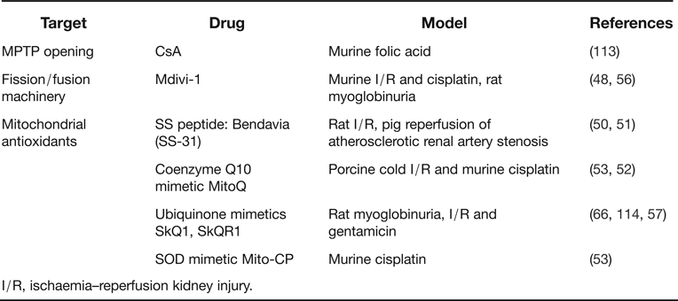
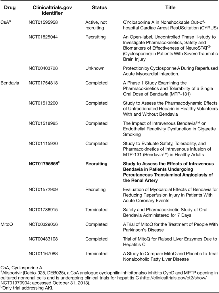
As a consequence of their involvement in the pathogenesis of renal cell injury mitochondria are therapeutic targets in the treatment of renal disease. However, it is often difficult to dissect the specific mitochondrial actions of therapeutic drugs from secondary mitochondrial function improvement as a consequence of an improvement in nonmitochondrial injurious mechanisms. Inhibition of mitochondrial pathways for apoptosis has been extensively studied and shown to be a successful prophylactic strategy in experimental AKI, although clinical translation has not yet occurred (Ref. Reference Ortiz110). During the course of these studies evidence emerged for a role of apoptosis regulatory proteins in the regulation of mitochondrial dynamics. Thus, Bak knockout mice were protected from ischaemic AKI and Bak deficiency prevented mitochondrial fragmentation (Ref. Reference Wei111). Bax and Bak promote mitochondrial fission by stimulating Drp1 sumoylation. This action is not prevented by anti-apoptotic Bcl-2 family members, thus dissociating mitochondrial fission–fusion dynamics and apoptotic events (Ref. Reference Sheridan112). Besides the strategies targeting mitochondrial apoptosis pathways, additional mitochondria-specific strategies include prevention of MPTP opening, targeting the fission/fusion regulatory system and mitochondria-targeted antioxidants (Table 2) (Fig. 4). Some of these agents are undergoing clinical trials in diverse clinical conditions, including prophylaxis of AKI (Table 3). However, to date all interventions have been prophylactic, that is, the medication was administered before the induction of kidney injury. This may be of interest in specific contexts, where the risk of AKI is very high, such as prevention of primary cadaveric kidney graft dysfunction or post-extracorporeal surgery AKI. However, the bulk of AKI patients present to clinicians with established AKI and therapeutic strategies where the drug is administered post-induction of injury should be explored.
Drugs targeting mitochondria that have been successfully explored in experimental AKI when used prophylactically (pre-induction of injury)
I/R, ischaemia–reperfusion kidney injury.
Clinical trials of drugs targeting mitochondria according to clinicaltrials.gov accessed on October 15, 2013
CsA, Cyclosporine A.
aAlisporivir (Debio-025, DEB025), a CsA analogue cyclophilin inhibitor also inhibits CypD and MPTP opening in cultured nonrenal cells and is undergoing clinical trials for hepatitis C (http://clinicaltrials.gov/ct2/show/NCT01970904; accessed October 31, 2013).
bOnly trial addressing AKI.
MPTP inhibitors
A variety of strategies have been used to prevent MPTP opening and, thus, mitochondria-driven cell death. Among them cyclosporine A (CsA) has reached clinical trial stage (Ref. Reference Piot115). CsA is an 11-aminoacid cyclic polypeptide immunosuppressant agent. CsA inhibits the Ca2+-activated phosphatase calcineurin, binds to the CypD component of the MPTP and inhibits MPTP opening (Refs Reference Mott116, Reference Elrod117). In addition, both CsA and tacrolimus, another calcineurin inhibitor, suppress Drp1 dephosphorylation and prevent mitochondrial fragmentation, Bax accumulation, cytochrome c release and apoptosis following ATP depletion in renal tubular cells (Ref. Reference Cho118). High cytosolic Ca2+ activates calcineurin to dephosphorylatie Drp1 at Ser637, thus promoting Drp1 translocation to mitochondria and mitochondrial fragmentation, sensitizing cells to apoptosis (Ref. Reference Cereghetti119). CsA is available for oral and intravenous use. CsA and tacrolimus have been key components of transplantation immunosuppressive regimes since the early 1990s. A proof-of-concept clinical trial showed that administration of CsA at the time of percutaneous coronary intervention-induced reperfusion was associated with a smaller infarct by some measures than that seen with placebo (Ref. Reference Piot115). However, both CsA and tacrolimus are intrinsically nephrotoxic and may promote tubular cell apoptosis, dedifferentiation and an inflammatory response (Refs Reference Gonzalez-Guerrero120, Reference Berzal121). This may limit their use as agents protecting mitochondria in the setting of AKI. In cultured tubular cells toxic concentrations of CsA increased the expression of Drp1 and decreased Mfn2 and Opa1 (Ref. Reference de122). Furthermore, a proteomic analysis disclosed that in proximal tubular cells CsA decreased electron-transfer flavoprotein-α subunit, which accepts electrons from dehydrogenases and transfers them to the respiratory chain and also of prohibitin, an IMM protein involved in the assembly of mitochondrial respiratory chain enzymes (Ref. Reference Puigmule123). Thus, alternative approaches that prevent MPTP opening are being explored in kidney cells. Inhibition of MPTP through genetic targeting of CypD protects from experimental ischaemia–reperfusion-induced AKI (Ref. Reference Devalaraja-Narashimha, Diener and Padanilam98) and from heart injury (Refs Reference Baines124, Reference Nakagawa125). CypD deficient cells were protected from necrosis induced by Ca2+-overload and oxidative stress but not from apoptosis (Refs Reference Baines124, Reference Nakagawa125). In this regard, alisporivir (Debio-025, DEB025) is an analogue of CsA that represents the prototype of a new class of nonimmunosuppressive cyclophilin inhibitors and is been developed clinically to treat hepatitis C virus infection (Ref. Reference Quarato126). Although it is thought that the anti-viral effect is mediated by inhibition of cyclophilin A, alisporivir also inhibits CypD and MPTP opening in cultured nonrenal cells (Ref. Reference Quarato126). However, we found no studies addressing nephroprotection or nephrotoxicity by alisporivir.
Mitochondrial division inhibitors
Mitochondrial movement and fusion are required to distribute ATP and mtDNA within the cell and to prevent the accumulation of damaged mtDNA, whereas mitochondrial division allows the redistribution of mtDNA during cell division, but may favour MPTP opening and apoptosis in the course of cell and mitochondrial injury (Refs Reference Ong and Hausenloy127, Reference Arnoult128, Reference Frezza129). Fission and fusion mitochondrial dynamics have also been successfully targeted for tissue protection (Ref. Reference Zhan130). Mitochondrial division inhibitor-1 (mdivi-1; 3-(2,4-Dichloro-5-methoxyphenyl)-2,3-dihydro-2-thioxo-4(1H)-quinazolinone, 3-(2,4-Dichloro-5-methoxyphenyl)-2-sulphanyl-4(3H)-quinazolinone) is a pharmacological inhibitor of Drp1. Two structural features are important for the efficacy of mdivi-1: an unblocked sulphydryl moiety on the 2-position of the quinazolinone and limited rotation about the 3-position nitrogen-phenyl bond (Ref. Reference Cassidy-Stone131). Mdivi-1 reduced cell death and inhibited MPTP opening after simulated ischaemia/reperfusion injury in cultured cardiomyocytes and reduced myocardial infarct size in mice in vivo (Ref. Reference Ong132). The i.p. administration of 50 mg/kg body weight mdivi-1 1 h prior to injury prevented mitochondrial fragmentation and tubular cell apoptosis and partially preserved renal function during AKI induced by ischaemia/reperfusion or cisplatin (Ref. Reference Brooks48). In cultured tubular cells ATP depletion or cisplatin caused Drp1 translocation to mitochondria and mitochondrial fragmentation prior to cytochrome c release and apoptosis. Mitochondrial fragmentation was inhibited by genetic or dominant negative targeting of Drp1 in cultured cells (Ref. Reference Brooks48). In rat rhabdomyolysis-induced AKI mdivi-1 suppressed the accumulation of Drp-1, inhibited the insertion of proapoptotic Bax in mitochondria and inhibited the release of cytochrome c apoptosis, but kidney function was not reported (Ref. Reference Tang56). Analogues of mdivi-1 have not been yet tried in human kidney injury. However, a note of caution is needed since brisk tubular cell division is required for recovery of renal structural and functional integrity following AKI and interference with mitochondrial division may have unexpected results on the number of mitochondria of daughter cells.
Mitochondria-specific antioxidants
Oxidative stress has long been suspected to be a key event in kidney injury and systemic antioxidants have been studied and are in clinical use for AKI prevention. Additionally, different approaches have focused on the mitochondria as a source of ROS and oxidative stress that promote sublethal or lethal cell injury (Ref. Reference Hall133). These include the SS peptides that accumulate predominantly in the IMM such as Bendavia (SS-31, MTP-131) (Ref. Reference Szeto49) and positively charged agents that selectively accumulate in the mitochondrial matrix by virtue of the mitochondrial membrane potential such as the quinone analogues MitoQ, SkQ1 and SkQR1 and the SOD mimetic Mito-CP. Of these, Bendavia and MitoQ are most advanced in clinical development.
Nonmitochondrially targeted antioxidants such as N-acetylcysteine (NAC) and vitamin E have undergone clinical trials as prophylactic agents for AKI. Despite the inconclusive evidence after 14 years of clinical research, NAC is frequently used to try to prevent radiocontrast-induced AKI (Ref. 4). NAC is a precursor to reduced glutathione that can regenerate gluthation stores. An initial proof-of-concept report of decreased incidence of AKI in NAC-treated patients undergoing contrast CT scanning were followed by a relatively small study showing a significant decrease in hospital mortality in patients undergoing primary angioplasty (Refs Reference Tepel134, Reference Marenzi135). However, disparate results have also been published (Ref. Reference Fishbane136), and a major trial, PRESERVE study (Prevention of Serious Adverse Events Following Angiography), enrolling 8680 patients is underway (http://clinicaltrials.gov/ct2/show/NCT01467466). Similarly, studies using ascorbic acid (vitamin C) have not demonstrated its benefit in reducing radiocontrast-induced AKI (Ref. Reference Brueck137). Vitamin E was antioxidative in animal model of cisplatin- or ischaemia–reperfusion-induced AKI. A proof-of-concept randomised clinical trial in 305 patients showed that vitamin E reduced the incidence of radiocontrast-induced AKI and another small study is ongoing (Ref. Reference Tasanarong138). Although these results are promising, the fact that the intended use is only prophylactic and the lingering doubts despite more than 14 years of research suggest that more targeted therapies should be explored. In this regard, there are many potential explanations for lack of consistency of results that range from issues in design and trial size to the molecular mechanisms (Ref. Reference Birk50). By not interfering with physiological ROS production outside the mitochondria, mitochondria-targeted therapies offer the chance of more specificity of the therapeutic intervention.
Bendavia
Bendavia (D-Arg-2′,6′-Dmt-Lys-Phe-NH2), from Stealth peptides (Newton Centre, Massachusetts), is a mitochondria-targeted tetrapeptide that binds to cardiolipin on the IMM (Ref. Reference Hall133). The anionic phospholipid cardiolipin is normally closely associated with cationic cytochrome c. Dissociation of cytochrome c from cardiolipin favours cytochrome c release into the cytosol and caspase activation. The Bendavia/cardiolipin complex protects cardiolipin from peroxidation by cytochrome c, thus protecting mitochondrial cristae. In vivo Bendavia preserved mitochondrial structure and function in proximal tubules, preventing cristae disorganisation and mitochondrial dysfunction in rat kidney ischaemia–reperfusion (Refs Reference Szeto49, Reference Birk50). Furthermore, Bendavia-reduced oxidative stress, preserved proximal tubular cell structure, decreased tubular cell apoptosis, partially preserved renal function and accelerated recovery of mitochondrial structure and ATP upon reperfusion (Refs Reference Szeto49, Reference Birk50). Bendavia was administered subcutaneously 30 min before onset of ischaemia, at the onset of reperfusion, and at 2 h after reperfusion and a dose-range of 0.5, 2.0 or 5.0 mg/kg was effective (Ref. Reference Szeto49). Pigs with chronic (6 weeks) atherosclerotic renal artery stenosis treated with percutaneous transluminal angioplasty of the renal artery (PTRA) + stenting (or sham) received Bendavia (continuous 4 h infusion, 0.05 mg/kg IV) or vehicle during PTRA. Bendavia did not immediately modify inflammatory mediators. However, 4 weeks later, stenotic kidney renal blood flow and glomerular filtration rate were higher (44 ± 0.2 and 36 ± 10%, respectively) in Bendavia-treated animals. Renal mitochondrial biogenesis was restored after PTRA + Bendavia, and microvascular rarefaction, apoptosis, oxidative stress, tubular injury and fibrosis decreased (Ref. Reference Eirin51). The EMBRACE-STEMI phase 2A randomised controlled trial is ongoing and aims to test the hypothesis that iv Bendavia at 0.05 mg/kg/h (60 ml/h) is superior to placebo for the reduction of myocardial infarction size among patients with first time, acute, anterior wall STEMI who undergo successful reperfusion with primary percutaneous coronary intervention and stenting (Ref. Reference Chakrabarti139). In culture Bendavia improved survival of myocytes exposed to hypoxia/reoxygenation, apparently through lowered ROS-mediated cell death during reoxygenation and sustainment of mitochondrial membrane potential. Postischaemic administration of Bendavia protected against myocardial reperfusion injury in distinct animal models (Ref. Reference Kloner140). An ongoing placebo-controlled proof-of-concept clinical trial is assessing the effects of intravenous Bendavia (30 min before and 3 h after PTRA) on change of measured and estimated glomerular filtration rate from baseline to 8 weeks after PTRA. Estimated completion date was November 2013, and estimated enrolment is 28 patients. Secondary outcomes include assessing changes in renal volume and perfusion, inflammatory markers and urinary osmolarity (http://clinicaltrials.gov/show/NCT01755858; accessed October 13, 2013). An oral formulation of Bendavia is also under development and was successfully tested in humans (http://clinicaltrials.gov/show/NCT01786915; accessed October 31, 2013). This formulation offers the possibility to treat chronic conditions such as chronic heart failure or CKD.
Quinone analogues: MitoQ, SkQ1 and SkQR1
MitoQ was the first antioxidant mitochondria-targeted drug to undergo clinical trials in humans. MitoQ synthesis was reported in 2001 as a ubiquinone analogue selectively targeted to mitochondria by addition of a lipophilic triphenylphosphonium cation (Ref. Reference Kelso141). MitoQ refers to a mixture of two redox forms, the mitochondrial-targeted quinol, 10-(6′-ubiquinolyl)decyltriphenylphosphonium (mitoquinol, reduced), and the quinone, 10-(6′-ubiquinonyl)decyltriphenylphosphonium (mitoquinone, oxidised) (Ref. Reference Kelso141). The antioxidant component of MitoQ is the same ubiquinone as found in Coenzyme Q10 (Ref. Reference Kelso141). In the mitochondrial matrix, MitoQ is continually reduced by the respiratory chain to its active form and protects mitochondria from oxidative damage. The oral MitoQ pharmacokinetic profile is favourable, with oral dosing at 80 mg (1 mg/kg) resulting in a plasma peak concentration of 33 ng/ml after ~1 h in humans (Ref. Reference Smith and Murphy142).
MitoQ administered i.p. 1 h before cisplatin at doses of 0.3–1–3–10 mg/kg prevented cisplatin-induced renal oxidative stress and AKI assessed as BUN, serum creatinine, histological tubular injury and apoptosis (Ref. Reference Mukhopadhyay53). A dose–response was observed and the highest dose was the most effective. MitoQ also prevented rat kidney injury following cold storage for 4 h. Kidneys were perfused with 100 µm MitoQ followed by cold storage in the MitoQ solution or in vehicle. MitoQ decreased mitochondrial superoxide generation, attenuated nitrotyrosine adduct formation, prevented mitochondrial respiratory complex inactivation and decreased renal injury and cell death (Ref. Reference Mitchell143). However, kidneys were not reimplanted and, thus, it is unclear whether these ex vivo observations translate into functional benefit in vivo. Cold storage may be required for human kidney transplantation.
SkQ1 (plastoquinonyl-decyl-triphenylphosphonium) and SkQR1 (plastoquinonyl decylrhodamine 19) are structural analogues of plastoquinone that contain a positively charged substituting group (Ref. Reference Antonenko144). SkQR1 has a better membrane permeability profile and access into cells (Ref. Reference Bakeeva114). At nanomolar concentrations SkQ1 possesses anti-oxidant while at micromolar concentrations has pro-oxidant activity. The SkQ1 ‘window’ between anti- and pro-oxidant concentrations was reportedly larger than for MitoQ (Ref. Reference Antonenko144). SkQ1 is reduced by the respiratory chain to SkQH2 and prevents oxidation of mitochondrial cardiolipin by OH*. SkQ1 may be administered orally and long-term studies (>20 months dietary supplementation with 250 nmol/kg/day SkQ1) have been performed in rats (Refs Reference Skulachev145, Reference Stefanova146).
Intraperitoneal 100 nmol/kg SkQ1 or SkQR1 3 h before ischaemia; 1 h after ischaemia, and then three times at 12 h intervals prevented ischaemia–reperfusion-induced AKI (Ref. Reference Plotnikov147). In ischaemia–reperfusion serum creatinine and urea increased at 48 h more than 4-fold in controls and less than 2-fold in SkQs-treated animals (Ref. Reference Plotnikov147). Intraperitoneal 100 nmol/kg SkQs 1 h after glycerol-induced rhabdomyolysis and then three times at 12 h intervals led to 2.5-fold lower creatinine and urea than in controls at 48 h (Ref. Reference Plotnikov147). In lethal single-kidney ischaemia/reperfusion in rats a single injection of SkQ1 or SkQR1 (1 µmol/kg a day before ischaemia) improved survival, but at this dose only SkQR1 decreased ROS in kidney cells and partially or completely normalised serum creatinine (Ref. Reference Bakeeva114).
SOD mimetics: Mito-CP
Mito-CP, a carboxy proxyl derivative of triphenyl phosphonium cation, is a mitochondria-targeted SOD mimetic antioxidant that accumulates in mitochondria because of the negative transmembrane potential. The antioxidant component of Mito-CP is a lipophilic cationic nitroxide (Ref. Reference Dilip148).
Mito-CP administered i.p. 1 h before cisplatin at doses of 0.3–1–3–10 mg/kg dose-dependently prevented cisplatin-induced renal oxidative stress and AKI assessed as BUN, serum creatinine, histological tubular injury and apoptosis (Ref. Reference Mukhopadhyay53). Interestingly Mito-CP did not decrease the anti-neoplastic effect of cisplatin in a cultured human cancer cell line (Ref. Reference Mukhopadhyay53). However, in vivo assessments to potential interference with cisplatin efficacy are needed.
Other agents with mitochondrial actions
PPAR agonists promote mitochondrial biogenesis mainly by inducing PGC-1α. There is much literature documenting the importance of PPARs and their agonists in the modification of AKI and CKD through effects on both glomerular and tubular cells. However, the specific role of mitochondrial biogenesis in these effects was not addressed, and sorting this out will be complex because of the multiple effects of these transcription factors (Ref. Reference Weinberg16).
Future research
Several therapeutic approaches have been shown to both prevent experimental AKI and to prevent changes in mitochondria structure and function usually associated with AKI. However, unless therapeutic interventions directly target mitochondria, it remains unclear whether prevention of mitochondrial changes was secondary to overall improvement of kidney injury or was the basis for prevention of kidney tissue injury. The demonstration of kidney protection by therapeutic approaches that have direct effects on mitochondria via known mitochondrial targets definitively settles the question of the pathogenicity of at least some of the mitochondrial changes and defines a novel therapeutic paradigm. The promising results require further study and validation. The road to clinical practice is long and uphill but potentially very rewarding and has to deal with efficacy and safety. If initial proof-of-concept trials suggest efficacy and safety in the prevention of AKI in patients at high risk for AKI, larger phase 3 trials are needed to validate the results and obtain regulatory approval. Additional challenges and opportunities may lie ahead. In this regard, in most clinical situations AKI is already established by the time it comes to medical attention. Thus, it is essential to explore whether targeting mitochondrial events provides a therapeutic effect (i.e. provides benefit even when treating after the kidney insult has already occurred rather than prophylactic use) through the design and implementation of clinical trials in patients with established AKI. Furthermore, mitochondria-targeting strategies that are successful in human AKI should be explored in the treatment of CKD. In this regard, urine metabolomics is increasingly being used to characterise metabolic derangements in injured kidneys and recently identified a signature of mitochondrial dysfunction in diabetic kidney disease associated to decreased kidney tissue PGC1α mRNA (Ref. Reference Sharma149). Diabetic kidney disease is the most common form of end-stage CKD. The long-terms safety of the drugs should be demonstrated to design trials with well-chosen effectiveness end-points in CKD. Ideally, mitochondria-targeted therapies should be guided by biomarkers that provide information on ongoing early mitochondrial injury or risk. Research into those biomarkers is also needed. The therapeutic rather than the prophylactic use of mitochondria-targeted strategies require procedures to noninvasively monitor the situation of kidney tissue mitochondria. The potential biomarkers to assess the functional status of the kidney mitochondria should provide information on whether a specific therapeutic strategy has still the potential to be effective or whether mitochondrial injury is beyond rescue. Potentially, different therapeutic strategies may be envisioned to prevent injury of to accelerate recovery of mitochondrial function.
Financial support
This work was supported by the Grants: ISCII and FEDER funds FIS PS09/00447, ISCIII-RETIC REDinREN/RD06/0016, RD12/0021 and Comunidad de Madrid/CIFRA/ S2010/BMD-2378. Salary was supported by FIS to MDSN, Programa Intensificación Actividad Investigadora (ISCIII/Agencia Laín-Entralgo/CM) to AO, FPU MINECO program to JP, and Fundacion Conchita Rabago to LCT.
Conflict of interest
None.