
Introduction
Paper is one of the oldest and most remarkable materials, and has greatly helped advance our civilization. Paper has a multitude of applications in everyday life including writing, printing, packaging, arts, and sciences. For instance, origami, the art of paper folding, is employed in various scientific and industrial applications such as in stent implants, space satellites, and car airbags.[ Reference Jamal, Kadam, Xiao, Jivan, Onn, Fernandes, Nguyen and Gracias 1 ] Paper, which is a cellulose-derived substrate, has been more recently employed as a material for tissue models. This is primarily due to its flexibility, ease of manufacturing, ability to shape into three-dimensional (3D) free-standing structures, low cost, wide availability, and accessibility.[ Reference Peraza-Hernandez, Hartl, Malak and Lagoudas 2 ] There are various types of paper with different physical and chemical properties that are suitable to study cell behavior such as cell migration, epithelialization, differentiation, and tumor metastasis.[ Reference Moreau, Anderson, Mauney, Nguyen, Kaplan and Rosenblatt 3 ]
The use of paper-based, multi-well plate designs for studying cell migration and co-cultures has increased over the past years due to the ability to generate and analyze samples in a high-throughput manner.[ Reference Derda, Tang, Laromaine, Mosadegh, Hong, Mwangi, Mammoto, Ingber and Whitesides 4 ] Resolutions from tens to hundreds of microns can be obtained using paper, which enables these platforms to be practical for studying the migration of cells in 3D.[ Reference Camci-Unal, Newsome, Eustace and Whitesides 5 ] Paper has also been utilized for fabrication of origami-inspired 3D scaffolds that can induce template-guided mineralization.[ Reference Camci-Unal, Laromaine, Hong, Derda and Whitesides 6 ] In this review, the use of paper-based platforms for culturing different type of cells is extensively discussed. More specifically, we focus on applications of paper-based platforms for tissue engineering, disease models, personalized medicine, and regeneration.
To mimic the in vivo tissue dynamics, it is important to control attachment, growth, proliferation, and differentiation of cells in 3D rather than 2D systems. With this in mind, researchers have developed 3D scaffolds from polymers, hydrogels, ceramics, and metals for culturing and analyzing cell behavior. The limitations of these materials and how paper-based platforms can overcome these hurdles are summarized below.
Although hydrogels are extensively used as 3D scaffolds,[ Reference Piraino, Camci-Unal, Hancock, Rasponi and Khademhosseini 7 – Reference Hsieh, Camci-Unal, Huang, Liao, Chen, Paul, Tseng and Khademhosseini 10 ] due to their weak mechanical properties it may not be easy to handle and manipulate them, especially when they have high aspect ratios. The use of paper in combination with hydrogels renders practical handling of hydrogel constructs.[ Reference Lantigua, Kelly, Unal and Camci-Unal 11 ] Cell-laden hydrogels in paper sheets can be easily stacked to form flexible and robust 3D scaffolds. This combination allows the hydrogels to have a sufficiently strong support structure and morphological integrity. Hydrogel-laden paper is not cytotoxic and does not elicit an immune response when implanted, thus making it an excellent material to be used in 3D models for tissue engineering applications.[ Reference Sapp, Fares, Estrada and Grande-Allen 12 , Reference Ng, Gao, Yong, Li, Shi, Zhao, Li, Zhang, Pingguan-Murphy, Yang and Xu 13 ]
Other polymers such as polystyrene, polyurethanes, polyanhydrides, and hydroxy acids have also been used as scaffold materials for 3D cell cultures.[ Reference Freed, Vunjak-Novakovic, Biron, Eagles, Lesnoy, Barlow and Langer 14 ] However, the use of synthetic polymers may require administration of immunosuppressants to avoid possible rejection reactions. Biological integration of the implanted constructs, made of polymeric scaffolds, into the body could also be an issue. Paper is made up of cellulose fibers that are not rejected by the body as shown by previous in vivo studies.[ Reference Chan, Huang, Guo, Ding, Kapur, Mai, Yuen and Huang 15 , Reference Park, Yu, Yang, Jin, Cho, Kim, Lee, Yang, Im and Cho 16 ] Because paper is non-biodegradable in the body, if in vivo degradation is desired, cellulase can be administered to degrade it without causing cytotoxic by-products.
Ceramics are widely used as grafting and scaffolding materials; however, they are very brittle. It was previously reported that the rigidity, non-degradability, and potential cytotoxicity of the ceramics-based scaffolds make them less than ideal scaffolds for tissue engineering applications.[ Reference Wang, Uemura, Dong, Kojima, Tanaka and Tateishi 17 ] In addition, implantation of ceramic scaffolds may demonstrate potential infection risks.[ Reference Wang, Uemura, Dong, Kojima, Tanaka and Tateishi 17 ] Paper is a bendable biomaterial; therefore it can be folded to achieve the desired 3D structure, and it is biocompatible.
Metallic biomaterials and alloys have also been used as 3D constructs for cell cultures. These scaffolds demonstrate lower biocompatibility compared to the other types of biomaterials that were mentioned previously.[ Reference Lantigua, Kelly, Unal and Camci-Unal 11 , Reference Agarwal and García 18 ] Even when coated with cell adhesive proteins, metallic biomaterials may face immune rejection upon implantation in vivo and require the use of immunosuppressants. Paper can be easily modified and coated with cell-interactive proteins to facilitate cell attachment and growth.[ Reference Ng, Gao, Yong, Li, Shi, Zhao, Li, Zhang, Pingguan-Murphy, Yang and Xu 13 ]
Paper as an emerging material for biomedical applications
To mimic the features of in vivo environments of tissues such as porosity, stiffness, and flexibility, it is essential to work with physiologically relevant 3D models. Paper is an alternative to conventional biomaterials that has been used as a tissue culture substrate in both 2D and 3D. More recently, origami-inspired paper folding approaches have led to the template-guided formation of tissue mimetics. To mimic tumor microenvironments, layer-by-layer paper stacking approaches have been developed by combining paper with cell-laden hydrogels.[ Reference Derda, Laromaine, Mammoto, Tang, Mammoto, Ingber and Whitesides 19 ] In their work, Derda et al. showed that breast cancer cells cultured in stacked paper scaffolds can be used as a disease model to study cancer metastasis. Multi-layered paper stacks that contain cells encapsulated in hydrogels allowed for precise control over the scaffold geometry and topography. This strategy could potentially be employed to direct differentiation of cells by modifying the physical and chemical properties of paper substrates.[ Reference Derda, Tang, Laromaine, Mosadegh, Hong, Mwangi, Mammoto, Ingber and Whitesides 4 ] In another work, Mosadegh et al. showed how multi-layered paper-based scaffolds can induce the formation of oxygen gradients similar to native tissues[ Reference Mosadegh, Dabiri, Lockett, Derda, Campbell, Parker and Whitesides 20 ].
Paper offers mechanical integrity to soft hydrogels that are laden with cells, thus offering structural robustness and support to the scaffolds. Due to their versatile structures and compositions, paper-based cell culture platforms have been used in drug screening assays, long-term cryopreservation, and ischemic disease models.[ Reference Mosadegh, Dabiri, Lockett, Derda, Campbell, Parker and Whitesides 20 ] For example, cardiac ischemia has been studied using several paper-based models.[ Reference Sapp, Fares, Estrada and Grande-Allen 12 , Reference Wang, Xu, Zhu, Yu, Sun, Zhang, Feng and Qin 21 ] The use of a multi-layered, stacked paper platform was demonstrated to model the mass transport of oxygen which induced an ischemic environment in the bottom of the stacks.[ Reference Mosadegh, Dabiri, Lockett, Derda, Campbell, Parker and Whitesides 20 ]
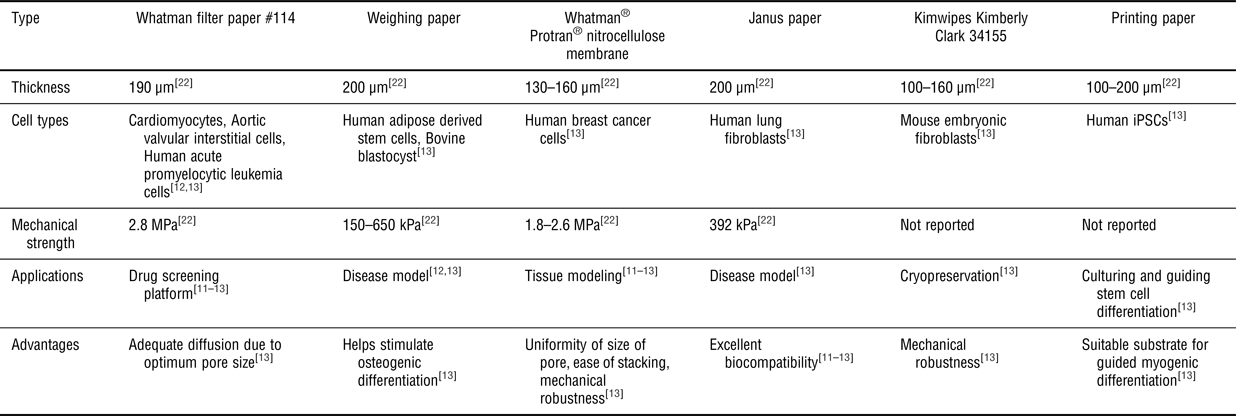
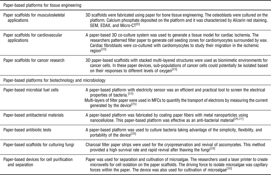
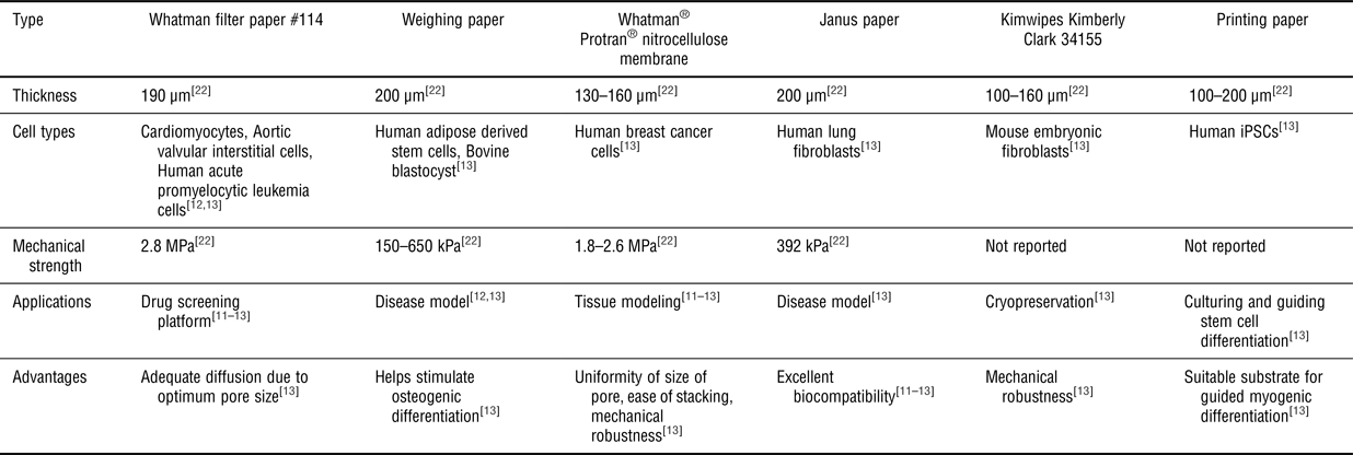
Table I summarizes the main types of papers that have been used in different biomedical applications, their major characteristics, and advantages. The following sections focus on the use of paper-based cell-culture platforms for musculoskeletal, cardiovascular, cancer, and microbiological applications. We also provide an overview of the use of paper-based cell culture platforms for biofilm formation, testing antibacterial agents, and biotechnology applications including separation and purification of cells. These applications are briefly summarized in Table II.
Common types of papers used in 3D cell culture platforms. Adapted with permission from Ref. Reference Ng, Gao, Yong, Li, Shi, Zhao, Li, Zhang, Pingguan-Murphy, Yang and Xu13, (Elsevier, 2017).[ Reference Ng, Gao, Yong, Li, Shi, Zhao, Li, Zhang, Pingguan-Murphy, Yang and Xu 13 ]
Applications of paper-based platforms in tissue engineering, biotechnology, and microbiology.

Paper-based platforms for tissue engineering
Paper scaffolds for musculoskeletal applications
To repair or regenerate damaged bone, tissue engineering approaches typically use scaffolds, cells, and growth factors separately or in combination.[ Reference Bhattacharya, Ghayor and Weber 31 ] 3D constructs with suitable biomimetic features, such as porosity, stiffness, swelling, degradation, and biological activity, need to be developed to guide the formation of new bone for damaged or diseased orthopedic and craniomaxillofacial tissues. Ideally, an engineered scaffold should be osteoconductive, porous, and biocompatible, as well as able to support adhesion, proliferation, and differentiation of bone cells. Additionally, mechanical properties of an ideal scaffold for bone regeneration provide adequate structural support for bone without causing stress shielding. The chemical functional groups, interconnected pores, and fibrous structure of paper are advantageous features compared to traditional scaffolding materials. Unlike the existing biomaterials, paper scaffolds are also much easier to fabricate in complex geometrical structures.
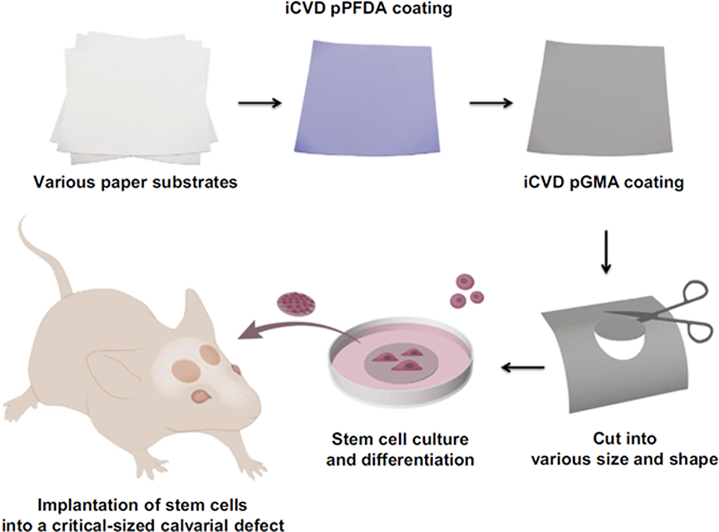
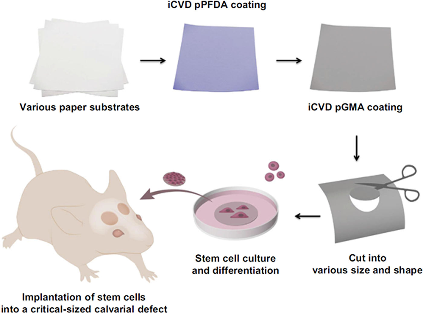
Traditional polymer-based scaffolds that are used in musculoskeletal applications are mostly biodegradable; however, 3D structures are difficult to fabricate in clinically-relevant sizes using these materials. To address this limitation, paper-based scaffolds have been employed for culturing adipose-derived stem cells.[ Reference Park, Yu, Yang, Jin, Cho, Kim, Lee, Yang, Im and Cho 16 ] It was found that osteogenic differentiation took place within the paper scaffolds (Fig. 1). After differentiating the human adipose-derived stem cells into the bone lineage, these paper constructs were implanted in a mouse model. The paper-based scaffolds demonstrated favorable properties for cell proliferation and differentiation.
Paper scaffolds were modified by initiated chemical vapor deposition (iCVD) of poly glycidyl methacrylate (pGMA). Stem cells were cultured on the paper scaffolds to induce osteogenic differentiation. Paper scaffolds were then implanted into a defect site in the cranium in vivo.[ Reference Park, Yu, Yang, Jin, Cho, Kim, Lee, Yang, Im and Cho 16 ] Reprinted with permission from Ref. Reference Park, Yu, Yang, Jin, Cho, Kim, Lee, Yang, Im and Cho16 (Elsevier, 2014).
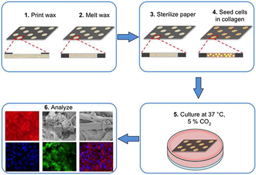
In a recent report, an origami-inspired approach was used to fabricate folded 3D constructs for bone tissue engineering. Paper scaffolds were generated using straightforward procedures at a low cost as demonstrated by Camci-Unal et al.[ Reference Camci-Unal, Laromaine, Hong, Derda and Whitesides 6 ] They fabricated 3D paper constructs for template-guided mineralization by mouse osteoblasts (Fig. 2).[ Reference Camci-Unal, Laromaine, Hong, Derda and Whitesides 6 ] This study demonstrated that calcium phosphate was deposited by osteoblasts within 3D paper scaffolds. Deposition of calcium was characterized by Alizarin red staining, scanning electron microscopy (SEM), energy dispersive x-ray spectroscopy (EDAX), and micro-computed tomography (Micro-CT). The researchers suggested that paper scaffolds would be suitable alternative materials for traditional scaffolds.
Filter paper was patterned using wax printing, and osteoblast cells were seeded on the wells of the paper scaffolds in a collagen matrix. After culturing the samples for 21 days, SEM, colorimetric assays, and immunostaining were performed to evaluate the deposition of minerals in the paper scaffolds.[ Reference Camci-Unal, Laromaine, Hong, Derda and Whitesides 6 ] Reprinted with permission from Ref. Reference Camci-Unal, Laromaine, Hong, Derda and Whitesides6 (Nature Publishing Group, 2016).

In another work, 3D paper scaffolds and 2D cell cultures on tissue culture plates were compared for osteogenic differentiation of adipose-derived stem cells.[ Reference Petersen, Hilbert, Trope, Kalle and Strappe 32 ] The paper scaffolds enhanced the deposition of calcium phosphate and increased the expression of osteogenic genes. These studies on paper-based scaffolds indicate that paper is a promising biomaterial for musculoskeletal applications.
Paper scaffolds for cardiovascular applications
Cardiovascular diseases are the leading cause of death globally.[ Reference Patnaik, Byers, DiGuiseppi, Dabelea and Denberg 33 ] Tissue engineering approaches are commonly utilized to address a wide range of these ailments. One major limitation for clinical translation of existing tissue engineering therapies is the inability to generate highly porous 3D scaffolds that prevent mass transport limitations.[ Reference Sapp, Fares, Estrada and Grande-Allen 12 , Reference Petsche Connell, Camci-Unal, Khademhosseini and Jacot 34 , Reference Camci-Unal, Alemdar, Annabi and Khademhosseini 35 ] To address this unmet need, different scaffolding materials have been tested.[ Reference Alemdar, Leijten, Camci-Unal, Hjortnaes, Ribas, Paul, Mostafalu, Gaharwar, Qiu and Sonkusale 36 ] Among those, paper-based scaffolds, in combination with hydrogels in multi-layered stacks, have been shown to be promising for cardiovascular tissue engineering applications. Paper is a suturable material and therefore could easily be used in applications where patches are needed.
A paper-based platform was employed to study the migration of aortic valvular interstitial cells (VICs).[ Reference Sapp, Fares, Estrada and Grande-Allen 12 ] Researchers layered filter paper sheets that were laden with collagen gels and VICs in a 96-well plate pattern obtained via wax printing. The stacked layers of paper provided a flexible and soft, yet structurally firm, 3D structure to study the factors that influence calcific aortic valve disease.[ Reference Sapp, Fares, Estrada and Grande-Allen 12 ]
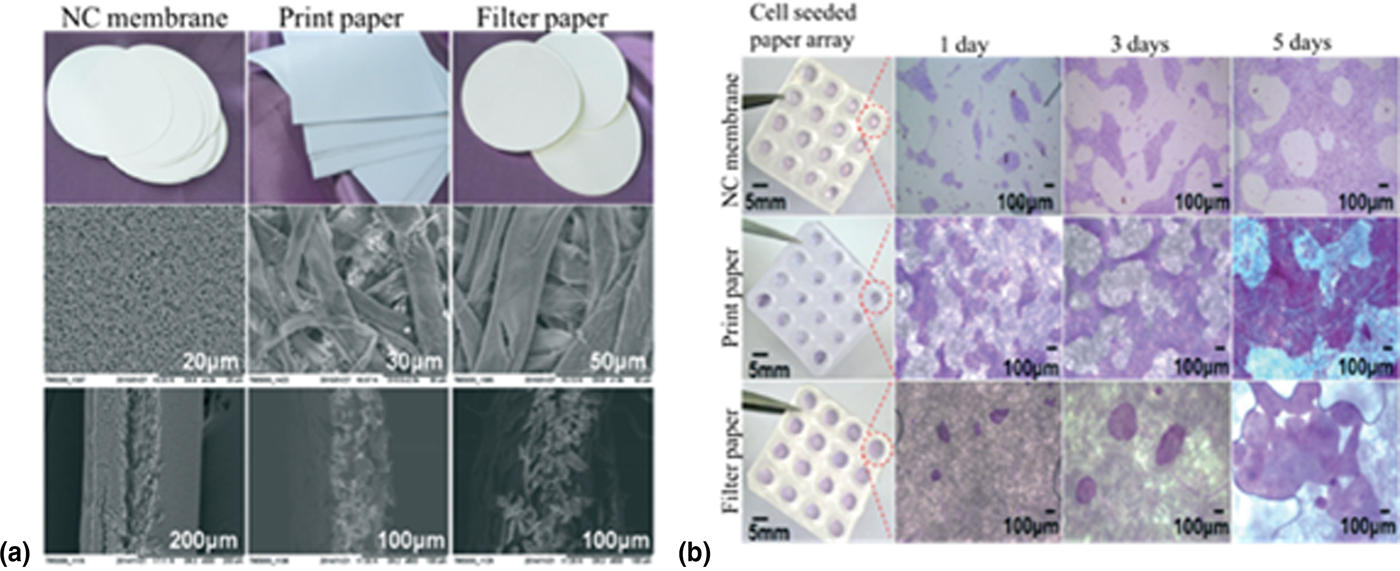
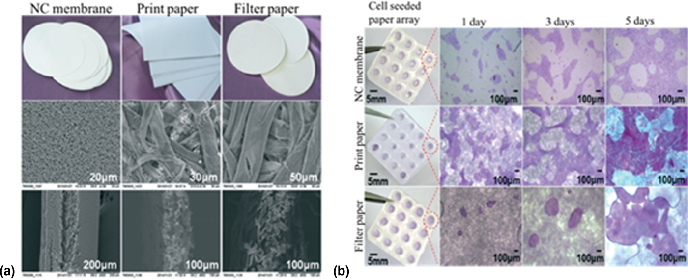
In another study, human-induced pluripotent stem cells (h-iPSCs) were cultured in a paper-based platform. The h-iPSCs differentiated into functional beating heart cells in these scaffolds. To generate the samples, paper was coated with Matrigel prior to the cell seeding step. The protein-based coat helped promote cell attachment to the paper substrate (Fig. 3).[ Reference Wang, Xu, Zhu, Yu, Sun, Zhang, Feng and Qin 21 ] The h-iPSCs derived cardiomyocytes were cultured in different paper substrates (nitrocellulose, filter paper, printing paper) that were fabricated by patterning poly dimethyl siloxane (PDMS) on the paper sheets.[ Reference Wang, Xu, Zhu, Yu, Sun, Zhang, Feng and Qin 21 ]
Different grades of paper were used to culture human iPSCs. (a) Paper substrates and their corresponding SEM images. (b) Human iPSCs cultured on three different types of paper up to 5 days in mTeSR1 medium. Hemotoxylin and Eosin (H&E) staining was then carried out. Modified from Wang et al.[ Reference Wang, Xu, Zhu, Yu, Sun, Zhang, Feng and Qin 21 ] Reprinted with permission from Ref. Reference Wang, Xu, Zhu, Yu, Sun, Zhang, Feng and Qin21 (Royal Society of Chemistry, 2015).
In their work, Mosadegh et al. generated an in vitro model for cardiac ischemia using a paper-based 3D co-culture system that included cell-encapsulated hydrogels stacked in multiple layers of paper sheets. They patterned Whatman filter paper to generate 20 circular cell-seeding zones surrounded by hydrophobic barriers generated by wax. Primary rat neonatal cardiomyocytes were then seeded in the designated culture zones. They also co-cultured cardiac fibroblasts with cardiomyocytes and showed how fibroblasts migrate towards the ischemic region in the 3D stacked paper-based model.[ Reference Mosadegh, Dabiri, Lockett, Derda, Campbell, Parker and Whitesides 20 ]
Paper scaffolds for cancer research
Cancer remains a major health problem that results in increasing rates of death each year across the world.[ Reference Lynch, Smyrk, Watson, Lanspa, Lynch, Lynch, Cavalieri and Boland 37 ] Prevention, diagnosis, and treatment of cancer are crucial for the survival of patients. To study and understand cancer, physiologically relevant cancer models should be developed. For screening drugs against tumors, cells are cultured and tested with different drugs and pharmaceutical products. Due to the unique properties of tumor microenvironments, many factors such as oxygen level, nutrient supply, and energy metabolism should be considered when designing biomimetic tumor models. Previously, 2D cell culture platforms were utilized in studying cancer cells. However, it was found that the microenvironment provided by these systems was different from that of the native ones in vivo. Additionally, it was shown that 3D models could mimic the native tumor niche better than 2D ones, as well as displaying distinct chemosensitivities[ Reference Karlsson, Fryknäs, Larsson and Nygren 38 ] and cellular interactions.[ Reference Luca, Mersch, Deenen, Schmidt, Messner, Schäfer, Baldus, Huckenbeck, Piekorz and Knoefel 39 ] Although 3D scaffolds can be fabricated using various materials and techniques, paper-based scaffolds have shown unique features beneficial to cancer migration studies.[ Reference Camci-Unal, Newsome, Eustace and Whitesides 5 ]
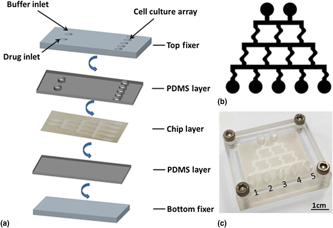
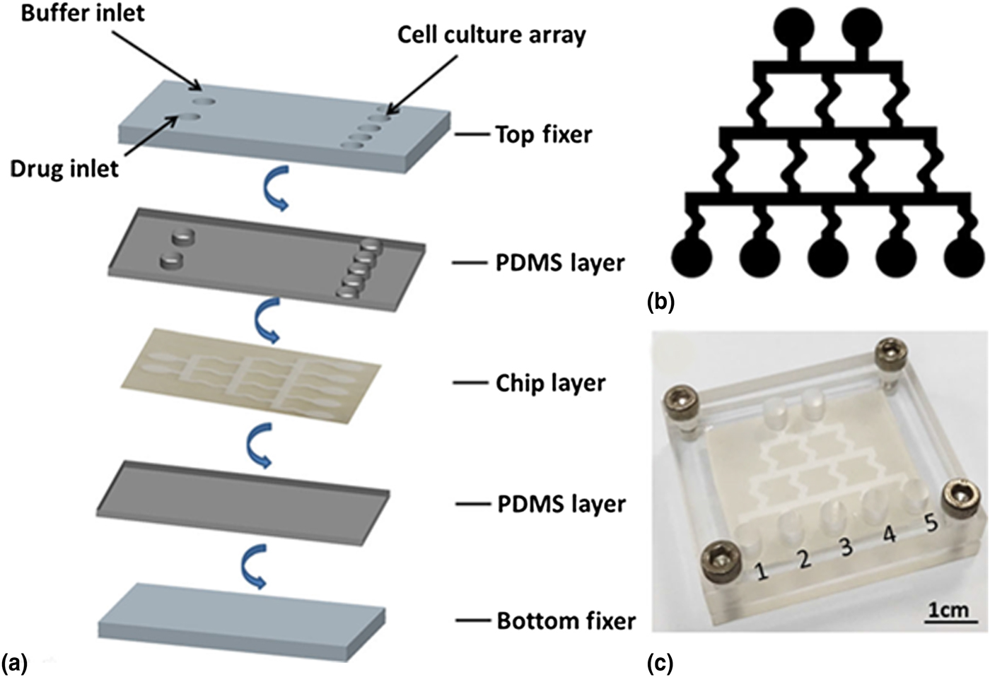
Investigating the influence of different types and concentrations of drugs on cancer cells is essential in developing anti-cancer drugs. In one study, a paper-based cell culture platform was developed to test the influence of a drug (doxorubicin) on HeLa cells.[ Reference Hong, Xue, Wu, Bao, Chuah and Kang 40 ] To test different concentrations of doxorubicin, a paper-based gradient generator was designed and fabricated. Drug diffusion through the paper fibers enabled the formation of a concentration gradient for doxorubicin (Fig. 4).[ Reference Hong, Xue, Wu, Bao, Chuah and Kang 40 ] The paper-based microfluidic device was then tested for cell response against different concentrations of doxorubicin. To carry out this task, HeLa cells in collagen were deposited in the designated cell-seeding reservoirs in the device and incubated for 8 h to evaluate cell viability upon exposure to doxorubicin. The paper-based device prevented an unstable flow of the drug and therefore reduced its quantity compared with a conventional PDMS device, which consumes larger amount of the drug to maintain a stable drug concentration gradient.
A paper-based microfluidic device for drug screening. (a) Multi-layered paper-based gradient generator. Patterned paper sheet is placed between two layers of PDMS and two layers of top/bottom fixers. (b) The channel patterns to create the gradients on the paper layer. (c) The assembled paper-based device. Cells were cultured in wells 1–5.[ Reference Hong, Xue, Wu, Bao, Chuah and Kang 40 ] Reprinted with permission from Ref. Reference Hong, Xue, Wu, Bao, Chuah and Kang40 (Springer, 2016).
Paper has also been used as a material for fabrication of diagnostic devices for detecting cancer biomarkers due to its high surface area and interconnected pores formed by cellulose fibers. Paper devices were fabricated by wax-printing and modified by chitosan to perform a chemiluminescent enzyme-linked immunosorbent assay for detection of α-fetoprotein, cancer antigen 125, and carcinoembryonic antigen.[ Reference Wang, Ge, Song, Yu, Ge, Huang and Zeng 41 ] These devices could potentially be used as cell culture and analysis platforms. For instance, cancer cells can be cultured in this 3D platform, and the biomarkers can be detected in situ by adding another layer in the scaffold.
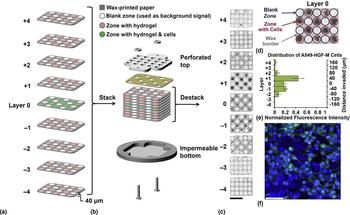
In contrast to healthy cells, cancer cells are commonly exposed to hypoxia (low levels of oxygen), which influences their cellular metabolism, proliferation, and radio-/chemo-resistance.[ Reference Erickson, Braun, Yu, Lanzen, Wilson, Brizel, Secomb, Biaglow and Dewhirst 42 – Reference Carmona-Fontaine, Bucci, Akkari, Deforet, Joyce and Xavier 45 ] One study described a 3D paper-based assay that could isolate sub-populations of cancer cells (A549 cells) based on their invasiveness under oxygen gradients (Fig. 5).[ Reference Mosadegh, Lockett, Minn, Simon, Gilbert, Hillier, Newsome, Li, Hall and Boucher 23 ] They found that oxygen acted as a chemoattractant for these cancer cells. The paper-based scaffolds created biomimetic environments for cancer cells in their stacked multi-layered structures. In this work, cancer cells responded differently to altered levels of oxygen. Another study also modulated the formation of oxygen gradients by adjusting the cell density in a paper-based device.[ Reference Boyce, LaBonia, Hummon and Lockett 46 ] The individual layers of the stacked paper platform were easily peeled off for individual analysis. To determine oxygen consumption rates of the cells, a luminescent thin film capable of quantifying the amount of oxygen was prepared in the paper-based scaffolds.[ Reference Boyce, Kenney, Truong and Lockett 47 ] The oxygen sensors demonstrated a linear response for oxygen tensions ranging from 0 to 160 mm HgO2. Researchers used this sensor to measure the spatial and temporal changes in oxygen for a breast cancer line (MDA-MB-231 cells).
Fabrication of paper scaffolds and results from cellular staining. (a) Multi-layered, wax-printed paper scaffolds. In layer 0, cells were encapsulated in a hydrogel matrix and added to the paper scaffolds. Only hydrogel was added to the paper in layers 1–4. (b) The layers were assembled using a perforated acrylic top, a liquid impermeable bottom, and screws. (c) After culturing for 24 h, samples were characterized by a high-resolution fluorescence scanner. The dark zones represent the cells, which were stained using a fluorescent reagent. (d) The cell culture area in layer 0. (e) The distance invaded by cells. (f) Fluorescent cells in layer 0 imaged by a confocal microscope. Scale bar represents 50 µm.[ Reference Mosadegh, Lockett, Minn, Simon, Gilbert, Hillier, Newsome, Li, Hall and Boucher 23 ] Reprinted with permission from Ref. Reference Mosadegh, Lockett, Minn, Simon, Gilbert, Hillier, Newsome, Li, Hall and Boucher23 (Elsevier, 2015).

In addition to the limited supply of oxygen and nutrients in tumors,[ Reference Chwalek, Bray and Werner 48 – Reference Truong, Lochbaum, Boyce and Lockett 50 ] the influence of other stromal cell types, including fibroblasts, endothelial cells, and immune cells are crucial in the progression of cancer.[ Reference Chwalek, Bray and Werner 48 , Reference Zhang and Liu 51 , Reference Bremnes, Dønnem, Al-Saad, Al-Shibli, Andersen, Sirera, Camps, Marinez and Busund 52 ] To develop flexible 3D scaffolds for studying the behavior of tumor cells, a multi-layered paper-based platform was developed. In this work, primary human lung tumor cells (VXN2s) and tumor fibroblasts from patient biopsies were co-cultured. It was demonstrated that the presence of fibroblasts enhanced the migration of human lung cancer cells.[ Reference Camci-Unal, Newsome, Eustace and Whitesides 5 ] The research team suggested that this finding might be mediated by the transforming growth factor-beta (TGF-β) pathway. This hypothesis was tested by blocking the activity of the TGF-β pathway, which significantly decreased the migration of the tumor cells.
Paper-based cell culture platforms[ Reference Derda, Tang, Laromaine, Mosadegh, Hong, Mwangi, Mammoto, Ingber and Whitesides 4 , Reference Derda, Laromaine, Mammoto, Tang, Mammoto, Ingber and Whitesides 19 ] were adopted by a number of researchers to generate 3D tissue-mimetics by stacking layers of paper impregnated with cells and hydrogels. This technique was utilized in a study[ Reference Deiss, Mazzeo, Hong, Ingber, Derda and Whitesides 53 ] for high-throughput analysis, which permitted the rapid assembly of arrays of 3D cell cultures. The paper-based system also allowed for isolation of cells from specific regions of these scaffolds. The constructs were generated by stacking paper sheets, each of which supported 96 individual spots of cell-hydrogel mixtures. This configuration conveniently enabled treatment of the cells in each individual well with different drugs. The 96-well format could possibly accelerate the development of new high-throughput assays for 3D cell cultures.[ Reference Deiss, Mazzeo, Hong, Ingber, Derda and Whitesides 53 ] Similarly, another study conducted by Derda et al.[ Reference Derda, Laromaine, Mammoto, Tang, Mammoto, Ingber and Whitesides 19 ] showed that arrays patterned on paper using Teflon can support peptide synthesis and cell attachment. The paper platform can also be used as a 3D model for cancer cells. The peptide array effectively promoted the adherence of MDA-MB-231 breast carcinoma cells in these paper scaffolds.[ Reference Deiss, Matochko, Govindasamy, Lin and Derda 54 ] Another paper-based biomimetic microenvironment for the study of the pathogenesis of cancer was created by Lei and Huang.[ Reference Lei and Huang 49 ] In this study, the wax-printed paper was found to be a convenient screening tool with reduced consumption of materials. This system allowed for a rapid turnaround compared with conventional materials, which are made of polymers and metals.
Radiation therapy remains a common strategy for treating tumor cells, which possess compromised DNA repair machinery and often proliferate at higher rates than normal cells.[ Reference Jackson and Bartek 55 – Reference Barnett, West, Dunning, Elliott, Coles, Pharoah and Burnet 57 ] Simon et al. utilized a paper-based platform to assess the metabolic response of lung cancer cells upon exposure to ionizing radiation. This paper-based system allowed for regulation of oxygen to cells within the 3D structure before and after irradiation.[ Reference Simon, Mosadegh, Minn, Lockett, Mohammady, Boucher, Hall, Hillier, Udagawa, Eustace and Whitesides 58 ] The platform could potentially be used for the isolation of a subpopulation of cells and for exposing cells to different doses of radiation or chemotherapeutics for patient-specific personalized treatment.
Invasion of cancer cells is widely studied using migration assays, such as transwell assays (e.g. Boyden chamber). These assays offer limited information about cellular responses to gradients in vivo because they oversimplify the 3D microenvironments of tissues. Other invasion assays commonly require specialized laboratory equipment or engineering expertise.[ Reference Kenney, Boyce, Truong, Bagnell and Lockett 59 ] Recently, a simple paper-based invasion assay was designed and implemented [ Reference Kenney, Boyce, Truong, Bagnell and Lockett 59 ] to monitor cellular movement in real-time with fluorescence microscopy. A similar study presented a paper-based device that could carry out polymerase chain reaction (PCR) for quantifying multiple cell types in a single culture. The PCR method detected, repeatedly and accurately, as few as 50 cells, allowing small populations of highly invasive cells to be differentiated from other cell types.[ Reference Truong, Lochbaum, Boyce and Lockett 50 ]
Investigation of cellular phosphorylation and signaling pathways has played an important role in studying the pathogenesis of cancer. A paper-based microreactor has been developed to integrate cell culture (human liver cancer cells and nasopharyngeal cancer cells) and subsequent immunoassay for phosphorylated kinases on a single paper scaffold.[ Reference Lei and Huang 49 ] This platform allows for the shortcomings of time-consuming and labor-intensive conventional assays to be overcome.
Closing the gap between in vitro experimental data and in vivo results is essential in the development of new cancer therapies. To achieve this goal, physiologically relevant tissue models should be designed and implemented. A multi-zone paper platform was developed to characterize cell behavior upon exposure to an anti-cancer drug, doxorubicin.[ Reference Derda, Tang, Laromaine, Mosadegh, Hong, Mwangi, Mammoto, Ingber and Whitesides 4 ] Cytocompatibility, proliferation, morphology, and chemosensitivity of the cells were evaluated in this biomimetic tissue model.[ Reference Tao, Xiao, Lei and Lee 60 ]
In addition to high-throughput applications of paper-based platforms, they also have been used to develop analytical tools. For example, in one study cell proliferation in paper scaffolds was quantified with an impedimetric method.[ Reference Lei, Huang and Tsang 61 ] The paper device with microchamber patterns was placed on a glass substrate with electrodes for the measurement of impedance. Cells in each microchamber were represented by an impedance magnitude, which allowed for the determination of cell proliferation up to 3 days. This paper device could potentially be used to evaluate other cell behavior including growth, differentiation, and protein expression.
Paper-based platforms for biotechnology and microbiology
Paper-based microbial fuel cells
Microbial fuel cells (MFCs) transform chemical energy stored in degradable organic materials into electricity by utilizing microorganisms as catalysts. A great deal of effort has been devoted to the efficient generation of electricity, which relies on the type of microorganism used as well as the design and manufacture of the device. Paper-based MFCs were recently utilized to form portable 3D culture systems for fuel cells.[ Reference Choi, Hassett and Choi 24 , Reference Choi and Choi 62 – Reference Desselberger 67 ]
To monitor liquid flow, and analyze the flow of cells or other particles in bodily fluids, Choi and Choi developed a new analytical approach based on electrochemical measurements in paper microfluidics using the principles of MFCs.[ Reference Choi and Choi 62 ] Although optical techniques such as the particle-imaging velocimetry can be used to monitor the flow of particles (e.g. cells) in the liquid medium, the porous and non-transparent nature of paper makes this method unsuitable. In this analytical tool, the electricity generated by bacteria was measured in real time. Oxidation of organic matter from the electron transfer in the anode facilitated the measurement of currents.
In another study, to provide an analytical tool for screening electrical properties of bacteria, a MFC array was generated using a paper-based platform.[ Reference Choi, Hassett and Choi 24 ] This rapid screening high-throughput sensor was able to monitor the generation of electricity from different genetically engineered bacteria. Eight mutant strains of Pseudomonas aeruginosa were analyzed under 20 min in the 48-well paper platform without the need for external tubes or pumps to drive the fluid flow. These efficient and practical tools could potentially be used to provide a fundamental understanding of the electrogenic potential of bacteria and to increase the performance of MFCs.
Paper platforms enable fluid transport through capillary action without the need for pumping devices.[ Reference Yetisen, Akram and Lowe 63 ] Liquid transportation in paper-based microfluidic devices occurs via passive capillary action.[ Reference Veerubhotla, Bandopadhyay, Das and Chakraborty 64 , Reference Zhang, Zhou, Wen, Bai, Lou and Dong 65 ] However, the flow of heterogeneous samples, including cells and particles is not yet understood in depth.[ Reference Shitanda, Kato, Hoshi, Itagaki and Tsujimura 66 ] A non-optical technique was employed to analyze and quantify the flow of particles in a paper matrix.
Although microbial fuel cells have been improved over the last decade,[ Reference Desselberger 67 ] MFC performance is still insufficient.[ Reference Cui and Boland 68 , Reference Martinez, Phillips, Carrilho, Thomas, Sindi and Whitesides 69 ] Conventional MFCs cannot achieve major performance breakthroughs using naturally existing species without the use of genetic engineering techniques.[ Reference Srimongkon, Ishida, Igarashi and Enomae 70 ] To address this need, a high-throughput and rapid screening tool for microbial biopower production was introduced. The designed MFC array displayed acceptable performance characteristics and identified genes in Pseudomonas aeruginosa that could trigger a higher power density. In another work, a simple, flexible, low-cost, and disposable paper-based MFC was fabricated to generate bioelectricity from P. aeruginosa and Shewanella putrefacient.[ Reference Veerubhotla, Bandopadhyay, Das and Chakraborty 64 ] The majority of the layers of the device were fabricated from filter paper. The hydrophilic and porous nature of paper allowed for the sufficient exchange of electrolytes maintaining the fluid content in the device and the elimination of external pumps to drive the liquid motion. Oxygen, which has a high oxidation potential, from the air was used as the catholyte to form water. The device generated 0.4 V from P. aeruginosa without the requirement of any catalysts. Similar paper-based platforms could potentially be used to fabricate the next generation of power devices.
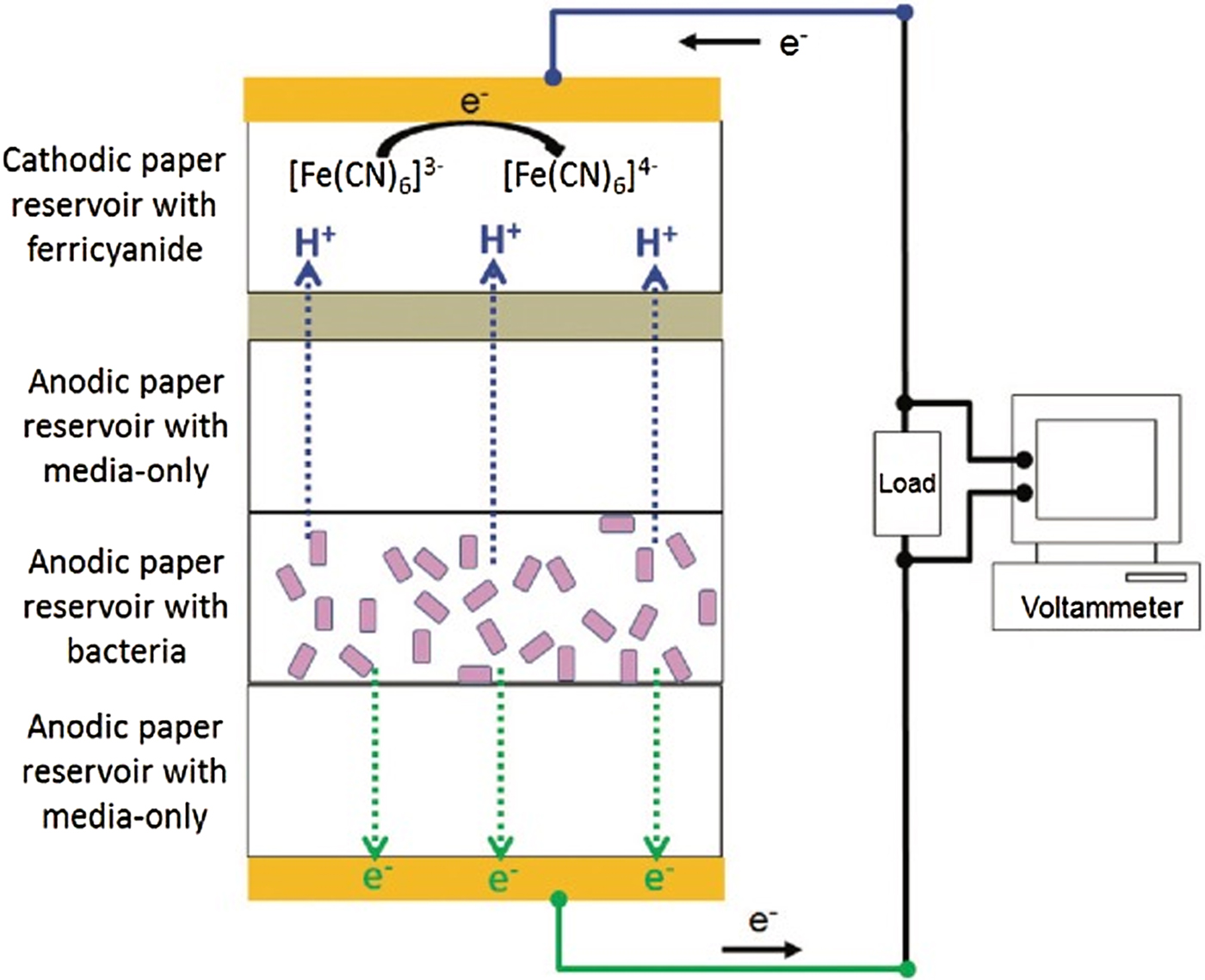
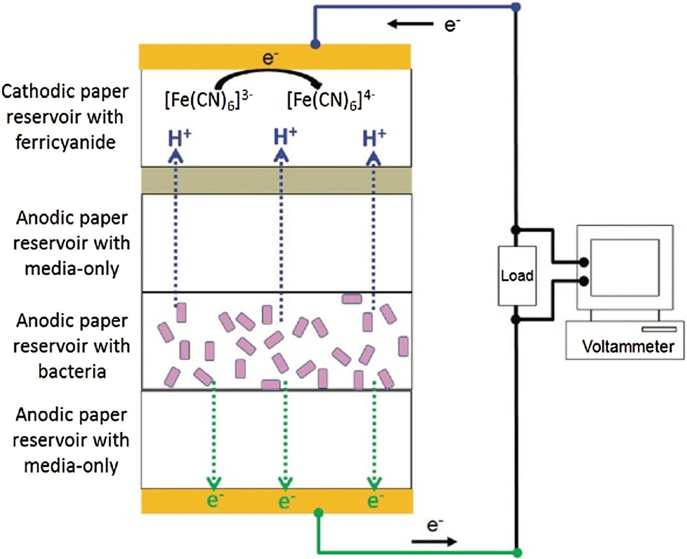
Key mechanisms that underlie electron harvesting from electroactive microorganisms are not fully understood. In addition, the optimization of fundamental factors responsible for the generation of electricity has been challenging. These hurdles have hindered the widespread use of MFCs in applications such as wastewater treatment and environmental power sources.[ Reference Choi and Choi 62 , Reference Marciano, Lima-Oliveira, Da-Silva, Diniz, Corat and Trava-Airoldi 71 , Reference Zhang, Jiang, Ding, Daskalakis, Jeuken, Povey, O'Neill and York 72 ] In particular, the major drawback of existing MFC systems is the difficulty in controlling the electrical communication between cells and electrode. To overcome the main limitations of these aspects,[ Reference Tankhiwale and Bajpai 73 – Reference Brayner, Ferrari-Iliou, Brivois, Djediat, Benedetti and Fiévet 75 ] a paper-based MFC stack was developed.[ Reference Fraiwan, Mukherjee, Sundermier, Lee and Choi 25 ] Multiple layers of filter paper, containing bacteria, were stacked to form a layered 3D model of biofilms. In addition, the mass transport of electrons/protons into this 3D system was quantified. In another study, a modular, and multi-layered paper-based MFC was used to study proton and electron diffusion within the Shewanella oneidensis biofilm by measuring the current generated from the 3D stacks of paper (Fig. 6).[ Reference Choi and Choi 76 ] The thickness of the biofilm was easily controlled by varying the number of layers of paper that were assembled. Current generation and maintenance were found to be higher for thicker biofilms compared to that of the thinner ones. This platform is promising for studying the effects of gradients of nutrients on the formation of biofilms, and for understanding the electrogenic potential of bacteria for power generation in MFCs. Transportation of bacterial cells in the paper-based microfluidic device was also quantified. These paper-based tools can be useful for clinical diagnostics, environmental monitoring, and control of food quality.
Schematic representation of the paper-based multi-stack microbial fuel cell. Whatman #1 paper was used as the anodic and cathodic substrate. Electricity generated by the device was dependent on the number of bacteria on the anode. The thickness of the biofilm was controlled by the amount of bacteria on the anodic paper. Reprinted with permission from Ref. Reference Choi and Choi76 (Royal Society of Chemistry, 2015).
Paper-based antibacterial materials
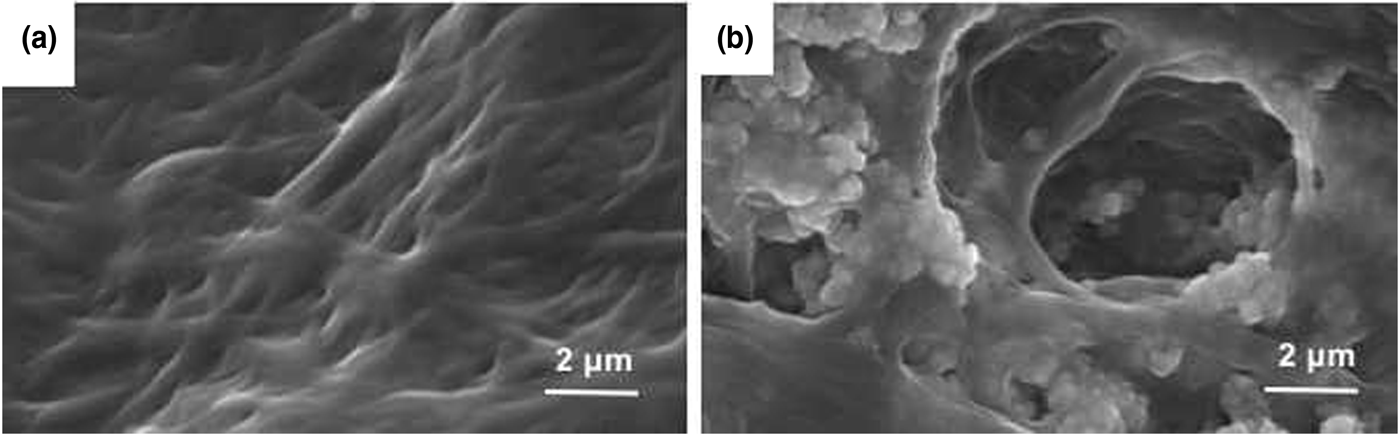
Metal oxide nanoparticles have been shown to demonstrate antibacterial activity. However, gel or agar plate-based platforms that include these nanoparticles have a number of limitations.[ Reference Azam, Ahmed, Oves, Khan, Habib and Memic 77 – Reference Sondi and Salopek-Sondi 79 ] Although gels and agar plates have a significant surface area from which to release the nanoparticles, the applications are still limited to research in the laboratory. This problem can be addressed by coating paper with metal nanoparticles using nanocellulose (Fig. 7).[ Reference Martins, Freire, Neto, Silvestre, Causio, Baldi, Sadocco and Trindade 26 , Reference Díez, Eronen, Österberg, Linder, Ikkala and Ras 80 – Reference Gottesman, Shukla, Perkas, Solovyov, Nitzan and Gedanken 85 ] The resulting modified paper devices are portable, and retain the desired surface area of gels and ager plates. The antibacterial tests revealed that the paper-based platform that included metal oxide nanoparticles was effective against both gram-positive and gram-negative bacteria.
SEM images of uncoated and antibiotic-coated paper scaffolds. (a) Untreated nanofibrillated cellulose fibers. (b) The cellulose fibers were coated using zinc oxide nanoparticles to explore their antibiotic properties. Reprinted with permission from Ref. Reference Martins, Freire, Neto, Silvestre, Causio, Baldi, Sadocco and Trindade26 (Elsevier, 2013).

Paper-based antibiotic tests
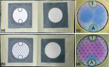
Paper is a promising platform for culture bacteria owing to its simplicity, flexibility, and portability.[ Reference Funes-Huacca, Wu, Szepesvari, Rajendran, Kwan-Wong, Razgulin, Shen, Kagira, Campbell and Derda 27 ] In one study, an inexpensive paper-based culture platform was used to screen antibiotics against bacteria. In this study, the standard Kirby–Bauer disk diffusion test was performed on the paper platform (Fig. 8).[ Reference Deiss, Funes-Huacca, Bal, Tjhung and Derda 28 ] The procedure was similar to the test that is traditionally performed on Petri dishes and agar plates. The paper-based device was able to indicate the bacteria as resistant, intermediate, or susceptible against the tested antibiotics. The results suggested that paper-based devices could also be used for environmental samples in the future due to the flexibility of the platform.
A paper-based culture platform for testing antibiotics. Two different patterns were used to culture bacteria in the presence of antibiotics (a–d). (a) and (c) Wax patterns without hydrophobic spots. (b) and (d) The addition of hydrophobic spots facilitated the uniform spreading of the bacterial inoculum. The antibiotics were added to zones T and A. Reprinted with permission from Ref. Reference Deiss, Funes-Huacca, Bal, Tjhung and Derda28 (Royal Society of Chemistry, 2013).

Paper-based scaffolds for culturing fungi
The prerequisite for microbiological research for fungi is the axenic cultivation. Fungal mycelia are frozen at a controlled rate and stored in liquid nitrogen.[ Reference Ryan and Smith 86 ] Some fungi species grow poorly under axenic conditions, and cryopreservation for these slow-growing microorganisms is difficult. Ascomycetes and basidiomycetes have been used in applications for the medical, pharmaceutical, and food industry. It is extremely challenging for eukaryotes to survive cryopreservation because they can hardly revive from the freezing temperature. Although new techniques have been used for eukaryotes such as basidiomycetes, these cryopreservation methods are not suitable for fungal family. A novel, simple, and cost-effective method based on charcoal filter paper strips (CFS) has been used for the cryopreservation and revival of ascomycetes, as well as ectomycorrhizal and non-ectomycorrhizal basidiomycetes. While neither the charcoal nor the filter paper alone provides higher survival rate for these species, the combination of the two types of paper makes it possible to achieve a favorable cryopreservation condition. This approach provided a greater survival rate and rapid revival after thawing the fungi.[ Reference Stielow, Vaas, Göker, Hoffmann and Klenk 29 ] The CFS was also used for cryopreservation, culture, and analysis of ectomycorrhizal, wood-decaying, and saprotrophic fungi.[ Reference Stielow, Vaas, Göker, Hoffmann and Klenk 29 , Reference Michaelsen, Pinzari, Ripka, Lubitz and Piñar 87 ]
Paper-based devices for cell purification and separation
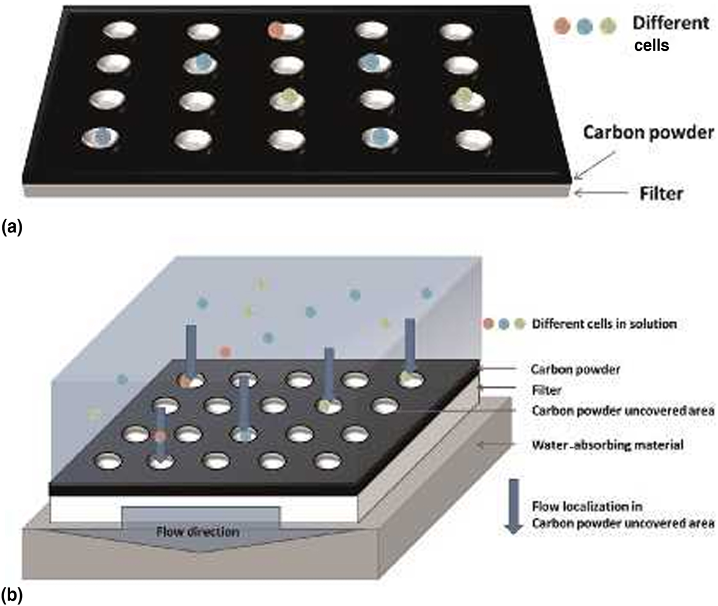
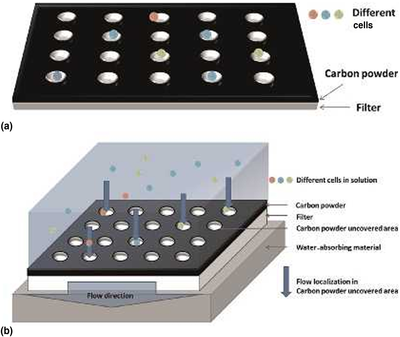
Microalgae are unicellular species that are commonly found in fresh water or marine systems.[ Reference Ben-Amotz, Fishler and Schneller 88 ] Most of microalgae produce valuable components such as carbohydrates, long chain fatty acids, and proteins, which are widely utilized in aquaculture, cosmetics, food supplements, and pharmaceuticals.[ Reference Yen, Hu, Chen, Ho, Lee and Chang 89 ] Microalgae species from aquatic environments are mixed heterogeneous populations that must be isolated and then cultivated for specific applications. The ability to select specific microalgae from a mixed heterogeneous cell population is one of the most useful techniques for biochemical research. Traditional methods for isolation of microalgae, such as serial dilution, are laborious and inefficient. Several other methods such as microfluidic devices and plastic microwell plates have been applied for the analysis of single cells.[ Reference Liu, Dechev, Foulds, Burke, Parameswaran and Park 90 , Reference Lindstrom and Andersson-Svahn 91 ] Although results are promising, the challenges associated with the high-cost and time required to complete these analyses are still not addressed. To meet this demand, the paper has been utilized for the separation and cultivation of single microalga (Fig. 9).[ Reference Chen, Liu and Yao 30 ] Major advantages of employing paper for microalga separation include the ease of fabrication of paper devices, as well as the inexpensive and disposable features of paper.[ Reference Chen, Liu and Yao 30 ] In this work, the paper-based device was fabricated using a laser printer to create microwell patterns on filter paper. Each well was designed to isolate one single cell. Most of the driving force to isolate microalgae with this device was the capillary forces within the paper. It took several minutes to construct the device, without the need for capital instruments or a cleanroom. The space between each microwell was 1 mm, which was visible in the macroscale and accessible by a micro-pipette. Moreover, the device was able to separate microalgae of different size and shape with a greater efficiency compared to the standard tests. The paper device also served as an incubator for microalgae cultivation. Algae growth medium was absorbed by the paper-based device, which allowed the microalgae to be cultured from a single cell to a population after the separation procedure without the need to transfer to a new container. The microalgae populations did not grow beyond the borders of the wells ensuring that there was no cross-contamination from well-to-well.
Fabrication process for paper scaffolds to grow algal species. (a) Carbon powder was used to generate patterns using a laser printer. The hydrophobic patterns defined cell seeding zones. (b) Growth medium with cells was deposited on the hydrophilic algae-seeding zones on the paper scaffolds. Reprinted with permission from Ref. Reference Chen, Liu and Yao30 (Elsevier, 2015).
Conclusion and future directions
With its unique advantages, paper has become a promising biomaterial for culturing mammalian cells, bacteria, fungi, and other microorganisms (e.g. algae). Such advantages include the hydrophilicity, porosity, and mechanical strength provided by the cellulose fibers. The highly porous nature of paper assists in the transfer of oxygen and nutrients to and from cells upon their culture, thus, these platforms are promising scaffolds for tissue engineering applications. The types of cells cultured on paper for the analysis of metabolites and the investigation of the mechanisms of life processes could be expanded to plant cells and viruses. The ease with which paper can be shaped and patterned using wax or inkjet printing helps with the fabrication of scaffolds, devices, or chips of different designs. These paper-based products can then be used for investigating co-cultures of cells, studying disease models, or as tools for diagnostics.
Studies which use paper-based designs for tissue engineering are expected to be conducted at a greater scale in the near future. Experiments such as the “beating cardiac tissues on paper” have been suggested recently.[ Reference Wang, Xu, Zhu, Yu, Sun, Zhang, Feng and Qin 21 ] It is also possible to incorporate various types of cells with the goal of forming functional tissues, and even more daring attempts may be proposed for making organ models on paper similar to current organ-on-chip designs.
Components of certain plants may be extracted as raw materials for synthesizing drugs and pharmaceutical products. The slow growth of a plant cannot meet the needs of the medical industry, which triggers the production of cells in bioreactors. Paper-based platforms can be employed as alternatives to the traditional approaches to grow plant cells. Some of the metabolites of bacteria possess pharmacological activities and are produced by standard germiculture. The output of these procedures could potentially increase if the paper is confirmed to provide better metrics.
Paper-based devices can be used in combination with analytical techniques for the analysis of cells and metabolites. One such technique is mass spectrometry, which has powerful identification and quantification functions while maintaining great sensitivity when it is applied for the detection of small molecules and peptides. The combination of this technique with paper-based cell culture would benefit the studies relevant to high-throughput testing of soluble compounds and high-throughput drug screening.
Paper-based platforms could also be used to study viruses that are co-cultured with cells. Production of a large number of adenoviruses for gene therapy is currently limited for clinical applications. Considering the advantages of paper as mentioned above, virus infection can be effectively improved and the kinetics of adenovirus production in cell lines can be controlled using paper-based co-culture of adenoviruses with cells.
Chemically modified paper substrates could be broadly used in conjunction with bioactive substances. For example, one could modify the hydroxyl group of cellulose, which could be a binding moiety for collagen, peptides, and small molecules with hydrogen bonds. Air permeability, pore size, and porosity of paper could also be modified for the growth of cells.
Another potential application of paper-based platforms would be the co-culture of bacteria and mammalian cells to mimic the in vivo environment when it is exposed to infections. There has been an increasing interest in the association of altered gut microbiota with human diseases and therapeutics to modulate microbiota to treat such diseases.[ Reference Khanna 92 ] In this context, it would be interesting to use paper-based platforms to co-culture intestinal cells and bacteria.
Although biocompatibility of paper scaffolds has been demonstrated in animal models in earlier studies, its further use for regenerative applications in vivo should also be shown.
The implants which serve as temporary fillers for tissues would need to be degradable in the body to allow for deposition of extracellular matrix (ECM) by the cells and regeneration of the tissue. Studies on cellulose degradation would result in the improvement of applications of paper constructs in tissue engineering. Although paper possesses numerous advantages as a scaffold for cells, if it cannot be degraded, some of its applications in tissue engineering may be limited. Therefore, studying enzymes produced chiefly by fungi, bacteria, and protozoans that catalyze cellulolysis, would benefit the applications of paper in this area.
In summary, paper-based platforms have been used in the fields of biology, medicine, engineering, materials science, biochemical analysis, and clinical diagnosis due to their numerous advantages mentioned above. Fabrication of paper-based devices for new applications may greatly benefit our society. Further work with paper has great potential for development and prospects of far broader applications.
Acknowledgments
This work was supported by the University of Massachusetts Lowell start-up funds.