Introduction
Sesquiterpene lactones are a large and structurally diverse group of plant metabolites (Heinrich et al., Reference Heinrich, Robles, West, Ortiz de Montellano and Rodriguez1998). Several biological activities of sesquiterpene lactones have been reported, including anti-tumour (Robles et al., Reference Robles, Aregullin, West and Rodriguez1995), anti-migraine (Beekman et al., Reference Beekman, Woerdenbag, van Uden, Pras, Konings, Wikström and Schmidt1997) and gastric cytoprotector effects (Giordano et al., Reference Giordano, Pestchanker, Guerreiro, Saad, Enriz, Rodriguez, Jauregui, Guzman, Maria and Wendel1992; Penissi et al., Reference Penissi, Fogal, Guzmán and Piezzi1998).
These activities are mediated principally by the alpha-methylene-gamma-lactone function. This chemical group is a powerful alkylating agent due to a Michael-type addition to suitable nucleophiles, e.g. nucleophilic attachment to cysteine sulfhydryl groups through the alpha,beta-unsaturated carbonyl group. However, this alkylating activity is non-specific, leading to the inhibition of a large number of enzymes or factors involved in key biological processes (Heinrich et al., Reference Heinrich, Robles, West, Ortiz de Montellano and Rodriguez1998).
Dehydroleucodine (DhL), a sesquiterpenic lactone, was isolated and purified from aerial parts of Artemisia douglasiana Besser, a medicinal herb used in Argentina (Giordano et al., Reference Giordano, Guerreiro, Pestchanker, Guzmán, Pastor and Guardia1990). DhL is an alpha-methylenebutyro-gamma-lactone ring connected to a seven-membered ring fused to an exocyclic alpha,beta-unsaturated cyclopentenone ring.
It has been previously shown that DhL selectively induces a dose-dependent transient arrest in G2 of both meristematic cells (López et al., Reference López, Giordano and López2002) and vascular smooth muscle cells (Cruzado et al., Reference Cruzado, Castro, Fernández, Gómez, Roque, Giordano and López2005). Treatment with DhL of Bufo arenarum fully grown oocytes arrested at G2, at the beginning of meiosis I, induces an inhibition of spontaneous and progesterone-induced maturation in a dose-dependent manner (Sánchez Toranzo et al., Reference Sánchez Toranzo, Giordano, López and Bühler2007). However, the nature of the mechanisms involved in the process is still unknown.
In amphibians, meiotic arrest is released under the stimulus of progesterone, a process termed oocyte maturation (Fortune et al., Reference Fortune, Concannon and Hansel1975; Schuetz, Reference Schuetz and Browder1985). Although progesterone is the established maturation inducer in Bufo arenarum, meiosis resumption also occurs spontaneously when follicular cells are removed and oocytes are cultured in vitro under suitable conditions. In this species oocytes competent or incompetent to undergo spontaneous maturation can be obtained according to the seasonal period in which animals were captured (Zelarayán et al., Reference Zelarayán, Oterino and Bühler1995). However, fully grown Bufo arenarum oocytes always respond to progesterone, independently of the time of year in which they are obtained (Zelarayán et al., Reference Zelarayán, Oterino and Bühler1996).
Meiotic maturation, which represents the transition from G2 to the M phase of the cell cycle, is controlled by the maturation promoting factor (MPF), a protein kinase composed of a catalytic subunit, p34 and a regulatory subunit, cyclin B. The activity of MPF is subject to a complex regulation that includes both specific phosphorylation events and protein–protein interactions.
In G2-arrested Bufo arenarum oocytes there is a preformed complex of cdc2 and cyclin B that accumulates during terminal oocyte growth to form a complex called pre-MPF. This complex is maintained in its inactive form by phosphorylation of cdc2 on Thr161 and Thr14/Tyr15 (Sánchez Toranzo et al., Reference Sánchez Toranzo, Bonilla, Zelarayán, Oterino and Bühler2006). These inhibitory phosphorylations are probably catalysed by Myt1 protein kinase (Gavin et al., Reference Gavin, Ni Ainle, Chierici, Jones and Nebreda1999; Patra et al., Reference Patra, Wang, Kumagai and Dunphy1999; Wells et al., Reference Wells, Watanabe, Tokusumi, Jiang, Verdecia and Hunter1999; Qian et al., Reference Qian, Erikson, Taieb and Maller2001; Inoue et al., Reference Inoue and Sagata2005) present in G2 oocytes and are hyperphosphorylated during oocyte maturation. As inhibition of the endogenous Myt1 can trigger meiotic maturation in the absence of progesterone stimulation, Myt1 could be responsible for maintaining the cdc2–cyclin-B complexes in an inactive form in G2-arrested oocytes (Palmer et al., Reference Palmer, Gavin and Nebreda1998; Peter et al., 2001; Furuno et al., Reference Furuno, Kawasaki and Sagata2003; Karaiskou et al., Reference Karaiskou, Leprêtre, Pahlavan, Du Pasquier, Ozon and Jessus2004).
Cdc2 Thr14/Tyr15 dephosphorylation is primarily responsible for MPF activation during oocyte maturation (Gautier & Maler, Reference Gautier and Maller1991; Minshull et al., Reference Minshull, Murray, Colman and Hunt1991; Ookada et al., Reference Ookada, Hisanaga, Okano and Kishimoto1992; Galas et al., Reference Galas, Barakat, Dorée and Picard1993) and dephosphorylations of these residues require cdc25 phosphatase activity (Perdiguero & Nebreda, Reference Perdiguero and Nebreda2004; Dekel, Reference Dekel2005).
In Bufo arenarum, the conversion of pre-MPF into active MPF, which occurs several hours after progesterone stimulation, is activated by dephosphorylation of these sites by the action of the protein phosphatase cdc25 (Sánchez Toranzo et al., Reference Sánchez Toranzo, Bonilla, Zelarayán, Oterino and Bühler2006). The exposure of Bufo arenarum oocytes to sodium metavanadate (NaVO3), which inhibits cdc25 phosphorylation, prevents release from G2 arrest by progesterone. These results suggest that cdc25 may be involved in the physiological activation of the MPF (Sánchez Toranzo et al., Reference Sánchez Toranzo, Bonilla, Zelarayán, Oterino and Bühler2006).
When oocyte maturation is initiated by progesterone, cAMP levels are reduced and protein kinase A (PKA) becomes inactivated. In G2-arrested Xenopus (Schmitt & Nebreda Reference Schmitt and Nebreda2002; Han & Conti Reference Han and Conti2006) and Bufo arenarum oocytes (Zelarayán et al., Reference Zelarayán, Oterino, Sánchez Toranzo and Bühler2000), cdc25 is probably maintained inactive by PKA-mediated phosphorylation. The induction of meiotic maturation by injecting PKI in the absence of progesterone stimulation suggests that PKA also plays an active role in the G2 arrest of oocytes. Although PKA has an important negative role in oocyte maturation, its substrate in the oocyte is still unknown. Interestingly, PKA and protein phosphatase 2A (pp2A) are both active in G2-arrested oocytes and become inactivated upon progesterone addition (Duckworth et al., Reference Duckworth, Weaver and Rudeman2002; Schmitt & Nebreda, Reference Schmit and Nebreda2002). PKA has been proposed as a regulator of the activity of okadaic acid (OA)-sensitive serine/threonine phosphatase (Grieco et al., Reference Grieco, Avvedimento and Gottesma1994) and can also potentiate the activation of pre-MPF by OA in cell free extracts obtained from Xenopus oocytes (Karaiskou et al., Reference Karaiskou, Leprêtre, Pahlavan, Du Pasquier, Ozon and Jessus2004).
Okadaic acid is an inhibitor of pp2A (Bialojan & Takai, Reference Bialojan and Takai1988). Microinjection of 50 nl of 2.5 μM OA into prophase oocytes induces rapid MPF activation and germinal vesicle breakdown (GVBD) (Goris et al., 1989). Thus, although the inhibition of cdc25 is probably an important G2 regulator, OA-sensitive phosphatases also are likely to be important regulators of G2 arrest. The identity of these phosphatases and their targets in oocytes are still unknown.
In amphibian oocytes, a property of MPF is its ability to undergo an autocatalytic activation in the absence of protein synthesis (Wasserman & Masui, Reference Wasserman and Masui1975; Sánchez Toranzo et al., Reference Sánchez Toranzo, Bonilla, Zelarayán, Oterino and Bühler2006) when a small amount of cytoplasm (50 nl) containing active MPF is introduced by microinjection into an immature oocyte. In Bufo arenarum oocytes, a microinjection of active cytoplasm is sufficient to activate a positive feedback loop that promotes the conversion of an inactive pre-MPF into an active MPF. Although little is known about the transduction pathway and the way in which this positive feed-back loop is initially triggered, previous results from our laboratory demonstrated that treatment with DhL had no effect on this process.
The aim of this work was to analyse whether the DhL's alpha-methylene-gamma-lactone function is responsible for the inhibition effect on meiosis reinitiation in Bufo arenarum oocytes as well as to study some of the possible transduction pathways involved using a derivative of DhL inactivated for alpha-methylenelactone, 11,13-dihydro-dehydroleucodine (2H-DhL).
Materials and Methods
Animals
Adult specimens of Bufo arenarum were collected in the northwestern area of Argentina from May to August (winter animals) and from September to December (summer animals) and kept at 15 °C until use.
Hormones and reagents
All hormones and reagents were purchased from Sigma. Progesterone was dissolved in ethanol and added directly to the culture medium to give a final concentration of 2.5 μM.
Dehydroleucodine (DhL) at 93% purity and 11,13-dihydro-dehydroleucodine (2H-DhL) at 95% purity were obtained according to Giordano (Giordano et al., Reference Giordano, Guerreiro, Pestchanker, Guzmán, Pastor and Guardia1990, Reference Giordano, Pestchanker, Guerreiro, Saad, Enriz, Rodriguez, Jauregui, Guzman, Maria and Wendel1992). DhL and its derivative were dissolved in DMSO and various doses were added to the culture medium.
The ratio of 2H-DhL methyl epimers at C-11, estimated from proton nuclear magnetic resonance spectral data, was 0.44:0.66 (2:3). Copies of the original spectra of compounds 1 and 2 can be obtained from the author for correspondence upon request.

In vitro denuded oocytes culture
Experimental manipulation and culture were performed at room temperature (22–25 °C) in amphibian Ringer solution (AR) (6.6 g NaCl/l, 0.15 g CaCl2/l and 0.15 g KCl/l) containing penicillin G-sodium (30 mg/l) and streptomycin sulphate (50 mg/l), pH 7.4.
Denuded fully grown oocytes were obtained according to Lin & Schuetz (Reference Lin and Schuetz1985). Follicle cells were removed by gentle shaking (100 oscillations/min) (Zelarayán et al., Reference Zelarayán, Oterino and Bühler1995).
Randomized samples of 20 oocytes were distributed into separate wells containing 2 ml of AR; the reagents were added (5 μl) directly to the culture medium.
Oocyte maturation was assessed 24 h after hormone or reagent addition. Meiosis reinitiation was scored both by the presence of a transient white spot in the animal pole and by the absence of germinal vesicle (GV) after dissection of the oocytes fixed in trichloroacetic acid.
Cytoplasm and germinal vesicle content
Cytoplasm from mature and immature oocytes was obtained according to Hedeimann's method, modified by Bühler & Petrino (Reference Bühler and Petrino1983). Oocytes matured with progesterone (2.5 μM) or immature oocytes were suspended at the interface of 60% Ficoll (w/v) in Ringer solution and then centrifuged for 30 min at 2500 g at 4 °C in a Sorval HB4 rotor. After this procedure the oocytes were stratified into four layers: yolk platelets, pigment granules, clear cytoplasm and oil cap. The content of the clear cytoplasm layer was used for microinjections.
To obtain the GV content, fully grown oocytes were isolated manually and pricked at the animal pole with a fine glass pipette. The GVs were squeezed out into AR Tris–HCl (pH 7.4). Forty GVs were packed into a capillary tube with AR to a final volume of 18 μl and centrifuged at 10 000 g for 10 min at 15 °C. The supernatant is referred to hereafter as GV content.
Microinjections
Denuded fully grown immature oocytes were microinjected with 50 nl of mature cytoplasm, with 50 nl of immature cytoplasm, or with 50 nl of the GV content. Following injection, oocytes were placed in AR and scored for GVBD after incubation for 24 h.
Microinjection was performed with a sharpened glass micropipette (outer diameter 40–50 μm) attached to a micromanipulator.
Results
Effect of 2H-DhL on the GVBD of denuded oocytes competent and not competent to mature spontaneously
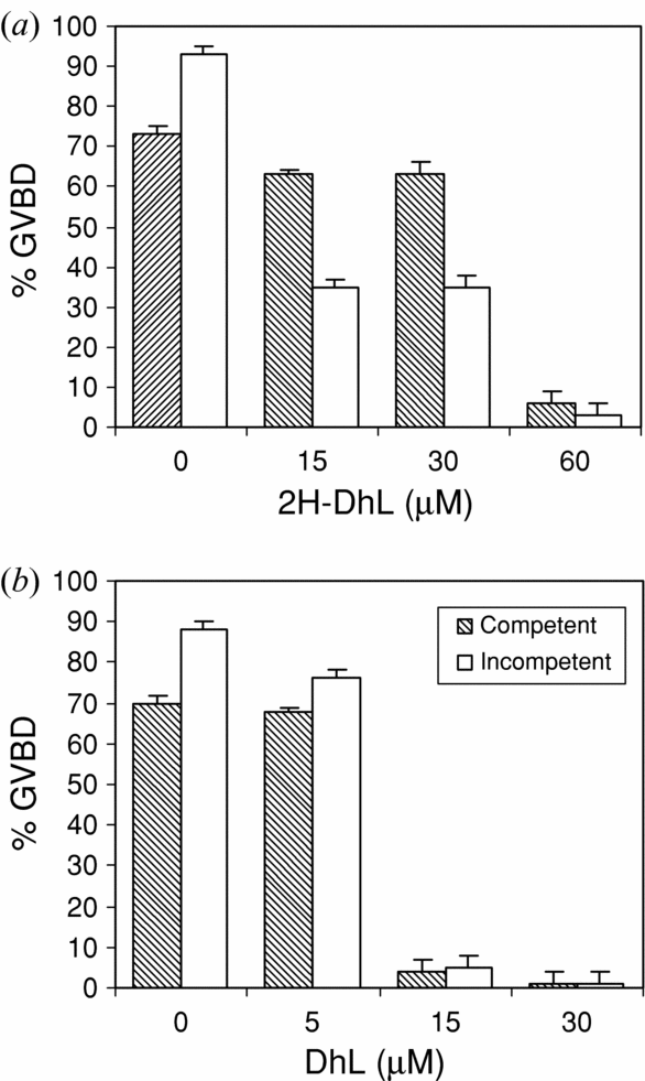
In order to determine whether a derivative of DhL inactivated for alpha-methylenelactone sesquiterpene lactones could inhibit oocyte maturation in the same way as DhL does, 2H-DhL was assayed in Bufo arenarum denuded oocytes competent and incompetent to mature spontaneously. Denuded oocytes competent and incompetent to mature spontaneously were cultured in AR for 60 min with or without different doses of DhL or 2H-DhL before the removal of follicular cells or the addition of progesterone (2.5 μM). GVBD was scored after 24 h of culture.
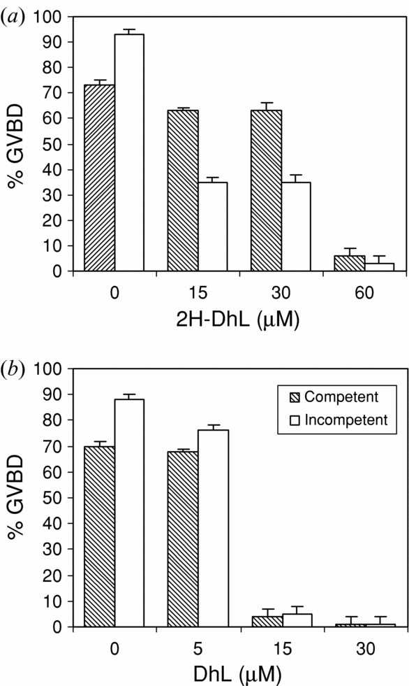
The results indicated that 2H-DhL was able to induce inhibition in GVBD percentages in a dose-dependent manner (Fig. 1a). However, 2H–DhL is less active that DhL and higher doses (60 μM) were required to obtain a significant GVBD inhibition (Fig. 1b).
Effect of 2H-DhL on the GVBD of denuded oocytes competent and incompetent to mature spontaneously. Denuded oocytes competent and incompetent to mature spontaneously were cultured in AR with different doses of: (a) 2H-DhL (15–60 μM) or (b) DhL (5–30 μM) 60 min before the removal of follicular cells or the addition of progesterone (2.5 μM). GVBD was scored after 24 h of culture. Values are the mean ± SEM (n = 4). Each experiment was performed on a different animal.
Effect of 2H-DhL on MPF amplification in oocytes not competent to mature spontaneously
When a small amount of cytoplasm taken from Bufo arenarum matured oocytes is microinjected into prophase oocytes, pre-MPF activation and GVBD occur. In order to determine the effect of 2H-DhL on this process, fully grown denuded oocytes incompetent to mature spontaneously were preincubated (60 min) in the presence of 2H-DhL (30–60 μM) before microinjection of the cytoplasm obtained from oocytes matured with progesterone (2.5 μM) (mature cytoplasm). As controls, oocytes were treated with DhL (30 μM) and without DhL or 2H-DhL and then injected with the same amount (50 nl) of the mature cytoplasm. Injected oocytes were cultured in AR for 24 h and GVBD was scored.
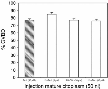
As shown in Fig. 2, injection of 50 nl of mature cytoplasm was enough to promote 90% of GVBD in recipient oocytes. When oocytes were preincubated with 2H-Dhl or DhL no change was observed in GVBD percentages, indicating that these lactones had no effect on MPF amplification.
Effect of 2H-DhL on MPF amplification in oocytes incompetent to mature spontaneously. Fully grown denuded oocytes incompetent to mature spontaneously were preincubated (60 min) in the presence of 2H-DhL (30–60 μM) before microinjection of the cytoplasm obtained from oocytes matured with progesterone (2.5 μM) (mature cytoplasm). As controls, oocytes were treated with DhL (30 μM) and without DhL or 2H-DhL and then injected with the same amount (50 nl) of the mature cytoplasm. GVBD was scored after 24 h of culture. Values are the mean ± SEM (n = 3). Each experiment was performed on a different animal.

Effect of 2H-DhL on GVBD in oocytes not competent to mature spontaneously injected with GV contents
Because GV content transfer is known to induce the events of meiotic maturation (Sánchez Toranzo et al., Reference Sánchez Toranzo, Giordano, López and Bühler2007) we thought it interesting to determine if 2h-DhL could interfere with this process.
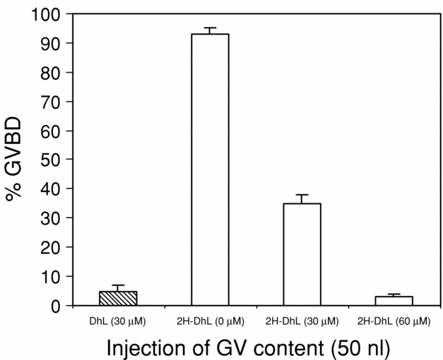
The GV contents of immature oocytes were obtained according to Materials and methods. Fully grown denuded oocytes incompetent to mature spontaneously were treated with different doses of 2H-DhL (30–60 μM) for 60 min before microinjection of 50 nl of GV content. Injected oocytes were cultured in AR with 2H-DhL (30–60 μM) for 24 h before GVBD scoring. As controls, oocytes were treated with DhL (30 μM) or cultured in AR without lactones and then injected with the same amount (50 nl) of GV contents.
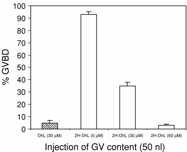
Results (Fig. 3) showed that 2H-DhL induced inhibition of GVBD at doses higher than 30 μM, indicating that this derivative is less active than DhL, which is able to significantly inhibit GVBD with a 30 μM dose.
Effect of 2H-DhL on GVBD in oocytes incompetent to mature spontaneously injected with GV contents. Fully grown denuded oocytes incompetent to mature spontaneously were treated with different doses of 2H-DhL (30–60 μM) for 60 min before microinjection of 50 nl of GV content. Injected oocytes were cultured in AR with 2H-DhL for 24 h before GVBD scoring. As controls, oocytes were treated with DhL (30 μM) or cultured in AR without lactones and then injected with the same amount (50 nl) of GV contents. Values are the mean ± SEM (n = 4). Each experiment was performed on a different animal.
Effect of 2H-DhL and DhL on GVBD induced by PKA inhibition
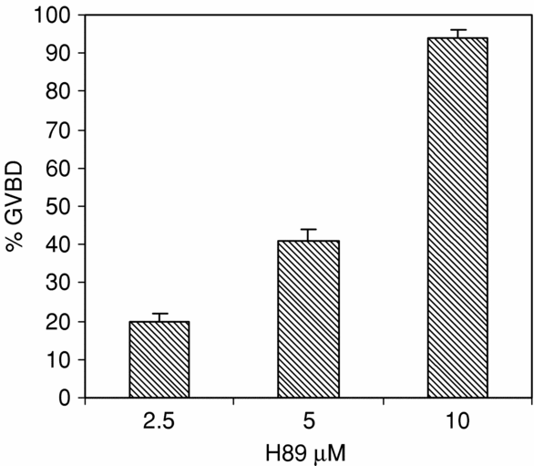
In order to characterize the signalling pathway by which DhL and 2H-DhL inhibited maturation, we analysed whether DhL and 2H-DhL required PKA activity using H89, a PKA inhibitor.
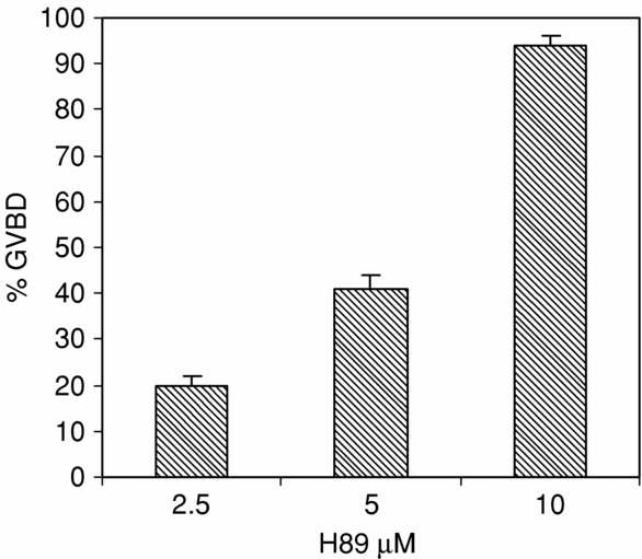
In order to determine the effect of H89 on GVBD in oocytes incompetent to mature spontaneously, a dose-dependent curve was performed. (Fig. 4). On this basis, the dose of 10 μM was selected for the following assays.
Dose–response curve of H89. Fully grown denuded oocytes incompetent to mature spontaneously were injected with different doses of H89 (2.5–10 μM). Injected oocytes were cultured in AR for 24 h at 25 °C before GVBD scoring. Values are the mean ± SEM (n = 3). Each experiment was performed on a different animal.
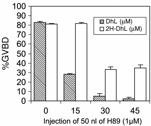
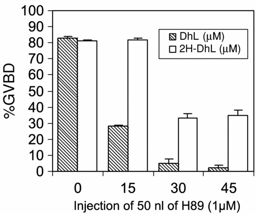
Fully grown denuded oocytes incompetent to mature spontaneously pretreated for 60 min with different doses of 2H-DhL or DhL were microinjected with 50 nl of H89 10 μM and subsequently incubated in 2H-DhL or DhL for 24 h. The results (Fig. 5) show that 2H-DhL and DhL inhibited the GVBD induced by PKA inhibition. Nevertheless, 2H-DhL is less active than DhL. These results suggest that the target of these lactones could be downstream in the pathway that induces GVBD.
Effect of 2H-DhL and DhL on GVBD induced by PKA inhibition. Fully grown denuded oocytes incompetent to mature spontaneously pretreated for 60 min with different doses of 2H-DhL or DhL were microinjected with 50 nl of H89 10 μM and subsequently incubated in 2H-DhL or DhL for 24 h. Values are the mean ± SEM (n = 5). Each experiment was performed on a different animal.
Effect of 2H-DhL and DhL on GVBD induced by inhibition of protein phosphatase 2A
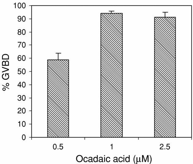
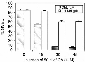
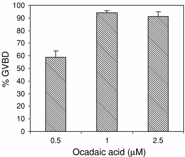
On the basis of a dose-dependent curve of the effect of the OA on the maturation of incompetent oocytes (Fig. 6), the dose of OA 1 μM. was selected. To determine the effect of 2H-DhL and DhL on GVBD induced by inhibition of pp2A, fully grown denuded oocytes not competent to mature spontaneously pretreated during 60 min with different doses of 2H-DhL or DhL were microinjected with 50 nl of an inhibitor of pp2A, OA 1 μM. Injected oocytes were cultured in AR with 2H-DhL or DhL for 24 h before GVBD scoring. The results in Fig. 7 indicated that although both lactones (2H-DhL and DhL) inhibited GVBD induced by OA, the 2H-DhL is less active. So that, DhL 30 μM induced a 70% inhibition of GVBD while the inhibition obtained with the same dose of 2H-DhL is about of 20%. This percentage of inhibition did not increase with the increase of 2H-DhL dose.
Dose–response of OA. Fully grown denuded oocytes incompetent to mature spontaneously were injected with different doses of OA (0.5–2.5 μM). Injected oocytes were cultured in AR for 24 h at 25 °C before GVBD scoring. Values are the mean ± SEM (n = 3). Each experiment was performed on a different animal.
Effect of 2H-DhL and DhL on GVBD induced by the inhibition of the 2A protein phosphatase (pp2A). Fully grown denuded oocytes incompetent to mature spontaneously pretreated during 60 min with different doses of 2H-DhL or DhL were microinjected with 50 nl of OA 1 μM. Injected oocytes were cultured in AR with 2H-DhL or DhL for 24 h before GVBD scoring. Values are the mean ± SEM (n = 4). Each experiment was performed on a different animal.

Discussion
The results of this work suggest that the alpha-methylene-gamma-lactone function of dehydroleucodine (DhL) would not be responsible for the inhibitory effect of this lactone on spontaneous or progesterone-induced GVBD in Bufo arenarum oocytes. Treatment with the hydrogenated derivative of DhL, 2H-DhL, in which the alpha-methylenelactone function was inactivated, continued to exert an inhibitory effect on spontaneous and progesterone-induced GVBD. However, 2H-DhL was less active than DhL as higher doses were required to obtain a significant inhibition.
The use of 2H-DhL in the MPF amplification experiments by injection of both cytoplasm with active MPF and of GV content showed results similar to the ones obtained with DhL, suggesting that the hydrogenated derivative would act in a similar way to DhL.
It has been proposed that in the amplification loop induced by the injection of mature cytoplasm a hyperphosphorylation of cdc25 phosphatase occurs that contributes to a rapid activation of the MPF and to the abrupt transition from phase G2 to M (Izumi & Maller, Reference Izumi and Maller1993). In Bufo arenarum, the amplification loop induced by injection of mature cytoplasm is dependent on phosphatase cdc25 activity (Sánchez Toranzo et al., Reference Sánchez Toranzo, Bonilla, Zelarayán, Oterino and Bühler2006). Pretreatment with 2H-DhL does not affect MPF amplification induced by injection of mature cytoplasm, suggesting that this compound would act in another step of the MPF activation process.
The decrease in cAMP concentration and the subsequent inactivation of PKA are the first early events known to be induced by progesterone (Maller & Krebs, Reference Maller and Krebs1977; Huchon et al., Reference Huchon, Ozon and Demaille1981; Zelarayán et al., Reference Zelarayán, Oterino, Sánchez Toranzo and Bühler2000). When oocyte maturation is initiated by progesterone, cAMP levels are reduced and PKA becomes inactivated. In G2-arrested Xenopus oocytes, cdc25 is probably maintained inactive by PKA-mediated phosphorylation (Qian et al., Reference Qian, Erikson, Taieb and Maller2001; Karaiskou et al., Reference Karaiskou, Leprêtre, Pahlavan, Du Pasquier, Ozon and Jessus2004). Previous studies have shown that inhibition of PKA is sufficient to induce GVBD (Noh & Han, Reference Noh and Han1998; Zelarayán et al., Reference Zelarayán, Oterino, Sánchez Toranzo and Bühler2000; Kovo et al., Reference Kovo, Kandli-Cohen, Ben-Haim, Galiani, Carr and Dekel2006; Wang et al., Reference Wang, Cao and Liu2006). PKA could regulate the activity of cdc25 because PKA phosphorylates ser287 of cdc25 in G2-arrested Xenopus oocytes (Duckword et al., 2002).
Pretreatment with DhL or 2H-DhL decreases the percentage of GVBD induced by H89, a PKA inhibitor, which suggests that these lactones would act on a later step of the signalling pathway that induces MPF activation.
In species with pre-MPF, this prefactor is kept inactive by the activity of the Myt1 kinase, which phosphorylates the Thr14 and Tyr15 residues of p34. In addition, the activity of MAPKs may directly or indirectly regulate some proteins that control MPF activity such Myt1 kinase or cdc25 phosphatase. MAP kinases are inhibited by pp2A (de Vantéry Arrighi et al., Reference de Vantéry Arrighi, Campana and Schorderet-Slatkine2000).
It has been proposed that the microinjection of OA, an inhibitor of pp2A by promoting phosphorylation/activation of cdc25 (Bialojan & Takai, Reference Bialojan and Takai1988), initiates the positive feedback loop that controls activation of p34 in oocytes (Izumi & Maller, Reference Izumi and Maller1993). In addition, initial studies reported that pp2A could be the phosphatase responsible for the dephosphorylation of cdc25 activatory residues (Karaïskou et al., Reference Karaïskou, Jessus, Brassac and Ozon1999).
The fact that both DhL and 2H-Dhl inhibit GVBD induced by OA microinjection suggests that they could act on the activity of the Myt1 kinase. This idea is supported by the experiments of injection of GV contents in which an inhibitory effect of these lactones on GVBD was also observed. The injection of GV contents in oocytes treated with NaVO3, an inhibitor of cdc25 phosphatase in which the GVBD occur (Sánchez Toranzo et al., Reference Sánchez Toranzo, Bonilla, Zelarayán, Oterino and Bühler2006) supported the idea that phosphatase cdc25 in not involved in this process.
It has been proposed that in the GV there would exist an inhibitor of pp2A (Picard et al., Reference Picard, Labbé, Barakat, Cavadore and Dorée1991; Okumura et al., Reference Okumura, Sekiai, Hisanaga, Tachibana and Kishimoto1996; Prigent & Hunt, Reference Prigent and Hunt2004). These results agree with the ones obtained with the injection of mature cytoplasm, suggesting that cdc25 phosphatase would not be the target of these lactones.
Interestingly, although in 2H-DhL the alpha-methylene-gamma-lactone function of DhL was inactivated, both lactones induced inhibition of GVBD, however 2H-DhL was less active than DhL as higher doses were required to obtain a significant inhibition.
In summary, our results indicate that the inhibitory effect on meiosis progression of DhL does not depend only on the activity of the alpha-methylenelactone function, as its hydrogenated derivative, 2H-DhL, in which this function has been inactivated, causes similar effects on amphibian oocytes. It seems possible that the effect would be due to the structure of the whole molecule. On the other hand, the analysis of the participation of certain mediators in some of the signalling pathways leading to MPF activation suggests that the Myt1 kinase could be a target of these lactones, while cdc25 phosphatase would not be affected. Besides, the PKA inhibition assays indicate that these lactones would act later in the signalling pathways.
Acknowledgements
This work was supported by a grant from the Science Council of the National University of Tucumán (CIUNT) and the National Agency for Promotion of Science and Technology (FONCYT).