Pulmonary venous stenosis is rare. In spite of significant improvements in the management and outcome of other congenital cardiac problems, the prognosis of this condition continues to be dismal. Such acquired stenosis is a known complication following surgery during childhood for correction of anomalous pulmonary venous drainage. Acquired stenosis has also been described in adults, especially after radiofrequency ablation for atrial arrhythmias, with generally a favourable outcome over the long term. Acquired stenosis in children has a very different outcome. When the pulmonary veins were initially normally connected, such stenosis is not well recognised, with non-specific symptoms and signs. We have recently encountered 7 infants, all with normal pulmonary venous connections, and all initially deemed to be normal on clinical assessment, who acquired pulmonary venous stenosis, with very significant resultant morbidity and mortality.
Patients and methods
We present a retrospective review of data culled from case notes and serial echocardiograms of seven infants with normal pulmonary venous connections on initial assessment, but who acquired stenoses of the pulmonary veins. The data was collected from two tertiary units for referral.
Results
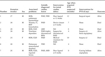
The demographic details are given in Table 1.
Table 1. Details of patients with acquired pulmonary venous stenosis.

First patient
Stenosis of the left pulmonary veins was diagnosed at 12 months of age by echocardiography, and subsequently confirmed angiographically. At surgery, the left upper pulmonary vein was identified, but only as a fibrous cord approximately 2 millimetres in diameter. No flow was demonstrated down this vessel. The left lower lobe pulmonary vein was identified, and noted to be of reasonable caliber proximally, but a probe of no more than 1 millimetre could pass through the stenosis. The vein was opened longitudinally, and the stenosing fibrotic material was excised. The postoperative course was relatively uncomplicated. Within a year of surgery, nonetheless, the left lower pulmonary vein had also become occluded. The left pulmonary artery was small in size, and the right lower vein small compared to the right upper vein. Subsequently, the patient continues to have elevated right-sided pressures, but some collateral flow has developed from the left-sided pulmonary veins to the territory drained by the left brachiocephalic vein, as demonstrated by angiography 3½ years after the initial presentation (Fig. 1).

Figure 1. Angiogram of the left pulmonary artery shows contrast agent spilling over into the right pulmonary artery. The arborisation pattern of the left pulmonary artery is abnormal (a). The pulmonary venous return from the right side is seen on follow through while there is still dye hold up in the left pulmonary artery territory (b). In some months time the same patient developed a pulmonary venous channel draining into the superior caval vein (c).
Second patient
This infant was being investigated for nocturnal hypoxia but developed respiratory syncitial virus positive bronchiolitis at 9 weeks of age, which required extracorporeal membrane oxygenation. A new heart murmur was audible at this time. Echocardiography showed a large, patent arterial duct, which was closed via the transcatheter route with an Amplatzer ductal occluder. Because of the lack of clinical improvement, he was again investigated 4 weeks after the ductal closure. Bronchoscopy and bronchography were normal, but pulmonary venous stenosis was diagnosed at trans-oesophageal echocardiography. Cardiac catheterisation showed right ventricular systolic pressure to be about 60% of corresponding systemic pressure, with a mean pulmonary arterial pressure of 30 millimetres of mercury. Angiography confirmed diffuse and severe stenosis of all pulmonary veins. This was not considered amenable to surgical repair, and the infant died at 10 months of age.
Third patient
This patient had been discharged with normal findings 4 months after birth. On follow-up by the cardiology team at 6 months, there was echocardiographic evidence of suprasystemic pressures in the right-sided chambers, and acquired stenosis of the right-sided pulmonary veins and the left upper pulmonary vein were seen. It was considered unlikely that the child would benefit from surgery. The infant died at 7 months of age. Post mortem examination showed endocardial fibrosis of the atrial insertions of all the 3 affected pulmonary veins. There were multiple areas of stenosis, and intrapulmonary extension of the stenotic lesions.
Fourth patient
In this infant, recanalisation and stenting of the arterial duct was undertaken at 10 days of age, but the pulmonary venous drainage was deemed to be normal. At 10 weeks of age, the right pulmonary artery was connected to the pulmonary trunk using a Gore-Tex® interposition graft of 6 millimetres diameter. On follow-up, the patient subsequently developed elevated pressures in the right heart, and acquired stenosis was noted of the left-sided pulmonary veins. Since then, he has undergone two surgical operations on the stenosed left-sided pulmonary veins. During a third procedure, a stent was implanted in one of the stenosed veins, the other having occluded in the interim. The pressure in the distal right pulmonary artery is normal, albeit with stenosis of the proximal portion of the vessel. The pulmonary veins on the right side have remained normal. The infant is alive, being dependent at home on a ventilator, and has significantly elevated right heart pressures.
Fifth patient
After initial discharge from hospital, this patient developed progressive wheezy symptoms, requiring repeated hospital admissions with suspected upper respiratory tract and chest infections. Transthoracic echocardiography revealed a grossly dilated right heart, with a poorly functioning right ventricle, and right-to-left shunting across the oval fossa. There was also continuous pulmonary venous flow at high velocity, consistent with pulmonary venous obstruction. Surgical relief of the pulmonary venous stenosis was attempted on two occasions, but she died with re-stenosis within a few months.
Sixth patient
This patient was diagnosed antenatally with a ventricular septal defect, and developed cardiac failure at 3 weeks of age, with suboptimal symptomatic relief provided by medication using captopril and diuretics. Echocardiography had initially shown a large perimembranous ventricular septal defect limited by tricuspid valvar tissue. A fenestrated atrial septal defect was also shown, along with normal pulmonary venous connections. As the infant continued to be more symptomatic than would have been expected from the now smaller ventricular septal defect, he was investigated further at 4 months of age. Echocardiography at this stage showed a perimembranous ventricular septal defect of 6 millimetres diameter permitting bi-directional shunting. The study also revealed continuous and turbulent flow at high velocity in the left lower pulmonary vein, suggestive of significant stenosis in that vessel (Fig. 2). An ultrasonic scan showed eventration of the right diaphragm, which was repaired without any symptomatic relief. Lung biopsy showed mesenchymal dysplasia, a rare developmental abnormality of the lungs. He was treated with systemic steroids, with some improvement initially, but deteriorated steadily, and died within a few weeks.

Figure 2. The trans-thoracic apical 4 chamber view (a) shows a narrow left pulmonary vein, with high velocity jet on colour flow mapping. Pulsed wave Doppler (b) shows a pattern of continuous flow without return to baseline.
Final patient
This male infant was discharged from hospital after a stay of nearly 6 months, having been treated for pulmonary hypertension with oxygen and sildenafil. On cardiac follow-up, he was shown, over a period of 3 months, to have developed progressive pulmonary venous stenosis. Angiography at a corrected age of 10 months showed the left upper vein to be completely occluded, albeit with some collaterisation. There was moderate-to-severe stenosis of the veins draining the left lower and right upper lobes. These were successfully dilated using cutting balloons. At the time of the procedure, the right lower vein was unobstructed.
Discussion
Pulmonary venous obstruction is not uncommon following surgery for abnormally connected pulmonary veins.1 Acquired stenosis of normally connected pulmonary veins is much rarer, may present later with a less specific clinical picture, and has been diagnosed even in adulthood.2 The symptoms and signs may be mimicked by chronic lung disease, which is common when following up neonates.3 There are limited data concerning the possible aetiology of this devastating condition, but it appears to be multi-factorial.
We have noted that, in our series of patients, additional flow in the pulmonary venous system may be a substrate for the acquired venous stenosis. This was usually the result of increased flow in consequence of a left-to-right shunt at the level of the arterial duct, the atrial or ventricular septum, or at multiple levels. This may be responsible for expression of pulmonary veno-constrictive factors. It is very unusual to develop such acquired stenosis when the pulmonary veins are normally connected and there is normal flow of blood to the lungs.4
In an otherwise structurally normal heart, a patent arterial duct is likely to produce higher flow in the left pulmonary artery, due to the so-called Coanda effect, and hence the left-sided pulmonary veins are those which typically become affected.5 If there is unilateral stenosis of one pulmonary artery, there is resultant additional flow in the contralateral artery. Should pulmonary venous stenosis develop in this setting, it is on the other side relative to the stenosed artery. Increased flow, however, cannot be the only factor contributing to acquired stenosis, since left-to-right shunts such as atrial septal defects have not been previously described to be associated with this problem even when continuing over prolonged periods of time.
Prematurity may be another associated factor, since extreme prematurity has the potential for disrupting normal organogenesis, and hence is a possible substrate for acquisition of the venous stenosis. Injury to the pulmonary venous ostial endothelium has also been implicated to be responsible for acquired stenosis,6 but could not be identified in any patient in our series.
It is also possible that pulmonary disease may be a trigger for pulmonary venous endothelial proliferation. Chronic lung disease associated with prematurity had been present in 3 of our patients, and another had severe bronchiolitis due to the respiratory syncytial virus, and required extracorporeal membrane oxygenation. It is possible, therefore, that such pulmonary factors may interact with excessive flow in susceptible infants, and lead to proliferation of tissues, and hence pulmonary venous stenosis. This particular form of stenosis is the most aggressive pattern of re-obstruction of any congenital obstructive disorders, and is almost certainly due to aggressive neoproliferation.7 When these patients have suffered turbulent perinatal courses, especially following extreme prematurity, there is a possibility of the release of high levels of inflammatory cytokines in the background of injury mediated by hypoxia, which may trigger such neoproliferation. Post-surgical stress may also be a trigger.7 Narrowed veins are more likely to be thrombosed, and it is likely that thrombosis in a stenotic pulmonary vein may further compromise the lumen, and contribute to the progression of obstruction.8
In view of the poor outcome of conventional surgery, newer techniques like sutureless pericardial marsupialisation are being used, with encouraging early results.9 Other neo-proliferative disorders have been treated with chemotherapy10 and gamma interferon.11 Such options might potentially offer better outcomes for this condition, but remain unproven. The beneficial effects of drug-eluting stents are well established in the setting of coronary arterial disease.12, 13 Similar treatments may become an option in the management of pulmonary venous stenosis. For reasons that are not yet clear, the condition is occurring with increased frequency. This may reflect the longer survival of premature infants, or the wider availability of echocardiographic imaging. It may also be possible that there may be substrates for stenosis prior to birth, which subsequently become more overt in response to various triggers and/or fall of pulmonary vascular resistance, leading to the observation of apparent “acquired” stenosis. As it is difficult to exclude this potentially fatal condition on the basis of a limited or single early normal echocardiogram, we suggest that repeat echocardiographic assessment should be undertaken of the infants considered to be at risk. Colour Doppler, and pulsed wave interrogation, of the all the individual pulmonary veins is important. Attention should be paid to those infants with chronic lung disease who fail to improve subsequent to treatment. Infants with left-to-right shunts and unilateral pulmonary arterial stenosis may be at particular risk, and merit detailed assessment. If treatment is to be successful, early identification of the venous stenosis is important before end-stage pulmonary hypertension ensues. All of our patients had an initial detailed echocardiogram that did not show any abnormality of the pulmonary veins. We believe, therefore, that ours is the first series of patients documenting the acquisition of stenosis in normally connected pulmonary veins.