Congenital coronary sinus anomalies are relatively uncommon. They can occur either in isolation or in conjunction with other congenital heart defects. The most common coronary sinus anomaly is dilation of the coronary sinus, either due to left-to-right shunt or due to persistent left superior vena cava draining into the coronary sinus. Less common anomalies include hypoplasia or absent coronary sinus. Reference Mantini, Grondin, Lillehei and Edwards1,Reference Shah, Teague, Lu, Dorfman, Kazerooni and Agarwal2 The left ventricle (LV) to coronary sinus fistula appears to be the rarest entity of this group. There have been only six prior reports in the literature which describe this condition. Reference Mcgarry, Stark and Macartney3–Reference Fetter, Backer, Alexander and Weigel8 This is the first case report that describes an association of the LV-coronary sinus fistula with Wolff–Parkinson–White Syndrome, and non-surgical management with successful transcatheter device closure of the fistula followed by radiofrequency ablation of the accessory pathway.
Case
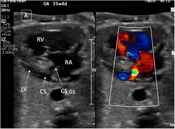
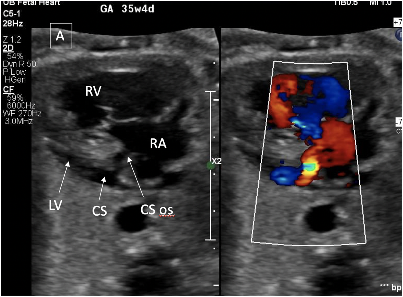
The cardiac defect was noted on prenatal echocardiogram and differential diagnosis was either a coronary-cameral fistula or left ventricle to coronary sinus fistula (Fig 1). A male infant was delivered at term to a 34-year-old mother. The baby weighed 3120 g at birth. The immediate postnatal course was uncomplicated. Physical exam was notable for a high frequency grade 3/6 continuous murmur at the base of the heart. Postnatal transthoracic echocardiogram confirmed a fistula between the base of the left ventricle and coronary sinus with associated dilation of the coronary sinus, right atrium, and right ventricle. There was moderate tricuspid valve regurgitation. A 12 lead electrocardiogram was notable for manifest pre-excitation with a negative delta wave in lead II concerning for a coronary sinus accessory pathway. Subsequently the child was started on propranolol due to multiple episodes of non-sustained supraventricular tachycardia.
A modified axial view on fetal echocardiogram showing the dilated coronary sinus (posteriorly) with a high-pressure fistulous connection of the left ventricle to the right atrium. The right atrium and ventricle are dilated.
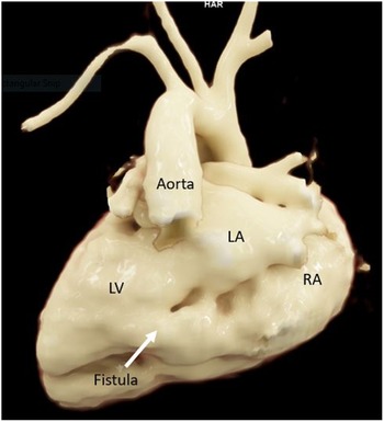
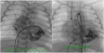
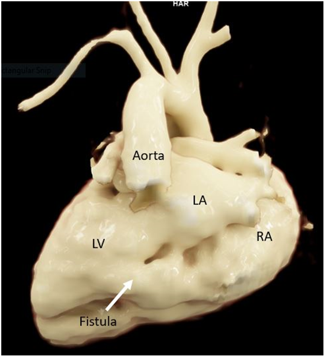
Intracardiac anatomy was confirmed with a cardiac Magnetic resonance imaging on day 2 of life which confirmed the presence of a fistula from the base of the LV to the right atrium via the coronary sinus with an approximately 5:1 shunt (Fig 2, Supplemental Video 1). The patient underwent electrophysiologic and cardiac catheterisation studies on day 4 of life. The transesophageal electrophysiology study showed a moderate risk accessory pathway with shortest preexisted RR interval of 260 mseconds with rapid atrial pacing. Angiography showed an LV to CS fistula (Fig 3a). The Qp/Qs was calculated at 2:1. No interventions were undertaken at that time. Propranolol was discontinued due to hypotension and Flecainide was started. The patient was started on furosemide to counter signs of heart failure and later started on milrinone due to right ventricular dysfunction, however severe pulmonary over-circulation persisted.
A 3D reconstruction of the heart from cardiac magnetic resonance imaging with posterior orientation of the heart showing the left ventricle to coronary sinus fistula (shown by arrow). LA: left atrium; LV: left ventricle; RA: right atrium.
( a ) Ventriculogram showing the left ventricle to coronary sinus fistula, which measured 6.8 mm by 4 mm at the origin with the left ventricle. ( b ) LV angiocardiogram showing the 8 mm Amplatzer Vascular Plug II device in place with no residual shunt.
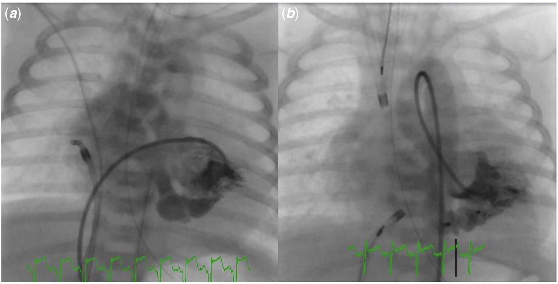
The patient underwent device closure of the fistulous connection of the LV to the coronary sinus diverticulum on day 12 of life. Initial attempts to advance the guiding sheath into the left ventricle via the tortuous fistulous connection were not successful despite utilising antegrade and retrograde approach (right internal jugular, right femoral vein, right femoral artery) as well as using a snare distally in the descending aorta to anchor the veno-arterial guidewire. Eventually, umbilical venous access was utilised to establish guidewire access from the right atrium to the LV across the tortuous fistulous connection using a 4 French Berenstein catheter and 0.014 inch Prowater guidewire inside the 5 French 45 cm long Cook Performer guiding sheath (Children’s Hospital of Boston design). The umbilical vein sheath was advanced over the catheter and guidewire across the fistula into the left ventricle (Supplemental videos 2 and 3), and an 8 mm Amplatzer Vascular Plug II device was implanted across the LV origin of the fistula (Fig 3b). Of note, every time a catheter or sheath was advanced across the coronary sinus orifice of the fistula, evidence of pre-excitation on the cardiac monitor ECG disappeared. Post-procedure transthoracic echocardiogram showed a well-positioned device with no residual shunt and no evidence of mitral valve regurgitation.
The patient recovered and was discharged on an antiarrhythmic regimen of amiodarone and flecainide. At the age of 13 months, the patient underwent diagnostic cardiac catheterisation with angiography. There was no evidence of any residual stunt and coronary angiography showed that the posterior descending coronary artery was not well developed and there was no major coronary artery branches traversing the CS. Intracardiac electrophysiology study was subsequently performed and the pathway was mapped to the neck of the coronary sinus diverticulum and successfully ablated using radiofrequency energy.
Discussion
We describe the first case of congenital LV to coronary sinus fistulae in association with Wolff–Parkinson–White Syndrome that was successfully treated with device closure and catheter ablation. Prior to our case there have been six reports of congenital LV to coronary sinus fistula in the literature. The first description of this entity was in 1981. Reference Mcgarry, Stark and Macartney3 All of these cases appeared to be postnatally diagnosed and the majority were haemodynamically insignificant, not requiring intervention. Two patients underwent surgical correction. Reference Mcgarry, Stark and Macartney3,Reference Fetter, Backer, Alexander and Weigel8
Embryologically, the coronary sinus arises from the left horn of the sinus venosus while the LV arises from the bulbus cordis. These are distinct embryological substrates. Hence, the coronary sinus should not have any communication with the left ventricle. It has been postulated that the pathogenesis of this lesion may be a primary coronary sinus aneurysm or diverticulum that may have ruptured into the LV. This would have resulted in an LV to coronary sinus fistula. Reference Gnanapragasam, Houston and Lilley5 In this case, the other differentials considered in fetal life were, congenital Gerbode ventriculo-atrial shunt or coronary artery to coronary sinus fistula. Reference Gerbode, Hultgren, Melrose and Osborn9 Hemodynamically, this lesion is most similar to a Gerbode shunt. Preexcitation with a negative delta wave in lead II is very suggestive of a coronary sinus accessory pathway and must prompt a thorough evaluation for coronary sinus pathology. Coronary sinus diverticula are often associated with accessory pathways and Wolff–Parkinson–White Syndrome, many of which require catheter ablation. Reference Saremi and Krishnan10,Reference Veloor and Lokhandwala11 However, there was no description of Wolff–Parkinson–White Syndrome in the previous cases of congenital LV to coronary sinus fistula reviewed; though two of the cases had a history of supraventricular tachycardia without manifest preexcitation.
The coronary sinus is important in electrophysiology as a site for left ventricular pacing or a location where accessory pathways are located. Reference Saremi and Krishnan10 Coronary sinus anomalies can impact cannulation of the coronary sinus for mapping, catheter ablation, or left ventricular pacing. Reference Shah, Teague, Lu, Dorfman, Kazerooni and Agarwal2 The coronary sinus diverticulum is a well-described entity that is seen in which an accessory pathway can be found in the neck of the diverticulum seen at the inferior aspect of the coronary sinus at its junction with the middle cardiac vein. Thankfully in our case, the percutaneously placed vascular plug across the LV origin of the fistula, did not cover the accessory pathway or complicate the ablation.
A coronary sinus to LV fistula often requires intervention as it would result in a pressure and volume load to the right atrium. Over time, there is potential for right atrial enlargement and atrial arrhythmia. The fistulous connection is a high-pressure shunt and the retrocardiac location would be difficult to access surgically. Surgical repair with a synthetic or pericardial material could be complicated by an aneurysm or pseudoaneurysm formation. In light of this, a percutaneous approach was chosen. In these cases, either umbilical venous or transhepatic access may provide more direct access to the coronary sinus and facilitate utilisation of larger sheath sizes for delivery of a device.
Conclusion
This is the first description of device closure and Wolff–Parkinson–White Syndrome in the cohort of congenital LV to coronary sinus fistulae. We report a percutaneous device closure with an excellent post-procedural result. Coronary sinus fistula should be on the differential diagnoses in a neonate born with Wolff–Parkinson–White Syndrome pattern on ECG. LV to coronary sinus fistula should be suspected in patients with a dilated coronary sinus. Device closure can be performed in congenital LV to coronary sinus fistula with a significant shunt and may be superior to surgical intervention. Device occlusion of this lesion did not preclude later ablation of an accessory pathway.
Supplementary material
To view supplementary material for this article, please visit https://doi.org/10.1017/S1047951122000853
Acknowledgements
We would like to thank the sonographers who obtained the images used in this manuscript.
Author contributions
PM compiled clinical data and wrote draft manuscript. SS, FC, and MA reviewed manuscript and edited it. AC and SP selected and edited accompanying images.
Financial support
This research received no specific grant from any funding agency, commercial, or not-for-profit sectors.
Conflicts of interest
None.
Informed consent
Informed consent was obtained from the family.