Our understanding of bipolar disorder (BD) in children and adolescents has advanced markedly since the early 1990s. Before then, few health professionals believed that children and adolescents could be diagnosed with, or treated for, mania and BD (Carlson & Strober, 1978). Yet, this was only opinion, not grounded in research. This situation mirrors that of the field of pediatric depression before the 1970s, when it was controversial as to whether or not pediatric patients could be diagnosed or treated for depression (Puig-Antich, Blau, Marx, Greenhill, & Chambers, 1978; Weissman & Myers, 1978). Now, clinicians and researchers alike are moving beyond debate about the existence of BD in youth toward greater understanding of its clinical presentation and pathophysiology. Specifically, because BD is differentiated from all other psychiatric disorders by marked, impairing fluctuations of mood and emotion, there is a mutually beneficial interplay between current affective neuroscience research investigating the development of emotion regulation in healthy youth, and pediatric BD research determining how this process is altered in BD youth.
Clinical Controversies in Pediatric BD
Three questions have fueled the diagnostic controversy about childhood-onset BD. First, which DSM-IV-TR “A” criterion for defining a manic episode, that is, elevated/expansive or irritable mood, is more diagnostically specific for, or more characteristic of, pediatric BD (American Psychiatric Association, 2000)? Second, to what extent are the symptoms of pediatric BD episodic or chronic? Third, how should one differentiate between symptoms of BD and those of disorders frequently comorbid with BD, especially attention-deficit/hyperactivity disorder (ADHD)?
Mania: Elevated, expansive versus irritable mood?
Both elevated/expansive (euphoria) and irritable mood are included in the DSM-IV-TR criterion A for mania; yet, there is a fundamental difference between these two mood states (American Psychiatric Association, 2000). Namely, elevated, expansive mood specifically differentiates mania from all other psychiatric disorders across the life cycle, while irritability is not specific to mania or BD (Geller et al., 2002). Instead, irritability is (a) a diagnostic feature of several psychiatric disorders, such as generalized anxiety disorder; (b) present in specific DSM-IV-TR diagnostic modifications for children, such as for major depressive episode; and (c) an associated feature, but not a DSM-IV-TR criterion, of several psychiatric disorders in children or adolescents, such as ADHD, oppositional defiant disorder (ODD), and even pervasive developmental delay-spectrum disorders.
Several issues have hindered research about irritability. To paraphrase Judge Potter Stewart, although clinicians and researchers alike “know [irritability] when they see it,” there is no well-accepted definition of irritability. Moreover, there is no consensus about the phenomenology of irritability, including what differentiates developmentally and contextually appropriate expressions of irritability and frustration from pathological irritability in children and adolescents (Leibenluft, Blair, Charney, & Pine, 2003). For example, semistructured psychiatric interviews commonly used to assess irritability rely upon markedly different prompts. These include the following: (a) Kiddie Schedule for Affective Disorders and Schizophrenia—Present and Lifetime version (K-SADS-PL): “anger, crankiness; bad temper; short-tempered; resentment or annoyance, whether overtly expressed or not; easily lose temper; touchy or easily annoyed” (Kaufman et al., 1997); (b) Diagnostic Instrument Schedule for Children: “cranky; angry toward people you had no reason to be; talk back; temper tantrum” (Shaffer et al., 1996); (c) Children's Depression Rating Scale: “grumpy, crabby, talked back, sassy, wouldn't do something your parents asked you to do” (Poznanski, Cook, Carroll, & Corzo, 1983). There is heterogeneity in what these instruments assess, as they mix notions of angry mood, externalizing behavior, and a lack of cognitive and emotional flexibility in adapting to parental/social expectations. Two things that are sorely needed are the development of reliable scales, rather than single items, to assess irritability and research comparing the neurobiological and phenomenological manifestations of irritability between typically developing youth and those with a variety of psychiatric diagnoses, including BD, ADHD, ODD, and depression.
Temporal course of mania: Episodic versus chronic?
A second controversial issue is whether childhood-onset BD is an episodic or chronic condition. Referring again to the DSM-IV-TR criteria, a manic episode is defined as a “distinct period of abnormally and persistently” altered mood for 4 or more days (hypomanic episode) or for 7 or more days (manic episode). Yet, there are several different ways of interpreting this statement. Some believe that childhood-onset BD, like that in adults, does manifest by episodes of mania that are distinguishable from a baseline, euthymic mood (Birmaher et al., 2006). If there is no clear-cut episode, then these children are not considered to be suffering from BD. In contrast, others believe that BD and mania has a developmentally unique presentation in children and adolescents, such that children are constantly ill either because clear episodes are not discernable, or because the episodes are extremely brief and frequent (Biederman et al., 2004; Geller, Tillman, Craney, & Bolhofner, 2004). Both viewpoints have been supported by large longitudinal studies, likely because different groups assess the presence of distinct episodes differently in both their inclusion/exclusion criteria and how they use their different forms of semistructured assessment devices. Two potential methods of resolving this issue include either collaborative multisite longitudinal studies or use of a phenotyping system, such as that of Leibenluft, Charney, Towbin, Bhangoo, and Pine (2003), which will be discussed later, that distinguishes children with an episodic course from those with a chronic course so that the two groups can be compared clinically and pathophysiologically.
Comorbidity: Diagnostic confound or co-occurring disorder?
There are two interrelated issues regarding comorbid psychiatric diagnoses in childhood-onset BD. First, BD rarely occurs in isolation, whether in adult or pediatric patients. Instead, patients with BD often have comorbid, meaning co-occurring, disorders, such as ADHD (Wozniak et al., 1995), anxiety disorders (Dickstein, Rich, et al., 2005; Harpold et al., 2005), or substance use disorders (Wilens et al., 1999). Second, the diagnostic criteria of these comorbid disorders overlap significantly with those of BD itself. This has led to several challenges for both researchers and clinicians. First, with so many BD children having comorbid ADHD, how should one distinguish whether the symptom of distractibility is due to BD or ADHD? For researchers who view pediatric BD (like adult BD) as an episodic illness, the answer is to diagnose comorbid illnesses on the basis of symptoms present during euthymia, whereas for those who see BD as a chronic illness the issue is more problematic. Second, how can one be certain that research studies demonstrating differences between BD and typically developing youth are not due to the presence of comorbid ADHD, rather than to BD itself? Research techniques to address this concern include stratifying samples based on the presence/absence of comorbid diagnoses as well as comparing youth with primary BD to those whose primary diagnosis is one that is commonly comorbid in BD, that is, primary ADHD or primary generalized anxiety disorders or primary conduct disorder (Dickstein, Garvey, et al., 2005; Geller et al., 1998; Olvera, Semrud-Clikeman, Pliszka, & O'Donnell, 2005; Moore et al., 2006).
Phenotypes of Pediatric BD: A Research Approach
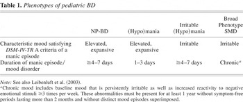
Although several groups are working on these controversies, there is no simple resolution (Biederman et al., 2004; Bhangoo et al., 2003; Carlson et al., 2003; Chang et al., 2003; DelBello, Zimmerman, Mills, Getz, & Strakowski, 2004; Geller et al., 2004). To facilitate hypothesis-driven research on the two principal controversies in pediatric BD (i.e., specificity of elevated/expansive vs. irritable mood, and presence and duration of episodes), we created a research classification system consisting of four phenotypes for juvenile mania (Table 1; Leibenluft, Charney, et al., 2003).
Phenotypes of pediatric BD

At one extreme in this system are so-called “narrow phenotype BD” (NP-BD) youth who meet criteria that are even more stringent than DSM-IV-TR because (a) they must have clearly defined episodes of euphoric mania accompanied by three “B” neurovegative symptoms and functional impairment and (b) symptoms of euphoric mania must be present most of the day, every day, for ≥4 days for hypomania or ≥7 days for mania (mirroring the DSM-IV-TR criteria for temporal duration of a major depressive episode). Many NP-BD subjects have irritable mood in addition to euphoria, but those with irritable mania only are excluded from this group. Such stringent criteria are necessary to ensure that the NP-BD sample is quite homogeneous, including satisfying the “cardinal,” that is, unique, features of mania (Geller et al., 1998). Increased sample homogeneity increases the power of neurobiological studies designed to differentiate emotion regulation mechanisms in typically developing youth, those with pediatric BD, and those with other forms of psychopathology.
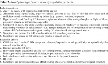
At the opposite end of the spectrum is the “broad phenotype.” Known as “severely mood dysregulated” (SMD) youth, these children have chronic, nonepisodic irritability that is operationalized as having a baseline sad or angry mood plus marked, developmentally inappropriate reaction to negative emotional situations accompanied by symptoms of hyperarousal (Table 2). By definition, these SMD subjects lack euphoric mania; that is, they have no history of of elevated/expansive mood. In fact, rather than having distinct mood episodes, as do NP-BD youth, SMD subjects have a chronic condition with continuous, uninterrupted symptoms. Children with SMD differ from those with ADHD in that only the former have an aberrant mood. These criteria were designed to capture a clinical presentation that some researchers suggest is the principal presentation of prepubertal mania (Biederman et al., 2004), a belief challenged by other researchers (Biederman, Klein, Pine, & Klein, 1998; Bhangoo et al., 2003; Geller et al., 2002).
Broad phenotype (severe mood dysregulation) criteria

Between the narrow and broad phenotypes are two intermediate phenotypes. “Hypomania not otherwise specified (NOS)” is defined as children with euphoric mania (like NP-BD) but whose manic episodes last 1 to 3 days. The second intermediate phenotype, known as “irritable (hypo)mania,” consists of children who have distinct episodes lasting at least 4 days (unlike SMD) characterized of irritability rather than euphoria. Of importance, although both intermediate phenotypes have clear-cut mood episodes, irritable (hypo)mania satisfies full DSM-IV-TR episode duration criteria for hypomania, whereas the hypomania NOS phenotype does not.
Studies using these pediatric BD phenotypes may address the key diagnostic dilemmas in the field by comparing these pediatric BD subpopulations to one another, to typically developing youth, and to children with other psychiatric illnesses. Researchers can test whether these phenotypic groups are distinct in longitudinal course, response to treatment, underlying pathophysiology, or genetics. An example includes findings from the Course and Outcome of Bipolar Youth (COBY) study. COBY is a multisite longitudinal study that enrolled over 400 BD youth, including BD type I and II as well as a group of BD NOS, the latter of whom met criteria similar to those of hypomania NOS with distinct episodes of elated mood of less than the 4 days required for DSM-IV-TR hypomania criteria. Recently, COBY investigators have demonstrated that 25% of BD NOS subjects converted to either BD type I or type II over the course of 2 years (Birmaher et al., 2006). Also, COBY has shown that more than 80% of BD type I and II subjects had both elevated/expansive mood and irritable mood as part of their illness, indicating that euphoria is indeed common in children with BD (Axelson et al., in press). Careful phenotyping will facilitate neurobiological research into the boundaries of emotion regulation between BD youth and healthy youth, including greater understanding of the mechanisms mediating euphoria and irritability and how they change with development.
Fundamental Psychological Constructs of Emotion Regulation
Before exploring differences between BD and healthy youth on measures of emotion and its regulation, it is important to start with the fundamentals of affective neuroscience. What is “emotion regulation”? Clinicians and researchers frequently describe youth with psychopathology, such as BD or BD-like symptoms, as suffering from a disturbance of emotion regulation, given their marked and sometimes rapid mood alterations. Yet, as stated by Forbes and Dahl, this is akin to saying that a febrile child suffers from “temperature dysregulation”; although it is true, it lacks specificity required to result in better understanding of disorder and its treatment (Forbes & Dahl, 2005; Thompson, 1994). Moreover, others have argued that the term is superfluous because emotions are inherently regulatory (Cole, Martin, & Dennis, 2004). Determining how BD children maintain and switch emotional state is crucial to understanding how youth with BD differ from those without psychopathology.
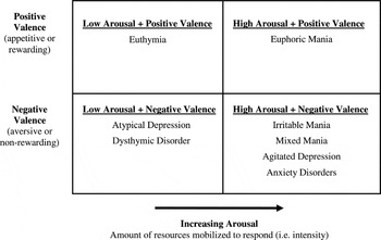
From a neuroscience perspective, “emotion” may be defined as an evoked response to an environmental stimulus with motivational salience (Lang, Bradley, & Cuthbert, 1998). This motivational salience may be characterized along two orthogonal dimensions: “valence” and “arousal.” The valence of an emotion is either positive, meaning that a stimulus is appetitively rewarding and an organism will expend energy to approach it, or negatively valenced and aversive, meaning that an organism will expend energy to avoid it. The term arousal connotes the amount of an organism's resources that are mobilized in response to an emotional stimulus.
Although this framework of Lang et al. is focused on emotion in general, it is useful in the conceptualization of psychopathology as well (Figure 1). Specifically, from this perspective, euphoric mania present in the NP-BD subjects is unique because, of all psychopathological states, it alone represents the extreme of the positively valenced, high arousal state. In contrast, both irritable mania and the clinical presentation of SMD youth would be classified as being negatively valenced, high arousal emotional states. Importantly, several other psychiatric conditions, including the agitated form of major depressive disorder and severe anxiety disorders, would also fall in the negatively valenced, high-arousal category.

Diagnoses characterized by valence and arousal. See also Lang et al. (1998).
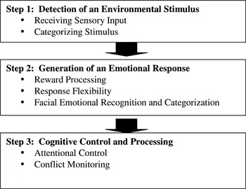
We have much to learn about the development of emotion regulation from infancy through adulthood (Dahl, 2004). What is known is that there is no single process or neuroanatomical area responsible for emotion regulation. Rather, emotion regulation results from separable but interrelated psychological processes, each mediated by integrated neural networks. At the most basic level, these processes are (a) gathering sensory input and categorize environmental situations, (b) initiating an emotional response, and (c) mediating higher order cognitive processing and control (Nelson, Leibenluft, McClure, & Pine, 2005; Thompson, 1994; Figure 2).

The steps of emotion regulation.
Emotion regulation Step 1: Detection of an environmental stimulus
Emotion regulation begins with the detection of an environmental stimulus. This involves receiving sensory input and categorizing the stimulus. Structures involved include those responsible for visual information processing, including the occipital cortex, the superior temporal gyrus, and the fusiform gyrus (Haxby, Hoffman, & Gobbini, 2002). These brain regions are responsible for determining an object's identity (important for subsequent determination if it should be approached or avoided), whether it is animate or not, and whether it is moving or not (and if moving, what activity it is engaged in). In particular, these regions are especially geared to process faces, because in humans, faces are one of the most important categories of emotional stimuli (Gauthier, Tarr, Anderson, Skudlarski, & Gore, 1999; Haxby et al., 2002; Kanwisher, McDermott, & Chun, 1997; Lehmann et al., 2004).
Emotion regulation Step 2: Generation of an emotional response
The generation of an emotional response, based principally on its categorization as being positively or negatively valenced, that is, rewarding or punishing, is the second step of emotion regulation. Several subprocesses are involved with this step, including reward processing, response flexibility, and facial emotional recognition and categorization. In turn, each of these psychological processes engages different neural networks.
Reward processing
Reward processing is the mechanism by which appetitive, pleasurable, and positively valenced stimuli evoke an emotional response (Baxter & Murray, 2002). Many of the same regions that mediate reward processing are also involved in processing punishment, that is, mechanisms by which aversive, negatively valenced stimuli create and shape behavior. The amygdala, the orbitofrontal cortex (OFC), and the ventral striatum (including the caudate, putamen, and the accumbens area) are the three primary brain regions most centrally involved in reward processing. In particular, the amygdala is well positioned neuroanatomically to have a central role in this process, as it receives sensory input from the cortex, thalamus, and hippocampus. Moreover, the basolateral amygdala has neuronal output connections to several key regions implicated in emotion regulation, including (a) the OFC (hypothesized to mediate response flexibility), (b) the hippocampus (thought to be involved in memory consolidation of emotionally salient events and spatial learning), (c) the striatum (mediating instrumental approach and avoidance behavior), and (d) the central nucleus of the amygdala and the lateral bed of the stria terminalis (modulating autonomic and somatic signs of anxiety; Davis & Whalen, 2001; LeDoux, Iwata, Cicchetti, & Reis, 1988).
A number of studies have established the amygdala's role in reward behavior. For example, nonhuman primate studies demonstrate that the amygdala, working in concert with the OFC, is essential in shaping behavior to respond to incentives, including food rewards (Baxter, Parker, Lindner, Izquierdo, & Murray, 2000; Izquierdo & Murray, 2004). Moreover, functional magnetic resonance imaging (fMRI) studies in humans indicate that the amygdala and OFC interact to encode the predictive reward value of a stimulus (Gottfried, O'Doherty, & Dolan, 2003).
Through its connection to the amygdala, the striatum is involved in generating emotional and behavioral responses to rewarding stimuli. For example, nonhuman primate studies demonstrate that dopaminergic neurotransmission in the striatum rapidly adapts via trial and error learning to fire only in response to trials during which the animal is certain to receive a reward, after initially responding to all trials regardless of reward probability (Pasupathy & Miller, 2005; Schultz, Tremblay, & Hollerman, 1998). Neuroimaging studies in healthy adults also demonstrate increased striatal activation in response to rewarding stimuli, including food (O'Doherty et al., 2004), money (Haruno et al., 2004; Zink, Pagnoni, Martin-Skurski, Chappelow, & Berns, 2004), positive feedback (Delgado, Stenger, & Fiez, 2004), or drugs of abuse, such as cocaine or alcohol (Breiter et al., 1997; Volkow et al., 2002). Moreover, positron emission tomography (PET) studies in healthy adults corroborate previous nonhuman primate studies by demonstrating increased striatal dopaminergic activity in response to monetary rewards (Zald et al., 2004). Monetary reward also has been shown to activate the striatum in healthy children and adolescents (May et al., 2004). In addition, a recent developmental fMRI study by Ernst et al. (2005) showed greater neural activation in the accumbens area in response to rewards and positive feedback in adolescents than in adults. Thus, one of the striatum's roles in emotion regulation may be to adjust behavior in response to pleasurable positively valenced experiences (Koob, 1996).
In sum, reward processing is mediated primarily by the amygdala, ventral striatum, and OFC. It is integral to the second step of emotion regulation, namely the generation of an emotional response. Moreover, reward processing is integral to the understanding of BD, whose hallmark clinical symptoms reflect aberrant reward processing, including excessive goal-directed activity and pleasure seeking during mania, and anhedonia during depression.
Response flexibility
In the context of emotion regulation, response flexibility connotes the process of adapting one's response to changing rewards and punishments. The primary brain region mediating response flexibility is the OFC, which, long before MRI was invented, was implicated in this process by case reports of patients with frontal lobe lesions. Perhaps the best known example is Phineas Gage, a 19th century railroad worker who was a hardworking affable man until an iron tamping rod was driven through his skull, entering below his left cheek and eye and exiting from the top of his head, and injuring his frontal lobes above his eyes, that is, the OFC. Miraculously, he survived this injury, but he subsequently experienced emotional dysregulation and lack of response flexibility, including lability, rage, and failure to comply with social conventions (Damasio, Grabowski, Frank, Galaburda, & Damasio, 1994).
More recent studies of response flexibility have used reversal learning paradigms, which require subjects to reverse a previously learned stimulus–reward association without an explicit warning. How readily a subject learns that the previously rewarded stimulus is now punished, and that the previously punished stimulus is now rewarded, is a measure of cognitive flexibility. Reversal learning is primarily mediated by the OFC, which regulates the reversal of stimulus–reward relationships (Clark, Cools, & Robbins, 2004; Cools, Clark, & Robbins, 2004; Rolls, 2000). Again, as in reward processing described above, reversal learning requires the interplay between the OFC and the ventral striatum, which mediates response to rewards and incentives (Knutson, Westdorp, Kaiser, & Hommer, 2000; Matsumoto, Hanakawa, Maki, Graybiel, & Kimura, 1999), and the amygdala, which encodes the salience of rewards (Baxter et al., 2000; Schoenbaum, Chiba, & Gallagher, 1999).
Reversal learning is linked to another important aspect of emotion regulation: the modulation of aggression. According to one theory, social response reversal is the process by which individuals learn to regulate their behavior in response to other peoples' negatively valenced emotional states, such as anger or disapproval (Blair, Colledge, & Mitchell, 2001). The OFC mediates social response reversal and is linked to a particular form of aggression, known as “reactive aggression,” occurring in reaction to emotionally evocative situations, such as frustration (Blair, 2004; Blair et al., 2001; Stone, Baron-Cohen, & Knight, 1998). Reactive aggression should be contrasted with “instrumental aggression” wherein aggression is actively used to accomplish something, for example, a planned theft of someone's handbag to obtain money. Thus, reversal learning is germane to how children learn to moderate their behavior in response to negative feedback from a parent or teacher. Moreover, it is also pertinent to the social–emotional deficits found in children with BD because these children react to social demands with irritability, frustration, and aggression when they are unable to adapt flexibly and apply social rules/conventions to novel situations (McClure et al., 2005; Rich et al., 2005).
Emotion recognition and categorization
In social situations, the generation of an emotional response requires yet a third process: the recognition of others' emotional states and subsequent categorization of them as positively (rewarding, stimulating approach behavior) or negatively (punishing, stimulating withdrawal behavior) valenced. This process is dependent upon face emotion recognition, which is integral to both emotion regulation and to the formation of social relationships. From birth, infants demonstrate gaze preference for human faces, such as that of their mother, rather than objects (Farroni, Csibra, Simion, & Johnson, 2002; Johnson, Dziurawiec, Ellis, & Morton, 1991; Striano, 2001). This behavior is thought to foster attachment between infant and caregiver while also alerting the infant to environmental cues (Koulomzin et al., 2002). This pattern continues into adulthood, with stronger neural activation associated with viewing faces of familiar, rather than unfamiliar, people (Gobbini, Leibenluft, Santiago, & Haxby, 2004), and with specific patterns of activation associated with viewing the faces of intimates, such as one's child or lover (Bartels & Zeki, 2004; Leibenluft, Gobbini, Harrison, & Haxby, 2004). Moreover, children with impaired social relatedness, such as those with autism, have impaired gaze preference, spending less time than controls looking at central face regions, including the eyes and mouth (Trepagnier, Sebrechts, & Peterson, 2002). In short, face processing is essential for emotion regulation and social relationship formation, and aberrant face processing is associated with social relationship dysfunction.
The primary region implicated in the recognition of others' emotions is the amygdala. Early work on the amygdala centered on its role in mediating negatively valenced emotions. This line of research includes Pavlov's investigations of fear conditioning, wherein an emotionally neutral conditioned stimulus elicits fear by itself after the animal is initially exposed to both the neutral conditioned stimulus and an aversive unconditioned stimulus (LeDoux, 2003; LeDoux et al., 1988). Moreover, in their classic 1939 article, Kluver and Bucy reported that bilateral temporal lobectomy, including amygdalectomy, in nonhuman primates caused a constellation of symptoms including hyperorality, hypersexuality, and reduced fear (Kluver & Bucy, 1997). More recent animal studies have demonstrated that bilateral amygdala damage maybe associated with blunted fear response to novel, unconditioned stimuli while leaving unconditioned/trait anxiety/fear responses intact (Kalin, Shelton, Davidson, & Kelley, 2001).
Many studies in humans, including those with amygdala lesions, align with these nonhuman primate studies by suggesting that the amygdala primarily mediates response to negatively valenced stimuli. For example, several studies of humans following either unilateral or bilateral surgical resection of the amygdala have shown impaired recognition of facial expressions of negatively valenced emotions, such as fear and sadness, but not of positively valenced emotions, such as happiness (Adolphs, Damasio, Tranel, & Damasio, 1996; Adolphs & Tranel, 2004). Neuroimaging studies of healthy adults and adolescents have also shown increased amygdala activation in response to negatively valenced emotional expressions, particularly those depicting fear, compared to neutral stimuli (Hariri, Bookheimer, & Mazziotta, 2000; Phillips et al., 2004; Williams, Morris, McGlone, Abbott, & Mattingley, 2004). A recent study demonstrating that healthy adolescents, compared to healthy adults, have greater amygdala activation in response to fearful versus neutral faces suggests that development plays a role in attenuating some of the response to negatively valenced stimuli (Monk et al., 2003). Finally, research suggests a link in healthy adults between a gene thought to convey susceptibility for mood and anxiety disorders (the so-called “short variant” of the serotonin transporter) and increased amygdala activation to fearful and threatening facial stimuli (Hariri et al., 2005).
However, there are also recent studies showing that the amygdala's role in emotion recognition and categorization may be more complicated than the previously held view that it only mediates negatively valenced emotions. For example, studies show increased amygdala activation to positively valenced stimuli, such as happy faces compared to neutral faces, in healthy adults (Breiter et al., 1996). Also, mothers have increased amygdala activation when viewing pictures of their own children, compared to viewing either unfamiliar children or familiar, but unrelated, children (Leibenluft et al., 2004). In addition, humans with amygdala lesions are more impaired in recognition of facial expressions of social emotions, such as guilt or admiration, rather than basic emotions such as sadness or happiness (Adolphs, Baron-Cohen, & Tranel, 2002). Thus, ample evidence shows that the amygdala is involved in the recognition of both negatively and positively valenced emotions, although further work is necessary to determine the specificity of its response.
Besides the amygdala, the OFC plays a role in the recognition and categorization of emotions. Studies demonstrate that the OFC has face responsive neurons (Thorpe, Rolls, & Maddison, 1983). Moreover, fMRI studies of healthy adults show increased OFC activation in response to negatively valenced stimuli, such as angry faces (Blair, Morris, Frith, Perrett, & Dolan, 1999), as well as in response to positively valenced stimuli, such as a mother looking at images of her newborn (Nitschke et al., 2004). Given that the OFC is also activated by both pleasurable and painful touch (Rolls et al., 2003), this suggests that this region may play a nonselective role in the recognition of emotions.
Emotion regulation Step 3: Cognitive control and processing
The third step of emotion regulation involves cognitive control and processing. This involves several psychological processes, including, but not limited to attentional control and conflict monitoring.
Attentional control
Top-down attentional control mediates two important aspects of emotion regulation. First, to which stimulus should I increase my attention, the rewarding one, for example, a chocolate bar, or the punishing one, for example, an upcoming test? Second, are there some stimuli that I should deliberately ignore, for example, intentionally not think about the creaking sound in my house because it makes me afraid the “boogie man” is coming? Thus, although it is important to study attention from a cognitive neuroscience perspective, it is equally important to study interactions between attention and emotion (Davidson, 2004).
The primary brain region implicated in top-down control of attention is the dorsolateral prefrontal cortex (DLPFC). The DLPFC has long been linked to attentional mechanisms, working memory, and cognitive control (Casey, Tottenham, & Fossella, 2002; Fuster, 2000; Goldman-Rakic, 1987). Adults with DLPFC lesions have impairments in planning, goal attainment, and problem-solving ability (Stuss & Levine, 2002). The DLPFC is also involved in visuospatial working memory, thus helping organisms remember the location of emotionally salient stimuli, that is, where the hungry tiger is located (D'Esposito & Postle, 1999; Ungerleider, Courtney, & Haxby, 1998). Neuroimaging studies document that the DLPFC plays a role in exerting top-down control in emotion regulation by directing attention to specific stimuli, some of which have emotional salience (Pessoa, McKenna, Gutierrez, & Ungerleider, 2002; Pessoa & Ungerleider, 2004). For example, the DLPFC demonstrates increased activation in healthy adults in response to negative versus neutral words (Compton et al., 2003). Recent developmental neuroimaging studies demonstrate that the PFC matures in the mid-20s, significantly later than subcortical structures (Giedd, 2004; Gogtay et al., 2004). Moreover, increased frontal gray matter thickness correlates with the progression of cognitive abilities across development (Sowell et al., 2004). Finally, an fMRI study demonstrated that children, in comparison to adults, failed to activate the PFC during successful inhibition (Bunge, Dudukovic, Thomason, Vaidya, & Gabrieli, 2002). Taken together, these findings suggest that maturation of the DLPFC is associated with the normal development of social and emotional maturity, including the emergence of sophisticated goal-directed behavior or the ability to delay gratification.
Conflict monitoring
The anterior cingulate cortex (ACC) is another frontal lobe structure that plays a role in the top-down control of attention to stimuli. In particular, research demonstrates that the ACC is involved in conflict monitoring, that is, adjusting behavior to override one response in favor of another (Botvinick, Cohen, & Carter, 2004). Conflict monitoring is relevant to emotion regulation because it enables people to choose between two competing emotional stimuli, for example, selecting between two rewarding stimuli, such as receiving either one cookie now or a plate of cookies in 5 min.
The Stroop paradigm is a classic paradigm assessing cognitive control in the face of interference. Subjects completing the Stroop task have greater difficulty (i.e., longer response time and more errors) naming the color in which a word is printed when the word and color are incongruous, for example, the word BLUE printed in green ink, compared to control items, for example, the word BLUE printed in blue ink (MacLeod & MacDonald, 2000). Another classic paradigm used to study cognitive control is the go/no-go paradigm, in which subjects develop a prepotent response to a stimulus, for example, press a button (go) if you see an “X,” and must inhibit this established prepotent response, for example, do not press the button (no-go) if you see an “X” followed by an “O.” Neuroimaging studies utilizing both Stroop and go/no-go paradigms have demonstrated increased ACC neural activation during conflict monitoring, that is, when evaluating conflicting responses (Carter et al., 2000), overriding prepotent responses (Braver, Barch, Gray, Molfese, & Snyder, 2001), or committing errors (which represents a cognitive conflict because, after committing the error, the subject subsequently realizes their mistake; Durston, Thomas, Worden, Yang, & Casey, 2002). Also, studies demonstrate increased ACC neural activation in response to positively valenced stimuli, such as the receipt of rewards or happy faces (Killgore & Yurgelun-Todd, 2004; Risinger et al., 2005) and in response to large, but not small, rewards (Rogers et al., 2004). Developmental studies have found that children ages 8 to 12 years have less activation of the ACC than adults during effective response inhibition on a go/no-go task (Bunge et al., 2002).
Thus, the ACC is involved in emotion regulation through its role in monitoring conflict between competing responses and shaping behavior accordingly (Barrett, Pike, & Paus, 2004). This role is facilitated by the ACC's functional connections to both other regions of the PFC (including both OFC and DLPFC) as well as the striatum (Casey et al., 2001; Koski & Paus, 2000).
Emotion Regulation in Phenotypes of Pediatric BD
Against this backdrop of emotion regulation in healthy individuals, what do we know about emotion regulation, and dysregulation, in children and adolescents with pediatric BD? To answer this question, our laboratory has utilized the pediatric BD phenotype construct to guide research addressing two important questions. First, how does emotion regulation in NP-BD children, who clinicians and researchers agree have BD by virtue of meeting very restrictive criteria for the illness, differ from that in typically developing youth? Second, what are the boundaries of emotion dysregulation between children with possible phenotypes of BD, that is, NP-BD and SMD, and typically developing children?
Since 2000, we have worked to address these questions with the following research strategy. First, as noted above, we delineated criteria to define NP-BD and SMD youth, in comparison to controls. Then, we created a battery of behavioral paradigms designed to capture a broad range of information about relevant psychological processes in children with BD. The paradigms included measures of reward processing, reversal learning, face emotion recognition and categorization, attentional control, and conflict monitoring. Having identified paradigms on which the behavior of patients with BD differed from that of controls, we began to build on the behavioral data with the addition of neuroimaging paradigms, especially fMRI, to identify the brain regions mediating the identified group differences in performance. Here, we focus largely on research in children with NP-BD because this line of research was begun first, at a time when the SMD criteria were being developed.
We are testing the following hypotheses: (a) NP-BD and SMD youth will share some patterns of emotion dysregulation in comparison to typically developing youth and (b) on a few select measures, NP-BD and SMD youth will differ both from one another and from typically developing youth. Moreover, it is likely that on many measures of emotion regulation, including both behavioral and brain imaging measures, there will be no difference between typically developing youth and those with either NP-BD or SMD because both patient groups have some preserved areas of function. At present, we are actively recruiting both NP-BD and SMD subjects to participate in research studies. What follows is a discussion of differences in the boundaries of emotion regulation between the two patient groups (NP-BD and SMD) and typically developing youth as revealed thus far.
Emotion dysregulation in NP-BD subjects
Currently, data from NP-BD subjects indicate that these subjects have deficits on behavioral tasks related to several processes involved in emotion regulation, including reward processing and reversal learning, face emotion recognition, and attention in emotional contexts. In addition, neuroimaging data suggest that NP-BD subjects have reduced gray matter volume in brain regions mediating these subprocesses of emotion regulation, including the amygdala, striatum, and PFC, as well as dysfunction in this circuit while performing tasks involving motor inhibition or face processing.
Reward processing and reversal learning
As previously stated, reward processing and response flexibility are two important aspects of emotion regulation. Moreover, both are integral to our understanding of BD, given the possibility that mania may represent a hyperhedonic period marked by clinical symptoms of excessive goal-directed activity and pleasure seeking, whereas depression may represent a hypohedonic period marked by clinical symptoms of anhedonia. A pilot study demonstrated that NP-BD youth have impaired reward-processing compared to typically developing youth on a decision-making task involving multiple levels of risk (Ernst et al., 2004).
Two subsequent studies have shown that NP-BD youth are impaired in reversal learning, which as previously discussed, evaluates how aberrant reward-related processes affect decision making (Clark et al., 2004; Cools et al., 2004; Rolls, 2000). Our first reversal learning study examined differences in neuropsychological performance between NP-BD youth and controls using the Cambridge Automated Neuropsychological Testing Battery (CANTAB). The CANTAB is a computerized battery of standard neuropsychological tasks. On one CANTAB measure, the interdimensional/extradimensional shift (ID/ED shift; a computerized task resembling the Wisconsin Card Sorting Task), we found that NP-BD youth were impaired on measures of attentional set shifting. Furthermore, when these results were deconstructed by type of set shifting, euthymic NP-BD youth were specifically impaired during a reversal learning stage of the ID/ED shift task, making more errors, taking more trials to complete this stage of the task, and having longer latency to complete this stage (Dickstein et al., 2004). This result aligns with the four previous studies using the CANTAB's ID/ED shift task in BD adults, which also found impaired reversal learning (Clark, Iversen, & Goodwin, 2001, 2002; Rubinsztein, Michael, Paykel, & Sahakian, 2000; Sweeney, Kmiec, & Kupfer, 2000). Importantly, these previous studies demonstrated impaired reversal learning during both mania and euthymia, suggesting that this may be a trait, rather than state, phenomenon.
Our second study of reversal learning utilized a task known as probabilistic response reversal (PRR). Unlike traditional response reversal tasks (including the reversal stages of the CANTAB's ID/ED shift task), PRR probes the role of reward processing in reversal learning by manipulating the probability of receiving a reward, or punishment. Specifically, the PRR paradigm involves two types of trials: 100/0 and 80/20. In 100/0 trials, the stimulus reward relationships are identical to those of the CANTAB's ID/ED shift task. During the initial acquisition trials, stimulus pair AB is presented, and stimulus A is rewarded 100% of the time (B is rewarded 0%). During subsequent reversal stages, stimulus B is now rewarded 100% of the time (A is rewarded 0%). However, the 80/20 trials are dissimilar to the ID/ED shift task because the reward is now probabilistic, that is, during 80/20 acquisition trials using stimulus pair AB, A is rewarded 80% of the time (B is rewarded 20%). Subsequently during a reversal trial, B is rewarded 80% of the time (and A is rewarded 20%). On the PRR task, pediatric NP-BD subjects were impaired on reversal trials with an 80:20 probability of reward (Gorrindo et al., 2005). NP-BD subjects' impairment on the 80:20 rather than 100:0 task has two possible interpretations. On the one hand, it could be that the 80:20 task is more difficult, and there is no difference on the 100:0 task because of ceiling effect. On the other hand, it is possible that NP-BD subjects may be more sensitive to the inconsistency of reward on the 80/20 vs. 100/0 condition of this decision-making task.
Thus, three studies have shown that NP-BD youth have impaired reward processing and reversal learning compared to healthy controls. These behavioral studies suggest that NP-BD youth have dysfunction in the distributed neural circuit, including the OFC, ventral striatum, and amygdala, mediating these functions (Alexander, DeLong, & Strick, 1986; Holland & Gallagher, 2003; Meunier, Bachevalier, & Mishkin, 1997). At present, we are conducting neuroimaging studies of reward behavior and cognitive flexibility in NP-BD youth to test whether dysfunction in the OFC–striatal–amygdala circuit does indeed mediate the behavioral deficits that we have observed. The only published neuroimaging study of decision making in BD is a PET study of adults demonstrating that OFC hypoactivation may mediate deficits observed during a complex decision-making task involving reversal learning, among other processes (Rubinsztein et al., 2001).
Emotion recognition and categorization
Recognizing the emotional state of others and categorizing it into either positively or negatively valenced categories is essential for emotion regulation. These interrelated steps allow an individual to begin to mentalize, that is, to understand another person's emotional state and intentions or, to develop a “theory of mind” (Fonagy, Steele, Moran, Steele, & Higgitt, 1993). In turn, this is an early step in the formation of social relationships. Face processing is therefore important to the study of pediatric BD for two reasons. First, previous work has demonstrated that adults with BD, whether depressed or manic, have impaired theory of mind abilities (Kerr, Dunbar, & Bentall, 2003) as well as impaired recognition of emotional faces (Chen et al., 2006; Lembke & Ketter, 2002; Lennox, Jacob, Calder, Lupson, & Bullmore, 2004). Second, neuroimaging studies demonstrate overlap in brain regions mediating BD and theory of mind abilities, including the PFC (including medial frontal and superior frontal cortex), anterior cingulate cortex, and anterior temporal pole (Calarge, Andreasen, & O'Leary, 2003; Fletcher et al., 1995).
We conducted two studies of facial emotion recognition in NP-BD youth using the Diagnostic Analysis of Nonverbal Accuracy (DANVA). In this paradigm, subjects view high- and low-intensity expressions of happiness, sadness, anger, or fear. Subsequently, they categorize the emotion that the subject was displaying. Both child and adult images are used (Nowicki & Duke, 1994). In our first study, we examined differences in facial emotion recognition in three samples of children and adolescents: NP-BD, anxiety disorders, and typically developing youth. We found that NP-BD youth differed significantly from both anxious and typically developing youth. In particular, NP-BD subjects were more likely to miscategorize child images of happy, sad, and fear as angry; however, this was not true of the adult images (McClure, Pope, Hoberman, Pine, & Leibenluft, 2003).
We have extended these findings in two subsequent studies. First, we conducted a second study using the DANVA in larger samples of both NP-BD and typically developing youth. Compared to controls, euthymic NP-BD youth made significantly more errors on both child and adult faces. Post hoc analyses revealed that NP-BD youth, compared to typically developing youth, were less sensitive to anger and happiness in children's faces and less sensitive to anger and sadness in adult's faces (McClure et al., 2005). Second, we have conducted a neuroimaging study of attentional processing of emotional faces discussed below.
Taken as a whole, these studies provide evidence that NP-BD subjects have impaired emotion recognition as measured by the DANVA and aberrant neural activation in the amygdala and striatum while attending to emotional aspects of neutral faces. These findings provide a possible biobehavioral substrate of the social dysfunction, which is a major source of impairment for children and adolescents with BD (Geller et al., 2000). Moreover, they suggest that future work is needed to determine the specificity of these deficits to BD versus other forms of psychopathology, the developmental trajectory of these deficits, and how these deficits are related to other aspects of face processing, including memory.
Attention–emotion interactions
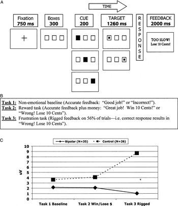
As discussed previously, the cerebral cortex plays a role in emotion regulation through its top-down control of attention to emotional stimuli. Yet, developing a paradigm that recreates in the laboratory emotionally evocative situations to study the interaction between attention and emotions is quite difficult. One such measure is the affective Posner paradigm, which enables the study of one of the most common, and most impairing, symptoms of pediatric BD, namely frustration and irritability (Derryberry & Reed, 1994; Perez-Edgar & Fox, in press). At its simplest, the affective Posner task is an attentional task, requiring participants to attend to targets and respond as to whether the target appeared on the left or the right side of the screen while attentional measures are collected. Then, contingencies, that is, winning or losing points or money based upon performance, are added to allow the examination of how emotions (reward/punishment) affect attention. Finally, by rigging feedback, that is, having a participant lose points/money even though they provided the correct answer, one can study the pathophysiology of one of the most impairing symptoms of BD, namely irritability and frustration.
An individual trial of this paradigm consists of the following, all displayed on a computer screen: (a) presentation of a fixation cross, (b) presentation of three horizontally arranged boxes (left, middle, and right) with one of them containing a blue orienting square (20% in the middle, 40% each in the left and right), (c) presentation of a target square in either the left or right box, (d) subjects indicate whether the target was on the left or right box by pressing a button on a stimulus response box, and (e) feedback on their performance (Figure 3A).

The affective Posner paradigm: (A) an individual trial, (B) tests, and (C) the P3 ERP at parietal sites NP-BD versus controls. Part C is adapted from “The impact of reward, punishment, and frustration on attention in pediatric bipolar disorder,” by B. A. Rich, M. Schmajuk, K. E. Perez-Edgar, D. S. Pine, N. A. Fox, and E. Leibenluft, 2005, Biological Psychiatry, 58. Copyright 2005 by Elsevier. Adapted with permission.
Three tasks using the same trial structure were administered in nonrandomized blocks (Figure 3B). The first task was a nonemotional baseline wherein accurate feedback was given for their performance, for example, “Good job!” or “Incorrect!” During the second task, contingencies were added to increase affect in response to reward and punishment. Specifically, not only was accurate feedback given, for example, “Great Job! Win 10 Cents” or “Wrong! Lose 10 Cents,” but also 10 cents was immediately awarded or deducted from the subject's cup placed in front of them. During the third task, rigged feedback was added to induce frustration. In other words, the feedback (words plus money) was the same as in Task 2, but on 56% of the trials where subjects had in fact given a correct, timely response, they were told that their response was too slow, and that therefore they would not be rewarded.
We recorded event-related potentials (ERPs) during this task, time locked to the time of stimulus presentation. ERPs were measured via electroencephalogram leads from temporal (T3, T4), frontal (Fz, F3, F4), central (C3, C4, Cz), and parietal (Pz, P3, P4) sites using the international 10/20 system, referenced to the right earlobe. P1, N1, and P3 amplitudes were used as measures of attention. Self-reported mood data, specifically in response to reward and punishment trials, was collected after each task using the Self-Assessment Manikin, which consists of line drawings of human manikins along a 9-point scale, with extremes of happy/unhappy (valence) or calm/aroused (arousal; McManis, Bradley, Berg, Cuthbert, & Lang, 2001).
In this study, our group found that pediatric NP-BD subjects, compared to healthy controls, had greater self-reported frustration, demonstrating that the affective Posner task can induce frustration and irritability in this patient population under laboratory conditions. Moreover, frustrated NP-BD subjects had significantly decreased P3 amplitude across sites in comparison to healthy controls (Rich et al., 2005; Figure 3C). Given that P3 is thought to be a measure of the deployment of attentional resources, this suggests that NP-BD have impaired attention, especially when experiencing the negatively valenced emotions of frustration and irritability (Polich & Herbst, 2000; Steger et al., 2001). Of note, when the NP-BD sample was divided into those with and without comorbid ADHD, there were no significant differences in P3 amplitude, reaction time, or accuracy.
These data are germane to several aspects of pediatric BD. First, they have high ecological validity, as they produced a self-described sense of frustration and negatively valenced emotion. Second, these data indicate that, despite the fact that many children with BD meet criteria for ADHD, in at least a subset of children with BD their attentional problems may be more marked in emotional, compared to nonemotional, contexts.
Neuroimaging data: Dysfunction in frontostriatal–limbic structures
Recent neuroimaging data complements behavioral data in the investigation of emotion dysregulation in pediatric BD. Taken as a whole, both structural and functional MRI data suggest that children and adolescents with BD are differentiated from healthy controls by alterations in a frontostriatal–limbic neural circuit (Blumberg et al., 2004).
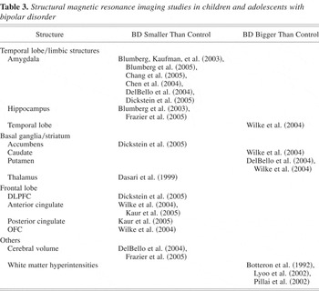
Thus, far, the primary focus of morphometric studies in pediatric BD has been on limbic structures (Table 3). Chief among these regions is the amygdala. Four out of six published pediatric BD studies show decreased amygdala volume compared to healthy controls (Blumberg, Kaufman, et al., 2003; Chang et al., 2005; DelBello et al., 2004; Dickstein, Milham, et al., 2005) and in the fifth it was a trend (Chen et al., 2004). Four of these studies used traditional morphometry methods, including regions of interest hand traced by human raters. The fifth study, by our group, used voxel-based morphometry (VBM), an automated technique that avoids the rater bias inherent in hand tracing and allows whole-brain volumetric analysis. The latter is important because it enables exploration of regions of interest beyond those for which standardized templates exist. Consistent with previous studies using both hand tracing and VBM techniques, we found that NP-BD subjects had decreased left amygdala volume compared to healthy controls. Recently, a seventh study demonstrated temporal stability of decreased amygdala volume in pediatric BD subjects, versus controls, after a mean 2-year follow-up (Blumberg et al., 2005).
Structural magnetic resonance imaging studies in children and adolescents with bipolar disorder

Decreased amygdala volume in pediatric BD subjects is especially interesting for two reasons. First, studies of BD adults have shown either increased or unchanged amygdala volume, compared to controls (Altshuler, Bartzokis, Grieder, Curran, & Mintz, 1998; Altshuler et al., 2000; Brambilla et al., 2003; Pearlson et al., 1997; Strakowski et al., 1999). The consistent and opposite direction of pediatric BD studies suggest a neurobiological difference between childhood and adult onset BD. Second, although decreased volume does not necessarily imply aberrant function, given the central role of the amygdala in emotion regulation, the consistency of these studies' results suggests that amygdala dysfunction might be important in the pathophysiology of pediatric BD.
Studies have also evaluated another limbic structure, the hippocampus, which is implicated in working memory for emotional events. At present, two studies have found decreased hippocampal volume in pediatric BD compared to controls, while three have not (Chang et al., 2005; Chen et al., 2004; Dickstein, Milham, et al., 2005). The first study found decreased hippocampal volume in a mixed sample of BD adolescents and adults compared to controls (Blumberg, Kaufman, et al., 2003). The second showed decreased hippocampal volume in BD adolescents compared to controls, an effect that the authors report was driven by the female BD subjects (Frazier et al., 2005). This inconsistency of hippocampal volume findings is also found in BD adults, with one study finding decreased volume (Altshuler et al., 1991) and several others finding no difference, although care should be taken in interpreting these negative studies because of the possibility of type II errors (Altshuler et al., 1998, 2000; Brambilla et al., 2003; Hauser et al., 2000; Strakowski et al., 1999).
Fewer studies have evaluated the frontal cortex in pediatric BD. Our abovementioned VBM study demonstrated that NP-BD youth, compared to controls, have decreased volume of the left DLPFC (Brodmann area 9), an area important in exerting top-down attentional control in response to emotionally evocative stimuli (Dickstein, Milham, et al., 2005; Posner & Rothbart, 1998). Studies have shown decreased DLPFC volume and density in BD adults, suggesting that there may be some similarities in brain structure between pediatric and adult BD (Lyoo et al., 2004; Sax et al., 1999). Other frontal lobe morphometry studies have shown that BD children and adolescents have decreased ACC volume, important in conflict monitoring (Kaur et al., 2005), but no between-group difference in volume of the subgenual PFC (Sanches, Sassi, et al., 2005), a region implicated in adult mood disorders (Drevets, Ongur, & Price, 1998). Thus, further work to evaluate frontal lobe structures is needed in pediatric BD given their importance in emotion regulation.
Like the frontal cortex, few morphometry studies have evaluated the striatum, a region important to reward processing, in pediatric BD. Using VBM, we found decreased volume in the left accumbens area in NP-BD youth compared to controls (Dickstein, Milham, et al., 2005). To the best of our knowledge, no such studies exist in adult BD subjects, likely due to the technical difficulty associated with hand tracing the accumbens. Studies of the caudate and putamen have yielded mixed results. Compared to controls, one study has demonstrated that BD youth have increased caudate volume (Wilke, Kowatch, DelBello, Mills, & Holland, 2004), whereas two have not (DelBello et al., 2004; Sanches, Roberts, et al., 2005), and two studies have shown that BD youth have increased putamen volume (DelBello et al., 2004; Wilke et al., 2004), whereas one has not (Sanches, Roberts, et al., 2005). Although the study by Sanches et al. did not find differences between BD youth and controls, they did find an inverse relationship between caudate (bilateral) and putamen (left) volume in BD subjects but not controls, suggesting that the neurodevelopmental process of pruning may function differently in patients with BD, versus controls (Sanches, Roberts, et al., 2005). Given the role of the striatum in reward processing, further work is necessary to determine the neurodevelopmental trajectory of striatal volumes.
In sum, morphometric MRI studies in pediatric BD have consistently demonstrated decreased amygdala volume and are beginning to evaluate frontal and striatal structures. Yet, what is the functional significance of these structural alterations? Size is not everything, particularly with regard to structure/function relationships of the human brain (Gur et al., 1999). Thus, one must use fMRI to examine changes in neural activation during tasks designed to probe aspects of cognition, including emotion regulation. However, at present, there are only two published, and two in press, fMRI studies in BD children or adolescents.
The first, by Blumberg et al., found that, during a Stroop color-naming task, BD adolescents, compared to controls, had increased activation in the left putamen and thalamus during incongruent versus congruent trials (Blumberg, Martin, et al., 2003). In contrast, previous studies in adults with BD demonstrated increased activation of the ventral PFC during tasks requiring conflict monitoring (Blumberg, Leung, et al., 2003; Rubinsztein et al., 2001). The second fMRI study in children with BD, by Chang et al., reported findings from two tasks: a visuospatial working memory task and a task with positive and negatively valenced pictures. Compared to controls, BD subjects had increased activation on the two-back working memory task in the bilateral ACC and left-sided putamen, thalamus, and DLPFC. BD subjects also had greater activation of the bilateral DLPFC, inferior frontal gyrus, and right insula while viewing negatively valenced nonface images. Finally, BD subjects had greater activation in the bilateral caudate and thalamus, left middle/superior frontal gyrus, and left ACC when viewing positively valenced pictures (Chang et al., 2004). Although these studies support the hypothesis that pediatric BD subjects have both structural and functional alterations of frontostriatial–limbic structures, more work is needed to understand the relevance of these alterations to emotion dysregulation and other symptoms in children and adolescents with BD.
Our group has recently conducted two fMRI studies comparing NP-BD youth to controls. The first (Leibenluft et al., in press) examined neural activation during motor inhibition using a stop-signal task that required subjects to inhibit a prepotent response (Logan, Schachar, & Tannock, 1997). This study demonstrated that NP-BD subjects had a reduced striatal “error signal” during failed motor inhibition compared to controls (Leibenluft et al., in press). The second (Rich et al., 2006), evaluated neural activation to emotional faces during an attention task. NP-BD subjects viewed neutral faces as more hostile, and feared these neutral faces more, than do controls. Furthermore, these subjective ratings were associated with increased activation of the amygdala and striatum in NP-BD versus control youth when subjects direct their attention toward emotional aspects of the faces (Rich et al., 2006).
Future neuroimaging projects by our group in NP-BD and SMD youth, compared both to one another and controls, will attempt to use fMRI to bridge the gap between structural MRI and behavioral data. At present, several projects are underway. These include an examination of cognitive control and flexibility. We are also continuing to use emotionally evocative faces to determine the functional neurocircuitry involved with encoding these faces. In addition, we have begun to use a version of the PRR paradigm as an fMRI task to determine if activity in the amygdala, OFC, and ventral striatum is different in NP-BD youth compared to controls during reversal learning. Finally, we are also investigating the specificity of these neuroimaging alterations relative to children with other forms of psychopathology, including unipolar depression, anxiety disorders, and ADHD.
Boundaries of emotion regulation between NP-BD youth and broad phenotype SMD youth
Clearly, NP-BD children and adolescents have specific impairments in emotion regulation, as evidenced by both behavioral and neuroimaging studies. What, then, is the boundary of emotion regulation between these NP-BD subjects and children with other forms of emotion dysregulation, specifically SMD youth?
This question must be answered on several levels. On the whole-patient level, is there a difference in clinical manifestations between NP-BD and SMD youth, beyond those dictated by the inclusion criteria themselves? Delving slightly deeper, do the two groups differ on behavioral measures of emotion regulation? If they do differ on some behavioral measures, using functional neuroimaging, are there differences in the neuroanatomical regions mediating these behavioral differences? Do NP-BD and SMD youth differ on the genetic substrates of behavior, and are these genetic differences associated with either behavioral or neural measures of emotion dysregulation? Thus, one must explore on all levels from the whole patient down to the genome how both NP-BD and SMD youth differ, and are similar, to typically developing youth.
We are presently gathering data to address these questions. Starting at the top, our first published work comparing NP-BD and SMD youth examines differences in clinical presentation, besides those related directly to the inclusion and exclusion criteria, between the NP-BD and SMD subjects. Toward this end, we compared these two samples to see if there were differences in comorbid anxiety disorders (Dickstein, Rich, et al., 2005). The rationale for this project was twofold. First, although much research has focused how to untangle the knot between BD and ADHD, few studies have examined how co-occurring anxiety disorders influence the course or presentation of BD (Biederman et al., 1998; Carlson, 1998), despite the fact that comorbid anxiety disorders are common in both adolescents (Birmaher et al., 2002; Harpold et al., 2005; Masi et al., 2001) and adults with BD, with rates ranging between 25 to 50% (Dittmann et al., 2002; Freeman, Freeman, & McElroy, 2002; Henry et al., 2003). The second rationale for examining rates of comorbid anxiety in phenotypes of pediatric BD is that comorbid anxiety diagnoses in patients with primary BD may be a marker for particularly severe emotion dysregulation, associated with greater functional impairment including suicidality (Dilsaver et al., 1997; Feske et al., 2000; Frank et al., 2002).
Therefore, we compared subjects with NP-BD to those with SMD in terms of clinical features, including rates of comorbid anxiety disorders and functional impairment. We used the K-SADS-PL administered by skilled graduate-level clinicians with high interrater reliability (κ ≥ 0.9), who interviewed the child and parent separately and then arrived at a best-estimate consensus for final diagnosis (Leckman, Sholomskas, Thompson, Belanger, & Weissman, 1982). It is important to note that all anxiety diagnoses were made relative to times of euthymia, to avoid counting symptoms of anxiety and either mania or depression twice and to avoid assigning anxiety diagnoses to patients who only exhibit such symptoms when acutely manic or depressed.
We found the following. First, age of symptom onset (either NP-BD or SMD) was significantly younger in the SMD group than in the NP-BD group (SMD = 5.85 ± 2.52, NP-BD = 10.25 ± 3.34). This shows that the SMD youth are experiencing functionally impairing emotion dysregulation at a very early age. In addition, it demonstrates that SMD and NP-BD youth are experiencing functionally impairing SMD or NP-BD symptoms at significantly different ages. However, this does not necessarily imply that these subjects might not be suffering from emotional dysregulation due to other forms of psychopathology, that is, anxiety disorders or ADHD, at even younger ages than when their SMD/NP-BD symptoms start.
Both groups had high prevalence of comorbid anxiety disorders (NP-BD, N = 31, 77.4% with anxiety disorder diagnoses; SMD, N = 32, 46.9% with anxiety disorder diagnoses). Furthermore, NP subjects had significantly more comorbid anxiety disorders per subject than did broad phenotype subjects (NP-BD mean number of anxiety disorders = 2.7 ± 1.3, SMD = 1.6 ± 0.8). In both groups, generalized anxiety disorder was the most common form of comorbid anxiety (NP-BD = 41.9%, SMD = 34.4%), followed by separation anxiety (NP-BD = 41.9%, SMD = 25.0%), and then by specific phobia (NP-BD = 45.2%, SMD = 9.4%).
When each phenotypic group was split between those with and those without comorbid anxiety, those with NP-BD plus comorbid anxiety had significantly earlier onset of BD and greater number of psychiatric hospitalizations than those with NP-BD without comorbid anxiety. Such differences were not present in the SMD subjects with versus without comorbid anxiety disorders. Neither phenotype of BD had evidence of greater suicidality based on the presence of comorbid anxiety disorders. Thus, this study suggests that comorbid anxiety disorders are common in both BD phenotypes, although they may be a marker of greater emotion dysregulation, as evidenced by greater numbers of hospitalizations, in the NP-BD group only.
In our second comparative study, a group of SMD subjects completed the affective Posner paradigm so that we could compare all three groups (NP-BD, SMD, and typically developing youth) on this paradigm (Rich et al., in press). This study was designed to answer two questions. First, do NP-BD and SMD children and adolescents differ from typically developing youth on this behavioral measure of attention/emotion interactions, particularly during frustration? Second, how do these two phenotypes of pediatric BD compare in performance on the affective Posner paradigm and the underlying physiology? We hypothesized that the Posner paradigm would yield interesting results because it taps into irritability and frustration that, although present in both NP-BD and SMD subjects, distinguish SMD subjects who have a chronic course of nonepisodic irritability. Therefore, it is important to determine if these children's different clinical course of irritability reflects different underlying brain/behavioral mechanisms.
We found that both pediatric BD phenotype groups reported significantly more negative arousal than typically developing youth during the frustration task, thus demonstrating that the affective Posner paradigm achieves the goal of inducing frustration and irritability in both NP-BD and SMD groups. Second, we extended the previous finding of decreased P3 amplitude (a measure of attentional resource allocation) in NP-BD youth compared to controls during frustration by showing that P3 amplitude was also significantly decreased in NP-BD versus SMD subjects during frustration. Third, SMDs had lower N1 amplitude than both NP-BDs and controls in all three tasks.
Because N1 is thought to indicate an initial orienting response, these results show that SMDs have deficient orienting attention independent of task manipulation (Loiselle, Stamm, Maitinsky, & Whipple, 1980; Mangun & Hillyard, 1991). Thus, this provides the first behavioral evidence that emotion dysregulation in SMD youth differs pathophysiologically from that of NP-BD youth. Moreover, it is interesting to note that SMD youth, who have a chronic course of irritability, are impaired on attentional measures (N1), regardless of task manipulations of reward, punishment, and frustration. Given both the high prevalence of ADHD in the SMD sample along with this psychophysiological evidence of impaired attention independent of task manipulation, it is possible that attentional impairments may be critical to the emotion dysregulation experienced by SMD youth (Jonkman et al., 2000; Perchet, Revol, Fourneret, Mauguiere, & Garcia-Larrea, 2001).
We recently completed a third comparative study examining cognitive flexibility in BD phenotypes compared to controls (Dickstein et al., 2006). Our behavioral measures included the simple and complex reversal stages of the CANTAB ID/ED shift task (see above) and the change task, which requires subjects both to inhibit a prepotent response (as in the stop signal paradigm) and to substitute a novel response. We showed that the previous impairment on the simple reversal in NP-BD youth, versus controls, was specific, with an enlarged sample of NP-BD youth making more errors and having longer latency to complete the stage than both SMD youth and controls. On the compound reversal, we found that both patient groups were impaired versus controls but on different outcome variables, with NP-BD youth requiring more trials and having longer latency than controls and SMD youth making more errors than controls. On the change task, NP-BD youth were slower to adapt than both SMD and control subjects. Taken as a whole, this study suggests that both BD phenotypes suffer from a lack of cognitive flexibility, although it may be more pronounced and specific to NP-BD than SMD youth.
Future Research
Work to compare and contrast NP-BDs and SMDs on measures of emotion regulation, including behavioral tasks, neuroimaging (both structural and functional MRI), and genetics, is ongoing. Elucidating the specific pathophysiological measures on which they differ either from one another or from typically developing youth may lead to greater diagnostic specificity, better targeted treatments, and a greater understanding of the boundaries between emotion dysregulation of pediatric BD and emotion regulation in healthy youth.
Thus far, we have shown that it is possible to operationalize criteria to identify children with what might be different phenotypes of pediatric BD. We have also demonstrated that NP-BD and SMD youth differ from one another in associated clinical features not directly related to their inclusion/exclusion criteria, including rates of comorbid anxiety disorders, age of symptom onset, and rates of psychiatric hospitalization and suicidality. With regard to work designed to discern underlying mechanisms of emotion regulation, we have demonstrated that NP-BD youth are impaired on measures of reward processing, reversal learning, emotion classification/recognition, and attentional control. We have also shown that NP-BD youth have decreased volume in areas relevant to these psychological constructs, including the DLPFC, amgygdala, and accumbens area.
Comparisons between NP-BD and SMD youth on measures of neural substrates underlying emotion regulation are ongoing. The affective Posner paradigm is the first of these efforts. In brief, this work demonstrates that NP-BD youth, whose illness is episodic, have impaired measures of attentional resource allocation only in the context of induced frustration. In contrast, SMD youth, whose illness is chronic, have consistently impaired measures of early attention, meaning across tasks regardless of emotional content of the task.
Future work will continue to examine the underlying basis of emotion regulation in these two phenotypes of pediatric BD. Moreover, to determine which of these measures are trait-related, we have embarked upon a study of children at high risk for developing BD by virtue of their first-degree relatives having BD. In this study, we use many of these same behavioral paradigms on which we have found differences between NP-BD patients and controls. In addition, future research will focus on determining which of these brain/behavior alterations are specific to pediatric BD and which are part of associated psychopathology, including comorbid mood, anxiety, and behavior disorders. All of our studies involve collecting genetic material for the twofold purpose of identifying which genes convey added risk for BD as well as which genes are related to the behavioral endophenotypes underlying the common DSM-IV-TR label of BD, which may, in fact, be more of a spectrum of disorders than was once thought.