Balloon-expandable stents and stent grafts have been developed and used safely in selected congenital heart disease patients. However, the use of self-expandable stents and stent grafts in congenital heart disease has been very limited. The self-expandable stents and stent grafts have been used mainly in the treatment of coarctation of the aorta and aortic aneurysm. The balloon-expandable Cheatham-Platinum stent and stent graft were reported to be good interventional tools for congenital heart disease.Reference Pedra, Fontes and Esteves 1 We now report successful implantation of a self-expandable stent and an overlapping self-expandable stent graft in a patient with Fontan circulation.
Case report
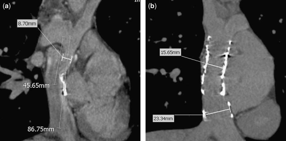
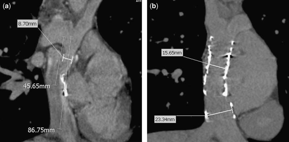
A 19-year-old boy with Fontan circulation with right isomerism, tricuspid atresia, double outlet of the right ventricle, and a large ventricular septal defect developed cyanosis that was prominent on exercise. Percutaneous oxygen saturation was 88% at rest and it diminished to 71% during exercise. He had undergone a fenestrated Fontan operation of the lateral tunneled type at 3 years of age. The cardiac catheterisation he underwent showed good pulmonary arterial pressure (12 millimetres of mercury) and good pulmonary vascular resistance. However, angiography revealed narrowing at the junction between the right atrium and the main pulmonary artery, and a lateral tunnel dehiscence with a right-to-left shunt. The fenestration had closed naturally. Balloon angioplasty was tried, but it was not effective. Chest computed tomography was performed, and we measured the dimension of the lateral tunnel. The length was 87 millimetres, and the diameter at the inferior caval vein junction was 19 millimetres; the narrowest diameter was 8.7 millimetres (Fig 1a).
(a) A coronal computed tomography image showing the narrowest diameter and the length of the tunnel for graft planning. (b) The computed tomography image 3 months after the implantation of a self-expandable stent and self-expandable stent graft showing an increased axial diameter at the narrowest point.
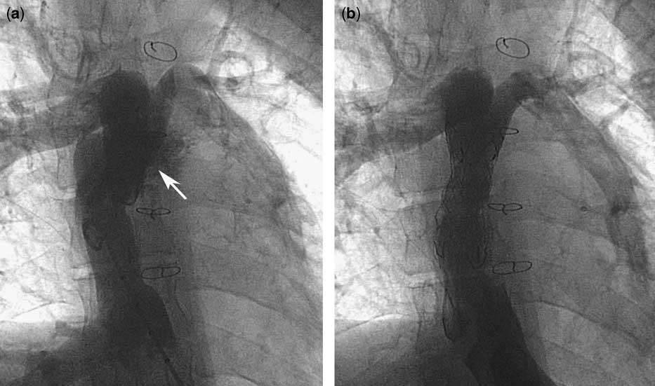
We decided to implant a self-expandable stent in the narrowing lesion and close the dehiscence with a custom-made, self-expandable stent graft. A HERCULES® self-expandable vascular stent and stent graft were made by SnG Biotech (Bundang, South Korea). After 6 months, we performed another catheterisation. We could check the low compliance of the lesion by test ballooning. With the patient remaining alert, the first self-expandable stent of 20 millimetres in diameter and 40 millimetres in length was introduced via the right femoral vein in its 18 French delivery system and implanted successfully. The next custom-made, self-expanding stent graft with a polytetrafluoroethylene membrane 24 millimetres in diameter and 65 millimetres in length overlapped with the first stent successfully. After the procedure, the angiogram showed no right-to-left shunt and increased diameter (Fig 2b). The procedure was performed smoothly and no significant blood loss occurred. The chest computed tomography performed on the next day showed an increase in the narrowest diameter to 13 millimetres and good positions of the stent and stent graft.
(a) The angiogram of the Fontan pathway showing a lateral tunnel dehiscence in the upper margin of anastomosis resulting in a right-to-left shunt. (b) The angiogram after the implantation of the graft stent showing disappearance of a right-to-left shunt.
At follow-up 3 months after the procedure, a chest computed tomography was performed and we found that the narrowest diameter of implanted stent had increased to 15 millimetres. The oxygen saturation at rest was 98% and during exercise was 97%. During the follow-up period, warfarin and aspirin were administered.
Discussion
Many interventional types of cardiac catheterisation have been introduced and shown to be safe for use in various congenital heart diseases. However, stent or stent graft implantation in congenital heart disease is not common owing to various limitations. First, there are difficulties in selection of the stent owing to the complexities of the vascular structures in congenital heart disease. The growth potential is also a crucial point in children, and usually the lesions in congenital heart disease contain not only stenosis but also dehiscence. Hijazi et alReference Hijazi, Ruiz and Patel 2 reported a catheter-based therapy for Fontan baffle obstruction and leak using a balloon-expandable covered stent. Recently, a Cheatham-Platinum stent was developed and used in congenital heart disease. A covered Cheatham-Platinum stent in a univentricular heart circulation due to the right-to-left shunt was also reported.Reference Gianfranco, Luciane and Massimo 3 However, reports of a self-expandable covered stent in a Fontan circulation are rare.Reference Uberoi, Roberts, Barnes and Wilson 4
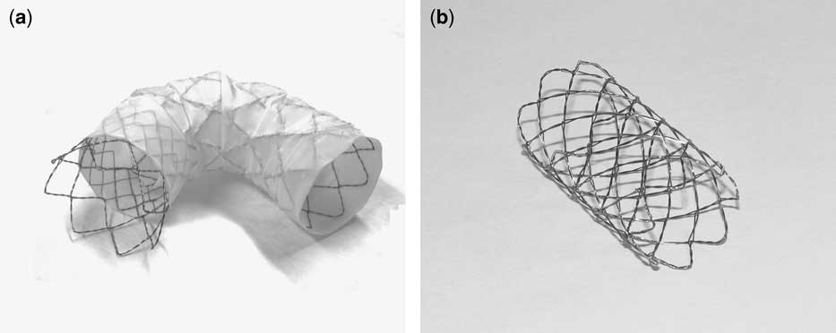
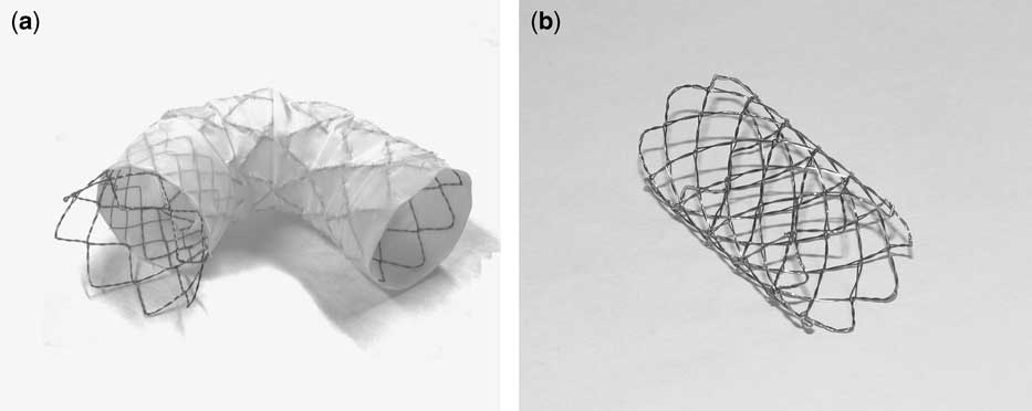
It is generally accepted that balloon-expandable stents are more accurate in placement than self-expandable stents. However, in our country, we had only a limited number of sizes of balloon-expandable stents, and no covered stent grafts were available. This patient had stenosis and a leak at the junction of the lateral baffle with the pulmonary artery. The pressure gradient at the front and rear of the stenotic portion was only 2 millimetres of mercury. However, we experienced a very low-pressure difference in the venous system, which is unusual in the arterial system. I think that low or absent pressure gradients across venous lesions have essentially no meaning in relation to the degree of stenosis. In general, a stent should not be implanted in lesions that are not balloon expandable. Therefore, test balloon angioplasty over the stenosis was necessary, and thus we performed the procedure. We were not concerned about rupture or further dehiscence of the lateral tunnel, because we expected the presence of strong peelings around the lateral tunnel due to post-operative adhesion. The HERCULES® self-expandable stent is made from nitinol with two meshed wires that are bent and welded to a cylindrical meshwork (Fig 3). It has substantial elasticity and a memory effect of nitinol following implantation, similar to other self-expandable stents. Owing to the weaker radial strength of the HERCULES® self-expandable stent graft due to an open-cell design, a HERCULES® self-expandable stent was implanted at first in the stenotic lesion. In addition, an additive self-expandable stent graft was introduced to close the baffle leak, even though self-expandable stents develop more neo-intimal hyperplasia than balloon-expandable stents.Reference Cheung, Sanatani, Leung, Human, Chau and Culham 5 The 18 French delivery sheath was used for our stent graft instead of the 24 French delivery sheath that is used for other commercial stent grafts.Reference Uberoi, Roberts, Barnes and Wilson 4 Considering the delayed additional expansion of the self-expandable stent and stable placement for a long time, an oversized stent was chosen. We used a somewhat long stent graft (65 millimetres) for stable positioning. If the patient needs an intracardiac electrophysiologic study or pacing lead, it is essential to change the whole Fontan tract. But I think that the stability of the graft stent is overbearingly important. Owing to variations in anatomy and vascular size in congenital heart disease, a custom-made stent is very helpful. We found an increase in target lesion immediately after the implantation, and another delayed dilatation 3 months later. There are concerns about application of self-expanding stents in children or adolescents with congenital heart disease. Neo-intimal proliferation is a very fatal complication, but the accurate pathophysiology remains unclear. There are some reports about significant intimal proliferation of self-expandable stents.Reference Cheung, Sanatani, Leung, Human, Chau and Culham 5 On the other hand, there are positive reports that show good patency and little complication.Reference Lou, Gu and He 6 , Reference Lastovickova and Peregrin 7
(a) HERCULES® self-expandable stent graft has a proximal bare stent and requires an 18 French introductory system. (b) HERCULES® self-expandable stent has soft ends and good flexibility. It is made of two nitinol wires, and the mean radial force is 0.94 Newton.
The positions of the stents were stable, and no thrombus was found on chest computed tomography. To our knowledge, this is the first case of the use of a self-expandable stent with a stent graft in a case with Fontan circulation.
Conclusion
In spite of the lack of data on long-term follow-up, the self-expandable stent and the stent graft are very useful tools for treating selected congenital malformations within special environment such as unserviceable balloon-expandable stent and stent graft.
Acknowledgement
The Institutional Review Board of the Sejong General Hospital approved this report.