Introduction
The use of cardiac MRI in pulmonary hypertension has grown rapidly, and it has been included as a Class I recommendation for initial diagnostic workup of pulmonary hypertension in children who do not require sedation or anaesthesia. Reference Latus, Kuehne and Beerbaum1 Although invasive catheterisation is the gold standard method for the diagnosis of pulmonary hypertension, some parameters, such as pulmonary artery to aorta ratio >1, mean pulmonary artery velocity <11.7 cm/sec, and ventricular mass index >0.37 (right-to-left ventricle mass ratio), obtained with cardiac MRI, could augment the evaluation. Reference Simpson, Kolb and Hsu2,Reference Sanz, Kuschnir and Rius3 In practice, however, the main use of cardiac MRI in pulmonary hypertension patients is to determine prognosis after diagnosis and perform risk assessment. The European Society of Cardiology cardiac MRI-based current risk stratification includes right ventricular ejection fraction, right ventricular end-systolic volume, and right ventricular stroke volume. 4 In addition, it is known that decreased left ventricular diastolic volume, increased right ventricular mass, ventricular mass index greater than 75%, increased right atrial volume, right ventricular shift to the left, and septal late gadolinium enhancement are associated with poor prognosis. Reference van Wolferen, Marcus and Boonstra5–Reference Roeleveld, Marcus and Faes8
Pulmonary arterial hypertension is defined as mean pulmonary artery pressure greater than 20 mmHg measured during right heart catheterisation including a pulmonary artery wedge pressure less than 15 mmHg and a pulmonary vascular resistance greater than 3 Woods units. By definition, to diagnose idiopathic pulmonary arterial hypertension, all other causes of elevated pulmonary artery pressures, such as familial pulmonary arterial hypertension, drugs and toxins, connective tissue disease, HIV, portal hypertension, and CHD, must be ruled out. 4
In this study, cardiac MRI findings between idiopathic pulmonary arterial hypertension and pulmonary arterial hypertension associated with CHD, which are two common causes of pulmonary hypertension in children, will be compared. We hypothesised that, in shunt defects, the pressure and volume balance in the ventricles may differ from that in patients with idiopathic pulmonary arterial hypertension. Our aim is to determine if there is a difference between cardiac MRI measurements in these two groups and to use the results in patient follow-up.
Material methods
Study design
This prospective cross-sectional study was conducted with 40 available patients who were diagnosed with idiopathic pulmonary arterial hypertension (n = 20) and pulmonary arterial hypertension associated with CHD (n = 20) at Ankara Bilkent City Hospital between 2020 and 2024 according to the above-mentioned criteria. 4 All patients with pulmonary arterial hypertension associated with CHD had an untreated heart defect with right-to-left shunting. The cardiac MRI was performed within one year of the diagnosis for all pulmonary arterial hypertension patients. One patient was excluded from the study due to poor image quality.
Data collection
The patients’ data was obtained from electronic medical records. Clinical and laboratory data included age at cardiac MRI, weight, height, body surface area, heart rate, the type of anti-pulmonary hypertension medication and its usage period, single or multiple anti-pulmonary arterial hypertension use status, six-minute walking test results (pre–post SpO2, oxygen saturation difference, and 6-minute walk test distance), brain natriuretic peptide level, current NYHA class, the presence of arrhythmia and its type, and the presence of syncope. Two of the patients could not do the six-minute walking test due to their age.
Cardiac magnetic resonance
The cardiac MRI protocol at our institution, which is routinely applied, is presented in Table 1. This protocol was created according to the Society of Cardiac Magnetic Resonance guidelines and the European Association of Cardiovascular Imaging textbook. Reference Dorfman, Geva and Samyn9,Reference Lombardi, Plein and Petersen10
CMR protocol applied in our institution for pulmonary hypertension

Table 1. Long description
The table is divided into four main sections: Anatomy, Function, Flow, and Viability. Each section lists different assessments, sequences, planes, and aims. The Anatomy section includes Scout images and Cine SSFP with specific planes and aims related to heart morphology. The Function section details SAX cine SSFP with aims related to heart function metrics. The Flow section lists Aa, MPA, RPA, LPA with aims related to flow measurement. The Viability section includes SAX, 4-C with aims related to scar assessment.
CMR = cardiac magnetic resonance; SSFP = steady-state free precession = Chamber; LVOT = left ventricular outflow tract; RVOT = right ventricular outflow tract; SAX = short axis; EF = ejection fraction; SV = stroke volume; EDV = end-diastolic volume; ESV = end-systolic volume; CO = cardiac output; 2D = two-dimensional; Ao = ascending aorta; MPA = main pulmonary artery; RPA = right pulmonary artery; LPA = left pulmonary artery; mvPA = mean velocity of pulmonary artery; PARF = pulmonary artery regurgitation fraction; LGE = late gadolinium enhancement.
All cardiac MRI studies were performed using a 1.5-Tesla system (GE Healthcare, Chicago, Illinois) in line with the above-mentioned protocol, and the CardiacVX software was used for post-processing. All cardiac MRI analysis was performed by the same person. Data was acquired during breath-holding or free-breathing if the patients were sedated. Breath-hold steady-state free precession sequence parameters were as follows: repetition time: 4.2 ms, echo time: 2 ms, flip angle 60°, slice thickness 6 mm, in-plane image resolution 1.6 mm × 1.6 mm × 6.0 mm, and temporal resolution 69 ms. The sequence parameters during free-breathing were as follows: repetition time 3.8 ms, echo time 1.6 ms, flip angle 60°, slice thickness 5 mm, and in-plane image resolution 1.6 mm × 1.6 mm × 5.0 mm. Short-axis cine images were used for ventricular volumetric analysis. The endocardial and epicardial borders of both ventricles were traced manually at end-systole and end-diastole. Then, the end-diastolic volume, end-systolic volume, stroke volume, ejection fractions, right and left ventricular mass, and cardiac output were calculated. All volumetric parameters were indexed to the body surface area. The ventricular mass index was calculated by dividing right ventricular mass by left ventricular mass. Four-chamber cine images were used for right atrial volumes and indexed to the body surface area. Whether the septum shifted to the right or the left was evaluated on short-axis images. In addition, the percentage of the entire cycle in which the septal shift is seen, known as the septal curvature duration index, was calculated by examining the short-axis slices.
Two-dimensional through-plane phase-contrast flow measurements were obtained from the ascending aorta, main pulmonary artery, and right and left pulmonary artery. Flow measurements were performed perpendicular to each targeted vessel using the double oblique technique. In all cases, encoding velocity was adjusted to avoid aliasing. Initial velocity encoding values were determined as 150 cm/s and 80–100 cm/s for the aorta and pulmonary arterial systems, respectively. The velocity encoding value is generally selected as small as possible for the pulmonary arteries, with a predefined upper velocity limit of 100 cm/s. In all cases, encoding velocity was adjusted to avoid aliasing. Imaging parameters that were used for the phase contrast imaging were as follows: repetition time/echo time, 6.7 ms/4.2 ms; flip angle, 25°; phase to reconstructed 30; temporal resolution, 107 ms; the number of signal averages, 1; and section thickness, 7 mm. Field of view and pixel size (1.6–2.3 mm) were adjusted individually according to a patient’s size. Through 2D-phase contrast flow, Qp:Qs, mean velocity of pulmonary artery, and pulmonary artery regurgitation fraction were obtained.
Respiratory navigator-gated, electrocardiography-triggered, 3-dimensional whole-heart balanced steady-state free precession MRI was acquired during free breathing. Through this, pulmonary artery and ascending aorta diameter in the diastole were measured, and the presence of aneurysm was evaluated. Lastly, after the injection of the gadolinium-based contrast agent (0.01 mmol/kg), late-phase (10–20 min) images were obtained. Then, the presence of late gadolinium enhancement and its localisation were evaluated.
Statistical analysis
All data were analysed by using the “SPSS (Statistical Package for Social Sciences) for Windows 25.0” programme (IBM Corp., Armonk, N.Y., USA). After descriptive statistics and normality analysis, normally distributed continuous variables were reported as mean ± standard deviation, non-normally distributed continuous variables were reported as median with range, and categorical variables were reported as count with percentage of total. Chi-square or Fisher’s exact test was used to compare categorical or ordinal variables, and Student’s T-test or Mann–Whitney U test was used to compare continuous variables.
Results
Of the patients, 28 (70%) were female, and the median age at diagnosis was 11.9 (0.5–23.5), and the cardiac MRI time was 12.5 (0.5–24) years, respectively. The primary diagnosis of the patients in the pulmonary arterial hypertension associated with the CHD group was aorto-pulmonary window in one patient and ventricular septal defect in 19 patients. All patients were using at least one anti-pulmonary arterial hypertension medication. Ventricular mass index >37%, pulmonary artery/ascending aorta ratio >1, and mvPA <11.7cm/sn used for pulmonary hypertension diagnosis were seen in 38 (95%), 38 (95%), and 35 (85%) patients, respectively.
Clinical findings of the study population based on groups
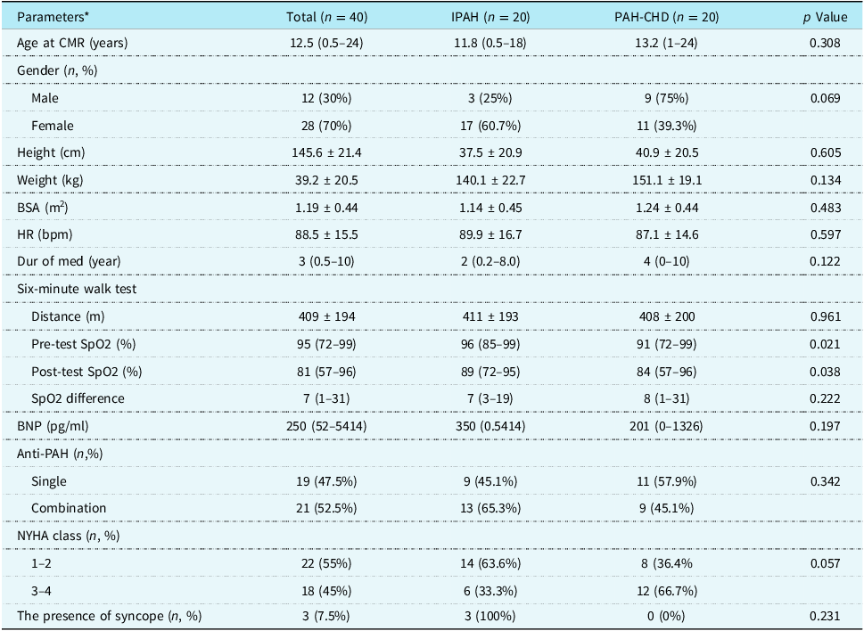
While 17 (60.7%) of the female patients were in the idiopathic pulmonary arterial hypertension group, 11 (30.3%) of them were pulmonary arterial hypertension associated with CHD patients. When comparing patients with idiopathic pulmonary arterial hypertension and pulmonary arterial hypertension associated with CHD, pre- and post-test SpO2 levels at the six-minute walk test were significantly higher in the idiopathic pulmonary arterial hypertension patients (p = 0.021, 0.038, respectively). However, no difference was observed between the groups in terms of pre–post test SpO2 differences and walking distance at the six-minute walk test. Likewise, no difference was observed between the groups in terms of other clinical findings. Although all three patients who experienced syncope were in the idiopathic pulmonary arterial hypertension group, no statistical difference was observed between the groups. No patient exhibited arrhythmia. Table 2 shows the comparison of clinical findings between the groups.
Clinical findings according to groups

Table 2. Long description
The table compares clinical findings between two groups of patients: idiopathic pulmonary arterial hypertension (IPAH) and pulmonary arterial hypertension associated with congenital heart disease (PAH-CHD). It has 40 rows and 10 columns. The columns are Parameters, Total (n = 40), IPAH (n = 20), PAH-CHD (n = 20), and p Value. The rows include Age at CMR (years), Gender (n, %), Height (cm), Weight (kg), BSA (m^2), HR (bpm), Dur of med (year), Six-minute walk test (Distance (m), Pre-test SpO2 (%), Post-test SpO2 (%), SpO2 difference), BNP (pg/ml), Anti-PAH (n,%), NYHA class (n, %), and The presence of syncope (n, %). Each row provides specific values for each group and the p Value indicating statistical significance. Notable trends include higher pre- and post-test SpO2 levels in the IPAH group compared to the PAH-CHD group, with significant p values of 0.021 and 0.038, respectively. No significant differences were observed in other clinical findings between the groups.
*Data were presented as n (%) for categorical variables and median (minimum-maximum) or mean ± standard deviation for continuous variables.
Significant difference = p < 0.05.
IPAH = idiopathic pulmonary arterial hypertension; PAH-CHD = pulmonary arterial hypertension associated with congenital heart disease; CMR = cardiac magnetic resonance; kg = kilogram; cm = centimetre; BNP = blood-natri-uretic peptide; PAH = pulmonary arterial hypertension; NYHA = New-York Heart Association.
Cardiac magnetic resonance findings of the study population based on groups
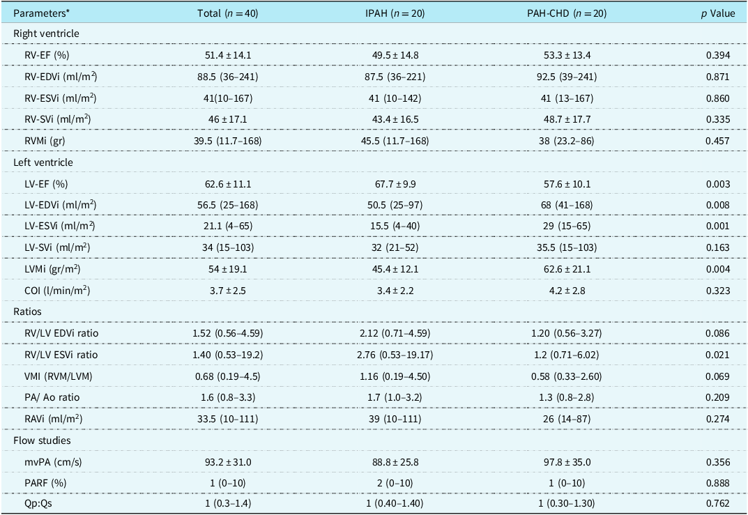
In this study, there was no statistical significance in right ventricular ejection fraction between groups. Similarly, there was no difference between the groups in terms of other right ventricle-related parameters such as indexed end-diastolic and end-systolic volumes, stroke volume, and mass. Similarly, cardiac index, indexed right atrial volume, pulmonary artery to ascending aorta ratio, and flow study parameters such as mean velocity of pulmonary artery, pulmonary artery regurgitation fraction, and Qp:Qs values were not different between the groups. However, while left ventricular ejection fraction was significantly higher (p = 0.003), indexed left ventricular end-diastolic and end-systolic volumes and left ventricle mass were significantly lower in patients with idiopathic pulmonary arterial hypertension than pulmonary arterial hypertension associated with CHD (p = 0.008, 0.001, 0.004, respectively). In addition, the right ventricle to the left ventricle end-systolic ratio was significantly higher in idiopathic pulmonary arterial hypertension than pulmonary arterial hypertension associated with CHD (p = 0.021). Moreover, ventricular mass index was higher in patients with idiopathic pulmonary arterial hypertension than pulmonary arterial hypertension associated with CHD; however, this did not reach statistical significance (p = 0.069). Ventricular mass index results did not differ significantly between groups, but of the 16 patients with a ventricular mass index greater than 75%, 12 (75%) were in the idiopathic pulmonary arterial hypertension group. This difference was found to be statistically significant (p = 0.009), as shown in Figure 1. Lastly, the left ventricular mass index was significantly greater in pulmonary arterial hypertension associated with CHD patients than idiopathic pulmonary arterial hypertension ones (p = 0.004). Table 3 shows functional and flow study results of the patients based on the groups.
Ventricular mass index comparison between groups (VMI = Ventricular mass index; IPAH = Idiopathic pulmonary arterial hypertension; PAH-CHD = Pulmonary arterial hypertension associated with congenital heart disease).

Cardiac magnetic resonance findings according to groups

Table 3. Long description
A table comparing cardiac magnetic resonance findings across different groups. The table has 23 rows and 5 columns. The columns are labeled Parameters, Total (n = 40), IPAH (n = 20), PAH-CHD (n = 20), and p Value. The rows are grouped into Right ventricle, Left ventricle, Ratios, and Flow studies. Each row lists specific parameters and their corresponding values for the different groups. Notable trends include significant differences in left ventricular ejection fraction, indexed left ventricular end-diastolic and end-systolic volumes, and left ventricle mass between the groups.
*Data were presented as n (%) for categorical variables and median (minimum-maximum) for continuous variables.
Significant difference = p < 0.05.
IPAH = idiopathic pulmonary arterial hypertension; PAH-CHD = pulmonary arterial hypertension associated with congenital heart disease; RV = right ventricle; LV = left ventricle; EF = ejection fraction; EDVi = end-diastolic volume index; ESVi = end-systolic volume index; SVi = stroke volume index; RVMi_= right ventricular mass index; LVMi = left ventricular mass index; COI = cardiac output index; VMI = ventricular mass index; RAVi = right atrial volume index; mvPA = mean velocity of pulmonary artery; PARF = pulmonary artery regurgitation fraction; TAPSE = tricuspid valve annular plane systolic excursion.
A total of 15 patients had a septal shift towards the left ventricle. Nine (60%) of them were in the idiopathic pulmonary arterial hypertension group, and six (40%) were in the pulmonary arterial hypertension associated with CHD group, and no statistically significant difference was found between them (p = 0.327). Of the nine patients with a septal curvature duration index greater than two-thirds of the cardiac cycle, six (66.7%) were in the idiopathic pulmonary arterial hypertension group; however, there were no statistically significant differences between the groups (p = 0.256). Moreover, 30 of the 40 patients (75%) had late gadolinium enhancement. Figure 2 shows the late gadolinium enhancement location. Additionally, the late gadolinium enhancement extended into the septum in five of 30 patients. There was no difference between the groups in terms of the presence of late gadolinium enhancement and its location. Figure 3 shows an image of a patient with a septal shift and late gadolinium enhancement extending into the septum. Lastly, as an additional finding, giant aneurysms were detected in two patients, and both of them were diagnosed with idiopathic pulmonary arterial hypertension, as shown in Figure 4. The giant aneurysm was located in the main pulmonary arteries, and one of them was extending to the right pulmonary artery.
Late gadolinium enhancement locations in the heart.

( a ) Arrow indicates a septal leftward shift. ( b ) Arrows indicate late gadolinium enhancement at the right and left ventricle inferior and superior hinge points.

( a ) The main pulmonary artery aneurysm. ( b ) The main pulmonary aneurysm extending to the right pulmonary artery.

Discussion
Our results showed that most of the prognostic parameters known in the literature, such as right ventricular volumes, late gadolinium enhancement, and septal curvature duration index, were not different between idiopathic pulmonary arterial hypertension and pulmonary arterial hypertension associated with CHD patients. However, the number of patients with ventricular mass index >75% and right-to-left ventricle volume ratio values was significantly higher, while left ventricular end-diastolic volume index was significantly lower in idiopathic pulmonary arterial hypertension compared to pulmonary arterial hypertension associated with CHD.
Lower SpO2 during the six-minute walk test in pulmonary arterial hypertension associated with CHD than idiopathic pulmonary arterial hypertension patients is the result of blood mixing due to right-to-left shunting, as expected. In a study comparing exercise capacity, lower predicted peak oxygen consumption was reported in patients with pulmonary arterial hypertension associated with CHD compared to those with idiopathic pulmonary arterial hypertension. Reference Chen, Chen, Hsu, Wu, Lai and Chien11 Similarly, in another study, patients with pulmonary arterial hypertension associated with CHD have impaired exercise capacity and substantially reduced quality of life compared to patients with idiopathic pulmonary arterial hypertension. Reference Kaemmerer, Gorenflo, Hoeper, Huscher, Ewert and Pittrow12 However, since cardiopulmonary exercise testing was not performed in this study, no conclusions can be drawn regarding the difference in oxygen uptake between the two groups. Nevertheless, the fact that no difference was observed in the six-minute walk test distance in our study may be significant in itself.
Right ventricular parameters obtained from cardiac MRI are used to determine the mortality risk of pulmonary arterial hypertension patients. In the current study, we did not find a difference between groups in terms of right ventricular volume parameters. Contrary to our results, in a study conducted in adults with a mean age of approximately 36 years, patients with idiopathic pulmonary arterial hypertension had worse baseline right ventricle function compared to patients with pulmonary arterial hypertension associated with CHD on echocardiographic evaluation. Reference Zhu, Wu and Tan13 Our suggestion is that no difference exists in right ventricular functional parameters between idiopathic pulmonary arterial hypertension and pulmonary arterial hypertension associated with CHD patients due to younger age.
The higher left ventricular end-diastolic and end-systolic volumes and the lower right-to-left ventricular end-diastolic and end-systolic volume ratios in pulmonary arterial hypertension associated with CHD patients compared to idiopathic pulmonary arterial hypertension, as shown in the current study, may be due to the deterioration of the configuration of the left ventricle as a result of increased right ventricular pressure. Increased left ventricular volume, probably due to the presence of a right-to-left shunt in pulmonary arterial hypertension associated with CHD patients, may prevent left ventricular compression. Furthermore, although no differences were observed between the groups in terms of right ventricular volume parameters, the lower right-to-left ventricular volume ratios in pulmonary arterial hypertension associated with CHD patients may be attributed to the higher left ventricular volumes. A study reported that the combined use of the right-to-left ventricular volume ratio and indexed right ventricular end-diastolic volume improves the detection of right ventricular enlargement in adult pulmonary arterial hypertension patients. Reference Altmayer, Patel, Addetia, Gomberg-Maitland, Forfia and Han14 However, in children, the right-to-left ventricular volume ratio may be less useful for monitoring, as it is more likely to reflect changes in left ventricular volumes.
The left ventricular ejection fraction in pulmonary arterial hypertension associated with CHD patients compared to idiopathic pulmonary arterial hypertension was found to be lower in the current study. We speculate that this decrease in pulmonary arterial hypertension associated with CHD patients may be related to the presence of increased volume load coupled with increased pressure load. Or, the higher left ventricular ejection fraction in the idiopathic pulmonary arterial hypertension group has more to do with the left ventricular compression by the right ventricle than diminished left ventricular contractility. Another reason why better ejection fraction is seen in idiopathic pulmonary arterial hypertension patients may be because their septum contribution to systolic function is greater compared to those with pulmonary arterial hypertension associated with CHD. Because, despite markedly different muscle mass and chamber geometry, both ventricles are bound together by spiral muscle bundles that encircle them in a complex manner, forming a highly interdependent functional unit. Reference Dell’Italia15 The presence of a ventricular septal defect may reduce the contribution of the septum to left ventricular function. Additionally, no patient had an ejection fraction below 40%, and no difference was observed between the groups in terms of NYHA class and 6-minute walking test distance. Therefore, this slightly lower left ventricular function does not seem to have a clinical significance.
Ventricular mass index is a relatively newly defined parameter used in pulmonary hypertension. To obtain this parameter, the mass of the right and left ventricles should be measured separately. While it is difficult to trace the epicardial borders as well as the endocardial borders of the right ventricle in the normal patient, this is easier in the pulmonary hypertension patient due to the hypertrophied right ventricle. According to a recent study conducted in children, ventricular mass index was correlated with transplant-free survival from the time of cardiac MRI in idiopathic pulmonary arterial hypertension patients. However, this correlation could not be confirmed in the CHD-pulmonary arterial hypertension patients. Reference Haarman, Coenraad and Hagdorn16 Additionally, van Wolferen SA et al. reported that a ventricular mass index greater than 0.75 is an independent predictor of mortality and adverse outcomes in idiopathic pulmonary arterial hypertension patients. Reference van Wolferen, Marcus and Boonstra5 In the current study, the ventricular mass index is higher, although not statistically significant, and the number of patients with ventricular mass index ≥75% was significantly greater in idiopathic pulmonary arterial hypertension than pulmonary arterial hypertension associated with CHD. Considering that the right ventricular mass index was not found to be significantly different in both groups and the left ventricular mass index was significantly higher in pulmonary arterial hypertension associated with CHD patients, it could be suggested that the determinant of this difference in ventricular mass index is left ventricular hypertrophy. Presumably, the pressure load is transferred to the left ventricle due to the right-to-left shunt, which may explain the higher left ventricular mass index in patients with pulmonary arterial hypertension associated with CHD. Or, the higher left ventricular mass index in the pulmonary arterial hypertension associated with the CHD group could be due to the CHD itself rather than pulmonary hypertension. As a result, lower ventricular mass index occurs in pulmonary arterial hypertension associated with CHD patients.
It is reported that leftward septal bowing occurs when the right ventricular pressure is ≥5 mmHg higher than left ventricular pressure. Reference Roeleveld, Marcus and Faes8 Although it is logical that septal shift would be less common and septal curvature duration index would be lower in pulmonary arterial hypertension associated with CHD patients with increased pressure in both ventricles, we could not demonstrate this. New studies on this subject can be considered by increasing the study population.
Late gadolinium enhancement is commonly seen in the right and left ventricular hinge point regions in pulmonary arterial hypertension patients. The incidence has been reported as 69–92% in adult studies. Reference Blyth, Groenning and Martin7,Reference Freed, Gomberg-Maitland and Chandra17 In our study, its rate was found to be 75%, similar to the literature. Also, we found no difference between groups in terms of the presence of late gadolinium enhancement and its location (hinge point or septal extending). Although studies have reported that late gadolinium enhancement is associated with a poor clinical course Reference Freed, Gomberg-Maitland and Chandra17 and late gadolinium enhancement mass index at right ventricular septal insertion may be suggestive of right ventricular dysfunction in pulmonary arterial hypertension patients, Reference Kazimierczyk, Małek and Szumowski18 there are also studies that show the opposite. Reference Kazimierczyk, Szumowski and Nekolla19,Reference Anderson, Sutton and Lie20 The same author, who reported that late gadolinium enhancement mass index predicts prognosis, showed in another, more recent study that late gadolinium enhancement is not associated with low metabolic activity in pulmonary arterial hypertension patients. Reference Kazimierczyk, Szumowski and Nekolla19 Anderson et al. described three different forms of cardiac fibrosis: reactive (interstitial; preserving cardiac structure and function), perivascular, and replacement fibrosis. Reference Anderson, Sutton and Lie20 Since low metabolic activity is not observed in fibrotic foci in pulmonary arterial hypertension patients, the authors considered that the late gadolinium enhancement observed in pulmonary arterial hypertension patients is of the interstitial type, and the presence of late gadolinium enhancement at right ventricular insertion points should not simply be interpreted as necrotic tissue in this group of patients. Reference Kazimierczyk, Szumowski and Nekolla19 Therefore, the prognostic value of late gadolinium enhancement in pulmonary arterial hypertension patients remains unclear, and longitudinal studies are needed to explore it.
Right ventricular failure is currently considered the most common cause of death in patients with pulmonary arterial hypertension. Reference Thenappan, Ormiston, Ryan and Archer21 However, it is a common clinical experience that pulmonary arterial hypertension patients often die suddenly or following an acute illness. In an adult study comparing pulmonary arterial hypertension associated with CHD and idiopathic pulmonary arterial hypertension patients, better survival was observed in pulmonary arterial hypertension associated with CHD patients as opposed to idiopathic pulmonary arterial hypertension patients. Also, the same study showed lower baseline right ventricular functions and less improvement in the six-minute walk test, World Health Organization functional class, and right ventricular functional parameters after anti-pulmonary arterial hypertension therapy in idiopathic pulmonary arterial hypertension patients compared to pulmonary arterial hypertension associated with CHD. Reference Zhu, Wu and Tan13 Similarly, in another study, pulmonary arterial hypertension associated with CHD patients has a better prognosis than idiopathic pulmonary arterial hypertension patients, and the higher functional class at initial diagnosis is an independent risk factor for poor prognosis. Reference Gu, Li, Gu, Xie and Liu22 The fact that right ventricular functional parameters, known to influence prognosis, did not differ between groups in our study may be due to our younger population compared to the study by Zhu et al. Of course, since this study is cross-sectional, longitudinal follow-up is necessary to understand the mechanism of cardiac deaths in paediatric pulmonary arterial hypertension patients.
Limitations
The most obvious limitation of this study is the small number of patients due to it being a single-centre and cross-sectional study. Also, the results of a single measurement were given in the study. Data from repeated measurements will provide more important information. Furthermore, we could not perform quantitative measurement of late gadolinium enhancement due to software-related deficiencies. For the parameters used in European Society of Cardiology risk stratification, no distinction is made regarding the patients’ primary diagnosis. However, prognostic markers related to ventricular mass studies have mostly been studied in idiopathic pulmonary arterial hypertension patients. Therefore, studies with prognostic parameters specific to pulmonary arterial hypertension associated with CHD patients would be beneficial. Since this study is cross-sectional, longitudinal studies are recommended to clarify this issue.
Conclusion
A comparison of cardiac MRI-based parameters in patients with idiopathic pulmonary arterial hypertension and pulmonary arterial hypertension associated with CHD showed that while most right ventricular functional markers did not differ between groups, several left ventricular key parameters, including left ventricular function, left ventricular end-diastolic volume, and mass index, were significantly lower in idiopathic pulmonary arterial hypertension patients. This suggests a distinct pathophysiological pattern in idiopathic pulmonary arterial hypertension, potentially related to pressure overload and altered ventricular interdependence.
Acknowledgements
None.
Financial support
The authors did not receive support from any organisation for the submitted work.
Competing interests
The authors have no relevant financial or non-financial interests to disclose.
Ethical standard
Written informed consent was obtained from the parents of all participants, and the study was approved by the institutional Ethics Committee of Bilkent City Hospital with the number TABED 1-24-174 on 24 April 2024.
The authors assert that all procedures contributing to this work comply with the ethical standards of the relevant national guidelines on human experimentation and with the Helsinki Declaration of 1975, as revised in 2013.