Although cardiomyopathy is a relatively rare disease, it is the primary reason for heart transplantation in children, particularly in those older than 1 year of age.Reference Andrews, Fenton, Ridout and Burch 1 – Reference Webber 4 Literature on the epidemiology of cardiomyopathy was negligible before the 1990s. Since 1994, the National Heart, Lung, and Blood Institute has funded the Pediatric Cardiomyopathy Registry Study Group, which has conducted a number of large, multi-centre observational studies of primary and idiopathic cardiomyopathies. At the time of its inception, it was primarily designed to determine the epidemiology and clinical course of cardiomyopathy in children. Since that time, the Pediatric Cardiomyopathy Registry has added studies of the aetiologies of paediatric cardiomyopathy, including gene discovery, genotype–phenotype associations, the utility of cardiac biomarkers in clinical assessment and outcome prediction, and the physical and psychosocial functional status of both affected children and their families.
Design of the Pediatric Cardiomyopathy Registry
The original Pediatric Cardiomyopathy Registry design included 98 paediatric cardiology centres in the United States and Canada, which collected clinical data regarding factors associated with the study’s primary outcomes: death or heart transplantation.Reference Grenier, Osganian and Cox 5 In addition, we attempted to enrol nearly 100% of cases at two Pediatric Cardiomyopathy Registry centres – Boston Children’s Hospital and its New England referral centres and Texas Children’s Hospital and its Central Southwest referral centres – to estimate cardiomyopathy incidence and other epidemiologic parameters.Reference Grenier, Osganian and Cox 5 , Reference Lipshultz, Sleeper and Towbin 6 Over the subsequent years, further Pediatric Cardiomyopathy Registry studies have focussed on 11–15 of the largest US–Canadian paediatric heart centres that could provide the largest number of eligible patients and the most complete data sets.
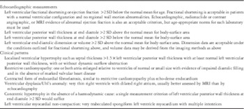
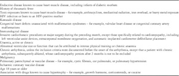
Since its inception, patients aged 18 years or less who were diagnosed with cardiomyopathy – via quantitative echocardiographic criteria, a pattern of cardiomyopathy matching a defined, semi-quantitative pattern; or tissue analysis – were eligible for inclusion to the Pediatric Cardiomyopathy Registry (Table 1). Each case was classified according to morphology as dilated, hypertrophic, restrictive, mixed, or other type of cardiomyopathy. Since 1994, new phenotypes of cardiomyopathy have been added to the Pediatric Cardiomyopathy Registry, such as left ventricular non-compaction. Patients with secondary causes of heart muscle disease were excluded from the study in order to focus this research on the aetiologies and outcomes of children with primary cardiomyopathies (Table 2).
Inclusion criteria for enrolment in the Pediatric Cardiomyopathy Registry.

Exclusion criteria for enrolment in the Pediatric Cardiomyopathy Registry.

At all times, the Pediatric Cardiomyopathy Registry has utilised a highly trained outreach team that travelled to the centres to enrol new cases and extract data from medical records at regular intervals to perform standardised data collection. Data collection included demographic descriptors, quantitative echocardiographic measurements, a brief family history, transplant status, and clinical findings.
Aims of the Pediatric Cardiomyopathy Registry
Since its establishment, the aims of the Pediatric Cardiomyopathy Registry have evolved over time. The original aims were primarily epidemiological in nature and intended to describe the incidence and clinical factors at presentation of cardiomyopathy in children as well as describe the clinical outcomes – death or heart transplantation and the predictors of those outcomes. In a subsequent study, the study aims were further expanded through collaboration with the Pediatric Heart Transplant Study Group’s data set to examine both pre- and post-transplant outcomes and associated risk factors.
At present, the Pediatric Cardiomyopathy Registry is the largest pediatric cardiomyopathy registry in the world containing data collected annually from 3549 children until heart transplant, death, or loss to follow-up. It provides the most comprehensive picture of this disease, and thus is the starting point for new diagnostic and treatment strategies. Analyses of the database have resulted in important findings regarding the incidence, aetiology, disease course, and estimates and determinants of mortality and transplant outcomes, with a new focus on the genetic determinants of cardiomyopathy in children.
Incidence of paeditric cardiomyopathy
A study by Lipshultz et alReference Lipshultz, Sleeper and Towbin 6 estimated that the annual incidence of paediatric cardiomyopathy in the United States was 1.13 cases per 100,000 children. This estimate is comparable with reports from similar registries in other countries including Finland and Australia.Reference Arola, Jokinen and Ruuskanen 7 , Reference Nugent, Daubeney and Chondros 8 The incidence of cardiomyopathy was significantly higher in infants under 1 year of age and was also found to be higher in boys, children who were black, and children who were from the New England region of the cohort, compared with those from the Central Southwest region.Reference Lipshultz, Sleeper and Towbin 6 Dilated cardiomyopathy was the most common phenotype among children in the Pediatric Cardiomyopathy Registry, accounting for 51% of all cases. After dilated cardiomyopathy, the most common phenotypes were as follows: hypertrophic cardiomyopathy (42%), restrictive cardiomyopathy and mixed hypertrophic and restrictive cardiomyopathy (3%), and idiopathic cardiomyopathy (4%).
Aetiology and outcomes by paeditric cardiomyopathy phenotype
An analysis of 916 children enrolled in the Pediatric Cardiomyopathy Registry showed that only one-third of patients in the Pediatric Cardiomyopathy Registry had a known cause of cardiomyopathy at diagnosis.Reference Cox, Sleeper and Lowe 9 Additional studies have confirmed that most cases of paediatric cardiomyopathy lack a causal diagnosis.Reference Lipshultz, Sleeper and Towbin 6 , Reference Nugent, Daubeney and Chondros 8 It is possible that this low rate of causal diagnoses may contribute to poor outcomes in children with this disease.
G4.5 gene variants found in 37 of 160 children enrolled in the Pediatric Cardiomyopathy Registry.Footnote *

DCM=dilated cardiomyopathy; HCM=hypertrophic cardiomyopathy; SNP=single nucleotide polymorphism
Source: Towbin et al.Reference Towbin, Sleeper and Jefferies 10
* The number of children with G4.5 gene variants as a proportion of all children with the same diagnosis was nine of 45 (20%) children with hypertrophic cardiomyopathy; 19 of 79 (24%) with dilated cardiomyopathy; three of 10 (30%) with restrictive cardiomyopathy; four of 22 (18%) with other or mixed forms of cardiomyopathy; and two of two with unknown forms
** Causes of cardiomyopathy in boys included two with Barth syndrome, two with probable myocarditis, one with Cori disease, one with Noonan syndrome, one with familial DCM, and six with idiopathic disease; causes in girls included two with familial HCM, two with confirmed myocarditis, and six with idiopathic disease. Children with Barth syndrome each had two variants, denoted here as hemizygous mutations, hemizygous SNPs were not counted
Aetiological results for all cardiomyopathy phenotypes
A preliminary analysis of the Pediatric Cardiomyopathy Registry examined both genetic and infectious aetiologies of the dilated, hypertrophic, restrictive, and mixed functional cardiomyopathy phenotypes in 160 children. This targeted genetic analysis found that the prevalence of variants of the G4.5 mutation of the taffazin gene was similar in both boys and girls (25 versus 22%, respectively; Table 3).Reference Towbin, Sleeper and Jefferies 10 G4.5 variants were found in 30% of children with restrictive cardiomyopathy, 24% with dilated cardiomyopathy, 20% with hypertrophic cardiomyopathy, and 18% with a mixed type of cardiomyopathy.
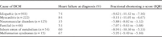
Prevalence of congestive heart failure and median fractional shortening z-scores for 1426 children with dilated cardiomyopathy at the time of diagnosis.
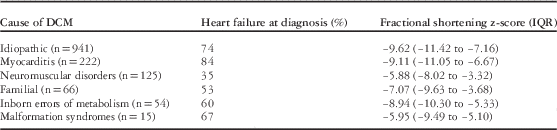
DCM=dilated cardiomyopathy; IQR=interquartile range
Data are from the Pediatric Cardiomyopathy Registry
In addition to examining the variants of the G4.5 mutation, the same study also analysed possible viral aetiologies using myocardial tissue samples from 44 children.Reference Towbin, Sleeper and Jefferies 10 Viral genome analysis of the tissues was positive for Epstein–Barr virus (4.5%) and for parvovirus B19 (13.6%). Tests for cytomegalovirus, adenovirus, and enterovirus were negative, which may indicate that these viruses are rarely seen in viral myocarditis.
Dilated cardiomyopathy
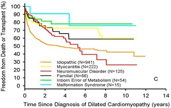
Outcomes for children with dilated cardiomyopathy depend on an aggregate of disease aetiology, age at diagnosis, echocardiographic parameters, and heart failure at presentation. A particular study of the Pediatric Cardiomyopathy Registry showed that children with dilated cardiomyopathy had a 31% death or transplant rate at 1 year, increasing to 46% at 5 years of age;Reference Towbin, Lowe and Colan 3 however, these rates showed wide variation based on the aetiology of the disease (Fig 1). Only 34% of children with dilated cardiomyopathy had a known aetiology: 16% myocarditis, 9% neuromuscular disorders, 5% familial, 4% inborn errors of metabolism, and 1% malformation syndromes. A separate analysis of the Pediatric Cardiomyopathy Registry found that a family history, older age at diagnosis, smaller left ventricular end-diastolic and end-systolic dimensions, higher left ventricular shortening fraction, viral serologic testing, and endomyocardial or skeletal biopsy were associated with the establishment of a causal diagnosis for children with dilated cardiomyopathy.Reference Cox, Sleeper and Lowe 9
Freedom from death or transplantation for 1423 children with dilated cardiomyopathy, by cause. Data are from the Pediatric Cardiomyopathy Registry collected between 1990 and 2002 (reproduced with permission from Towbin et alReference Towbin, Lowe and Colan 3 ).

Overall, 71% of children with dilated cardiomyopathy presented with congestive heart failure at diagnosis, although the percentage of those with congestive heart failure varied by aetiology (Table 4).Reference Towbin, Lowe and Colan 3 In addition, children with dilated cardiomyopathy who were under the age of 6 had better survival rates than older children (p<0.001). We found that congestive heart failure at the time of diagnosis and lower left ventricular fractional shortening z-scores were predictors of worse outcomes in patients with idiopathic dilated cardiomyopathy.Reference Alvarez, Orav and Wilkinson 11
Although children with dilated cardiomyopathy often have poor outcomes, normalisation of systolic function and ventricular size was demonstrated in 22% of children with dilated cardiomyopathy within 2 years of diagnosis in the Pediatric Cardiomyopathy Registry.Reference Everitt, Sleeper and Lu 12 Predictors of normalisation of ventricular function included younger age and lower left ventricular dilation at diagnosis; however, a small percentage of children who experienced recovery later died or were transplanted, which may indicate the need for close follow-up of these patients.
Analysis of the Pediatric Cardiomyopathy Registry data at the time of diagnosis and up to 12 months later showed that children with dilated cardiomyopathy who survived at least for 1 year had a significant decrease in left ventricular end-diastolic dimension and an increase in left ventricular thickness-to-dimension ratio, consistent with favourable left ventricular re-modelling.Reference Kantor, Orav and Wilkinson 13 In addition, children with dilated cardiomyopathy who survived at least for 1 year had a significant improvement in left ventricular function compared with those who died or received a heart transplant during the 12 months after diagnosis.
Before the Pediatric Cardiomyopathy Registry, little was known regarding risk factors for sudden cardiac death in children with dilated cardiomyopathy. We found that the 5-year cumulative incidence of sudden cardiac death in children with dilated cardiomyopathy in the Pediatric Cardiomyopathy Registry was only 2.4%.Reference Pahl, Sleeper and Canter 14 Factors that increased this risk included presence of congestive heart failure and anti-arrhythmic medications. The same study helped develop a model to aid clinicians in identifying children who may be at high risk for sudden cardiac death (sensitivity 86%, specificity 57%). Based on this model, children with dilated cardiomyopathy who have a left ventricular end-systolic dimension z-score greater than 2.6, an age at diagnosis <14.3 years, and a left ventricular posterior wall thickness-to-dimension ratio <0.14 are considered to be at high risk for sudden cardiac death.
A competing risks model, stratified by aetiology, was used to identify differential predictors of death or transplantation in children with dilated cardiomyopathy.Reference Alvarez, Wilkinson and Lipshultz 15 For children with neuromuscular disorders and dilated cardiomyopathy, decreased left ventricular fractional shortening was associated with both death and heart transplantation. In addition, higher left ventricular end-diastolic dimension predicted transplantation in the same group. In children with myocarditis and dilated cardiomyopathy, an increased risk of death or transplantation was associated with an older age at diagnosis, the presence of congestive heart failure, and an increased left ventricular end-diastolic dimension. When comparing children with familial dilated cardiomyopathy, the risk of death or transplantation was increased in those who had congestive heart failure at diagnosis and lower left ventricular fractional shortening z-scores.
Overall, 26% of children with dilated cardiomyopathy in the Pediatric Cardiomyopathy Registry with a known cause of cardiomyopathy had an underlying neuromuscular disorder.Reference Towbin, Lowe and Colan 3 The primary neuromuscular disorders observed in the cohort were Duchenne muscular dystrophy and Becker muscular dystrophy. In comparing survival rates for children with each type of muscular dystrophy, we found that children with Becker muscular dystrophy had better survival rates at 5 years than those with Duchenne muscular dystrophy (57 versus 100%, respectively). On the other hand, patients with Becker-type muscular dystrophy had a higher transplantation rate than patients with Duchenne muscular dystrophy (25 versus 0%, respectively).Reference Connuck, Sleeper and Colan 16 The same analysis also compared patients with a neuromuscular disorder – either Duchenne- or Becker-type muscular dystrophy – and patients with non-neuromuscular disease-related dilated cardiomyopathies to examine differences in outcomes. Survival did not differ significantly between children with a neuromuscular disorder and children with other non-neuromuscular dilated cardiomyopathies (59 versus 71% at 5 years, respectively).
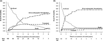
Myocarditis accounts for almost 50% of cases of dilated cardiomyopathy with a known cause of disease in the Pediatric Cardiomyopathy Registry.Reference Cox, Sleeper and Lowe 9 An analysis of the Pediatric Cardiomyopathy Registry showed no differences in the outcomes for children with probable myocarditis and biopsy-confirmed cases of myocarditis (Fig 2).Reference Foerster, Canter and Cinar 17 In children with myocarditis and dilated cardiomyopathy, normalisation of echocardiographic findings occurred in nearly half the patients, and the mortality and transplant rates were much lower, when compared with idiopathic dilated cardiomyopathy. Lower left ventricular fractional shortening at presentation predicted mortality, whereas a higher left ventricular posterior wall thickness predicted transplantation in this group.
Overall, 14% of children with a known cause of dilated cardiomyopathy were found to have familial isolated cardiomyopathy, defined as having two or more family members with a history of cardiomyopathy.Reference Towbin, Lowe and Colan 3 The Pediatric Cardiomyopathy Registry found that 1-year survival was significantly better in patients with familial dilated cardiomyopathy (87%) compared with idiopathic dilated cardiomyopathy (60%).Reference Rusconi, Wilkinson and Sleeper 18 In addition, patients with familial isolated cardiomyopathy had higher left ventricular ejection fractions, lower end-diastolic and end-systolic dimensions, and lower left ventricular mass than those with idiopathic dilated cardiomyopathy. Left ventricular end-diastolic dimension z-scores predicted an increased risk of death or transplantation in children with familial isolated cardiomyopathy.
Hypertrophic cardiomyopathy
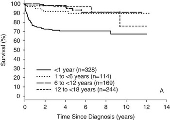
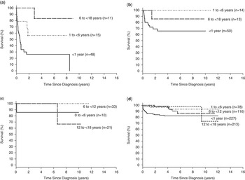
Outcomes for children with hypertrophic cardiomyopathy varied widely with age and cause of diagnosis (Figs 3 and 4).Reference Colan, Lipshultz and Lowe 19 In one study of the Pediatric Cardiomyopathy Registry, only 26% of children with hypertrophic cardiomyopathy had a known aetiology.Reference Colan, Lipshultz and Lowe 19 For the 26% of patients with a known aetiology, causes of diagnosis included malformation syndromes (9%), inborn errors of metabolism (9%), and neuromuscular disorders (8%). Decreased height and weight for age, sex (female), and higher posterior wall thickness were associated with establishing a causal diagnosis.Reference Cox, Sleeper and Lowe 9 Left ventricular fractional shortening and prevalence of congestive heart failure varied greatly by the aetiological group (Table 5).Reference Colan, Lipshultz and Lowe 19
Crude cumulative incidences of echocardiographic normalisation, cardiac transplantation, and death among children with myocarditis – combined biopsy-confirmed myocarditis and probable myocarditis groups – and abnormal function at presentation with left ventricular end-diastolic dilation at diagnosis ( a ) or no left ventricular end-diastolic dilation at diagnosis ( b ). The two groups differed in the incidence of cardiac transplant (p=0.02) and echocardiographic normalisation rates (p<0.001) but not mortality (p=0.45). Curves are truncated at 8 years (reprinted with permission from Foerster et alReference Foerster, Canter and Cinar 17 ).

Freedom from death or transplantation for 855 children with idiopathic hypertrophic cardiomyopathy, by age at diagnosis. Data are from the Pediatric Cardiomyopathy Registry collected between 1990 and 2002 (reproduced with permission from Colan et alReference Colan, Lipshultz and Lowe 19 ).

Survival rates from the date of diagnosis of cardiomyopathy in children with ( a ) inborn errors of metabolism (n=74, logrank p<0.001); ( b ) malformation syndromes (n=77, logrank p=0.07); ( c ) neuromuscular disease (n=64, logrank p=0.22); and ( d ) idiopathic hypertrophic cardiomyopathy (n=634, logrank p<0.001), by age at diagnosis (reproduced with permission from Colan et alReference Colan, Lipshultz and Lowe 19 )

Prevalence of congestive heart failure and mean fractional shortening z-scores for 849 children with hypertrophic cardiomyopathy at the time of diagnosis.

HCM=hypertrophic cardiomyopathy; IQR=interquartile range
Data are from the Pediatric Cardiomyopathy Registry
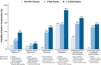
The poorest outcomes for children with hypertrophic cardiomyopathy were seen in those with a concurrent diagnosis of hypertrophic cardiomyopathy and inborn errors of metabolism (2-year rate of death or transplantation, 57%).Reference Lipshultz, Orav and Wilkinson 20 Children with mixed hypertrophic and dilated cardiomyopathy or mixed hypertrophic and restrictive cardiomyopathy also had high rates of death or transplantation at 2 years (45 and 38%, respectively). Although risk factors for death or transplantation differed by the aetiology of hypertrophic cardiomyopathy, in general, these included younger age, lower weight, presence of congestive heart failure, increased left ventricular posterior wall thickness, decreased left ventricular fractional shortening, increased left ventricular septal thickness, or the presence of any two or more of these risk factors (Fig 5). The risk of death or transplantation increased significantly as the number of risk factors increased.
Risk of death or transplantation in 882 children with hypertrophic cardiomyopathy, by number of cause-specific risk factors. Data are from the Pediatric Cardiomyopathy Registry collected between 1990 and 2002 (from Lipshultz et alReference Lipshultz, Orav and Wilkinson 20 ).

Results from the Pediatric Cardiomyopathy Registry showed that children with Noonan syndrome and hypertrophic cardiomyopathy were more likely to present in the first 6 months of life, were three times more likely to present with heart failure, and had a significantly higher mortality in the 1st year of life compared with other children with hypertrophic cardiomyopathy.Reference Wilkinson, Lowe and Salbert 21 This increased mortality rate was at least partially attributable to the younger age and increased prevalence of congestive heart failure in the children with Noonan syndrome compared with other children with hypertrophic cardiomyopathy. Children with Noonan syndrome and hypertrophic cardiomyopathy who presented with congestive heart failure in the first 6 months of life were at the highest risk for mortality. Children with Noonan syndrome and hypertrophic cardiomyopathy in the Pediatric Cardiomyopathy Registry were rarely listed and none of them received a heart transplant. This group should be considered for listing for heart transplantation at the time of diagnosis in the absence of contraindications for listing.
Inborn errors of metabolism accounted for 27% of the cases of hypertrophic cardiomyopathy with a known cause of disease in the Pediatric Cardiomyopathy Registry.Reference Cox, Sleeper and Lowe 9 Almost 50% of children with cardiomyopathy caused by inborn errors of metabolism had an unspecified disorder. Across all cardiomyopathy phenotypes, the most common mitochondrial disorder was Barth syndrome (27%). Poor survival rates were observed in children with hypertrophic cardiomyopathy caused by a metabolic gene deficiency, with a 2-year survival rate of only 51%. In addition, a diagnosis of cardiomyopathy before 1 year of age was associated with an increased risk of death in patients with inborn errors of metabolism who present with hypertrophic cardiomyopathy.
Restrictive cardiomyopathy
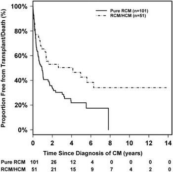
Restrictive cardiomyopathy is the least common form of all phenotypes, accounting for only 4.5% of all cardiomyopathies, and can occur as an isolated phenotype or as a mixed phenotype with hypertrophic cardiomyopathy.Reference Webber, Lipshultz and Sleeper 22 The mixed restrictive and hypertrophic cardiomyopathy phenotype comprised 33% of all restrictive cardiomyopathies in the Pediatric Cardiomyopathy Registry. Children with restrictive cardiomyopathy were found to have the poorest outcomes compared with other phenotypes of cardiomyopathy (Fig 6). The 5-year survival was 68% for children with both the mixed restrictive and hypertrophic cardiomyopathy and pure restrictive cardiomyopathy phenotypes. In addition, transplant-free survival was only 28% for pure restrictive cardiomyopathy vsersus 43% for the mixed phenotype. Risk factors for poor survival included the presence of congestive heart failure at diagnosis, a decreased left ventricular fractional shortening z-score for both phenotypic groups, as well as an increased left ventricular posterior wall thickness z-score for children with the mixed phenotype.
Left ventricular non-compaction
Left ventricular non-compaction is one of the rarest phenotypes of cardiomyopathy in children with an incidence of only 4.9%, according to an analysis of the Pediatric Cardiomyopathy Registry.Reference Jefferies, Colan and Sleeper 23 Left ventricular non-compaction in children can present in isolation or with other cardiomyopathy phenotypes. Diagnosis of this phenotype is often made on the basis of echocardiographic findings, although at present there is no clear consensus on diagnostic criteria. A decreased left ventricular fractional shortening z-score was a strong predictor of death or transplantation in children with left ventricular non-compaction; however, when the left ventricular systolic function was preserved, children with left ventricular non-compaction had better outcomes than children with dilated or restrictive cardiomyopathy.
Treatment of cardiomyopathies in children
Medical management
Pharmacological treatment patterns were compared between Pediatric Cardiomyopathy Registry patients diagnosed with idiopathic dilated cardiomyopathy between 1990 and1995 and those diagnosed between 2000 and 2006.Reference Harmon, Sleeper and Cuniberti 24 We found that the use of heart failure medications, including digoxin, a diuretic, or both (84% of children diagnosed between 1990 and 1995 and 87% of children diagnosed between 2000 and 2006), as well as angiotensin-converting enzyme inhibitors (66 and 70%, respectively) did not change significantly between the two time periods; however, β-blocker usage increased from 4 to 18%. Anti-heart-failure therapy remains the most common treatment, and was reported in 84% of all children with idiopathic dilated cardiomyopathy. Anti-heart-failure medications were reported in 60% of children with asymptomatic heart failure and in 93% of children with an NYHA class greater than II. Angiotensin-converting enzyme inhibitors were reported in 74% of children; however, in contrast to anti-heart-failure therapies, angiotensin-converting enzyme inhibitor therapy was often not initiated in children with asymptomatic left ventricular dysfunction in the absence of echocardiographic evidence of more advanced disease.
Surgical management
For many children with severe or end-stage cardiomyopathy, heart transplantation remains one of the only long-term surgical solutions.Reference Kirklin, McGiffin and Dabal 25 The Pediatric Heart Transplant Study Group collects morbidity and mortality data on children under 18 years of age beginning at the date of listing for heart transplantation.Reference Hsu, Naftel and Webber 26 As the Pediatric Cardiomyopathy Registry does not collect data on patients after transplantation, collaboration with the Pediatric Heart Transplant Study Group gave the Pediatric Cardiomyopathy Registry Study Group the ability to examine patient outcomes before and after transplantation.
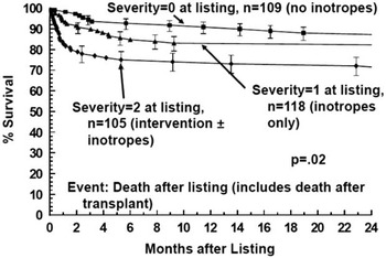
An analysis of the combined Pediatric Heart Transplant Study and Pediatric Cardiomyopathy Registry data sets examined mortality in children with varying levels of support before transplantation (Fig 7).Reference Larsen, Canter and Naftel 27 For children with no pre-transplant circulatory or mechanical support, mortality primarily occurred after transplantation. Although we observed no difference in pre- and post-transplant mortality rates in children who received only inotropic support, for children requiring both circulatory and mechanical support, mortality occurred most frequently while on the waiting list for heart transplantation. This suggests that patients with cardiomyopathy requiring higher degree of support at listing might have a survival benefit with transplantation compared with children who require no support or only inotropic support before transplantation.
Heart transplantation was found to be most common among children with dilated cardiomyopathy in the combined Pediatric Cardiomyopathy Registry and Pediatric Heart Transplant Study data set.Reference Canter and Kantor 28 For patients with dilated cardiomyopathy who were listed for transplant, the median age was 3.4 years at diagnosis and 4.4 years at transplantation; 30% of these patients were ventilator-dependent at the time of listing, and pre-transplant mortality was 11%.Reference Pietra, Kantor and Bartlett 29 Mechanical ventilation and older age at diagnosis – without mechanical ventilation – were predictors of mortality while waiting for a heart transplant. Post-transplant outcomes were worse for children with myocarditis, which suggests that infectious or immune mechanisms affected patient mortality.
A separate analysis of the combined Pediatric Cardiomyopathy Registry and Pediatric Heart Transplant Study data set examined the association between measures of left ventricular dilation or function and the risk of death in children with dilated cardiomyopathy after listing for transplantation or within 6 months of transplantation. Increased left ventricular end-diastolic dimension close to the time of listing was independently associated with an increased risk of death after listing or up to 6 months after transplant for children with dilated cardiomyopathy who were under 5 years of age at diagnosis.Reference Singh, Sleeper and Lipshultz 30 This association was not observed in children who were older than age 5 at diagnosis; however, the association was strongest for those diagnosed before 6 months of age. Left ventricular function and mass were not associated with an increased risk of death after listing or within 6 months of transplantation.
Functional status of children with cardiomyopathy
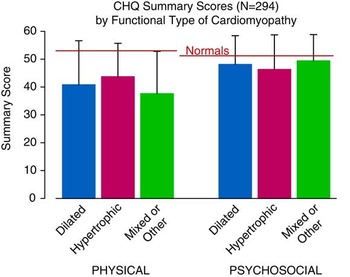
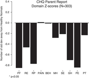
Few studies have systematically studied the functional status in children with cardiomyopathy and their corresponding quality of life in comparison with healthy children. We collected parent-reported functional status of children with cardiomyopathy above 5 years of age using two validated surveys: the Child Health Questionnaire and the Functional Status II(R) Reports Instrument. On average, children with cardiomyopathy had impaired physical and psychosocial functioning compared with healthy children (Figs 8 and 9).Reference Sleeper, Colan and Towbin 31 Several socioe-conomic variables were associated with impaired functional status – for example, lower total household income, lower parental education level, etc. Predictors of poorer physical function specifically included increased left ventricular size and higher left ventricular thickness-to-dimension ratio; however, improved functional status was directly associated with time since diagnosis, suggesting that functional status in children with cardiomyopathy may improve with time.
Probability of freedom from death, censored at transplantation, among 152 children with RCM stratified by phenotype, pure RCM versus mixed/overlapping phenotype RCM/HCM. CM=cardiomyopathy; HCM=hypertrophic cardiomyopathy; and RCM=restrictive cardiomyopathy (reprinted with permission from Webber et alReference Webber, Lipshultz and Sleeper 22 ).

The Pediatric Cardiomyopathy Registry in the present era
At present, the Pediatric Cardiomyopathy Registry is funded by the National Heart, Lung, and Blood Institute with supplemental funding from the Children’s Cardiomyopathy Foundation to conduct two new studies. The first study, PCM Genes – Genotype–Phenotype Associations in Pediatric Cardiomyopathy – has at present enrolled more than 400 children with dilated cardiomyopathy, hypertrophic cardiomyopathy, restrictive cardiomyopathy, or left ventricular non-compaction, with an enrolment goal of 600, who will receive next-generation whole-exome sequencing for both gene discovery as well as genotype–phenotype associations. The phenotypes of interest include cardiomyopathy functional type, age at onset, as well as 2-year echocardiographic outcomes. The PCM Genes study is also enrolling parents and affected relatives of the probands to better understand inheritance patterns and moderator gene influences.
The second study named PCM Biomarkers – Cardiac Biomarkers in Pediatric Cardiomyopathy – which is at present enrolling up to 400 children with dilated cardiomyopathy or hypertrophic cardiomyopathy has the following three aims: to determine the association of specific cardiac biomarkers and echocardiographic parameters in accurately assessing newly diagnosed dilated cardiomyopathy patients’ clinical status; to determine the association of biomarkers of cardiac fibrosis with fibrosis as measured on cMRI in children with hypertrophic cardiomyopathy; and to determine which cardiac biomarkers are associated in children with either dilated cardiomyopathy or hypertrophic cardiomyopathy who continue to be clinically stable up to 2 years after diagnosis to better inform transplantation listing decisions.
Both of these studies in progress will end enrolment by 2016, and their results will be disseminated for the next generation of paediatric cardiomyopathy research proposals, which will focus on early diagnosis and disease screening, targeted treatment interventions via randomised clinical trials, and improved patient and family support interventions. We are also proud that during its more than two decades of study, the Pediatric Cardiomyopathy Registry has also helped train the next generation of paediatric cardiomyopathy and heart failure investigators who will carry on this important research.
In addition, the Pediatric Cardiomyopathy Registry Study Group is at present conducting ancillary analyses of the Pediatric Cardiomyopathy Registry database to determine the importance of renal function, growth and body composition, electrophysiological parameters, and pharmacological interventions as determinants of clinical outcomes in this population.
Pediatric Cardiomyopathy Study Group-sponsored international conferences on paediatric cardiomyopathy
The first International Workshop on Idiopathic and Primary Pediatric Cardiomyopathies was organised by the Pediatric Cardiomyopathy Registry investigators in 2007. More than 30 participants attended the conference, which was co-sponsored by the Children’s Cardiomyopathy Foundation and the National Heart, Lung, and Blood Institute. The results of the conference were published in three issues of the journal Progress in Pediatric Cardiology.Reference Alvarez, Wilkinson and Lipshultz 15 , Reference Canter and Kantor 28 , Reference Alcalai, Arad and Depreux 32 – Reference Young and Hare 63 The Second International Conference on Cardiomyopathy in Children was held in May, 2010, with more than 50 participants in attendance. The Children’s Cardiomyopathy Foundation and the National Heart, Lung, and Blood Institute were also the principle co-sponsors for this conference. Similar to the 2007 conference, the results from the second conference were published in three issues of Progress in Pediatric Cardiology.Reference Kirklin, McGiffin and Dabal 25 , Reference Bernstein, Fajardo and Zhao 64 – Reference Wilkinson, Zebrowski and Hunter 98 The Third International Conference on Cardiomyopathy in Children was held in May, 2014 and was attended by more than 60 world experts. Again, the principle co-sponsors for this conference were the Children’s Cardiomyopathy Foundation and the National Heart, Lung, and Blood Institute. The third conference added new themes focussed on paediatric heart failure, the next generation of left ventricular support devices, novel cardiac imaging, and personalised medicine for children with cardiomyopathy and heart failure. The main outcome of this third conference was to propose a “future research agenda” in paediatric cardiomyopathy and heart failure to be presented to the National Heart, Lung, and Blood Institute and patterned on the recently published results from a National Heart, Lung, and Blood Institute Working Group on paediatric heart failure.Reference Burns, Byrne and Gelb 99 The results of the third conference will be published in the current and future issues of Progress in Pediatric Cardiology in 2014 and 2015, including the proposed future research agenda for paediatric cardiomyopathy and heart failure.Reference Asimaki, Kleber, MacRae and Saffitz 100 – Reference Wittlieb-Weber, Lin and Zaoutis 111
Future directions of the Pediatric Cardiomyopathy Registry
With the continual collection of prospective follow-up data from enrolled Pediatric Cardiomyopathy Registry patients, Pediatric Cardiomyopathy Registry Study Group researchers will be able to provide a more complete description of the clinical course of paediatric cardiomyopathy and the associated risk factors. This will also allow for a more thorough examination of previously identified risk factors and a better understanding of their long-term utility in the diagnosis, prognosis, and care of these children. This type of registry data and their usefulness in guiding clinical decision-making is continuing to gain appreciation from research methodologists and is becoming increasingly useful as advances in analytical and statistical theory are made.Reference Dreyer and Garner 112 Pediatric Cardiomyopathy Registry investigators are at present working to proposing to leverage the Pediatric Cardiomyopathy Registry infrastructure to identify the genetic aetiologies of paediatric cardiomyopathy and the usefulness of cardiac biomarkers in the evaluation of these patients. The ultimate goal of the Pediatric Cardiomyopathy Registry is to identify robust diagnostic strategies, aetiology-specific treatments informed by genotyping, and the most clinically effective evidence-based approaches for these children. An associated goal is to recruit and develop the next generation of paediatric cardiomyopathy and heart failure researchers and clinicians.
Survival after listing for heart transplantation among children with cardiomyopathy by heart failure severity score. Severity score 2=children on mechanical ventilatory or circulatory support; 1=children on intravenous inotropic support without mechanical support; 0=children on neither intravenous inotropic nor mechanical support (reprinted with permission from Larsen et alReference Larsen, Canter and Naftel 27 ).

Physical and psychosocial function of 294 children with cardiomyopathy, by functional type, compared with scores for healthy children. Data are from the Child Health Questionnaire administered by the Pediatric Cardiomyopathy Registry between 1990 and 2002 (from Sleeper et alReference Sleeper, Colan and Towbin 31 ).

Mean Child Health Questionnaire (CHQ) z-scores for 303 children with cardiomyopathy, by domain. Data are from the Pediatric Cardiomyopathy Registry and were collected between 1990 and 2002. PF=physical functioning; RE=role/social limits-emotional; RP=role/social limits-physical; PAIN= bodily pain; BEH=behaviour; MH=mental health; SE=self-esteem; GH=general health perception; PE=parental impact-emotional; PT=parental impact-time (from Sleeper et alReference Sleeper, Colan and Towbin 31 ).

Acknowledgements
The work of the Pediatric Cardiomyopathy Registry would not have been possible without the collaboration of many physicians and other healthcare professionals, scientists, and research staff from the United States and Canada. Special acknowledgement should be given to the Pediatric Cardiomyopathy Registry investigators: Steven D. Colan, MD; Gerald Cox MD, PhD; Jane Messere, RN; Lynn Sleeper, ScD; Jeffrey A. Towbin, MD; Stephanie Ware, MD, PhD; John Lynn Jefferies, MD, MPH; E. John Orav, PhD; Daphne Hsu, MD; Steven Webber, MBChB; Charles Canter, MD; Linda Addonizio, MD; Beth Kaufman, MD; Melanie Everitt, MD; Elfriede Pahl, MD; Paul Kantor, MBBCh; Paolo Rusconi, MD; Robert E. Shaddy, MD; Paul R. Lurie, MD; David Connuck, MD; April Lowe, MS; Bonnie Salbert, DO; Tajinder Singh, MD, MSc; Jorge Alvarez, AB; Biagio Pietra, MD; Ranae Larsen, MD; Jacqueline Lamour, MD; Renee Margossian, MD; Beth Kaufman, MD; Susan Foerster, MD; Wendy Chung, MD; Rakesh Singh, MD, MS; and Juanita Hunter, MD. The authors acknowledge Mrs Lisa Yue and the Children’s Cardiomyopathy Foundation for their continuing support of the Pediatric Cardiomyopathy Registry. They also acknowledge the Parker Foundation for their support of the Registry. In addition, the authors acknowledge the Myocarditis Foundation, GeneDx, and the Department of Pediatrics of the Miller School of Medicine at the University of Miami for their support of the Second International Conference on Cardiomyopathy in Children, as well as the Macquarie Group Foundation, Global Productivity Solutions, MyoKardia, Berlin Heart, and the Children’s Hospital of Pittsburgh of the University of Pittsburgh Medical Center for their support of the Third International Conference on Cardiomyopathy in Children. Finally, the authors express their most sincere gratitude to the children with cardiomyopathy and their families whose participation has made the Pediatric Cardiomyopathy Registry possible.
Financial Support
This work was principally supported by the National Heart, Lung, and Blood Institute (S.E.L., grant numbers R01HL53392, R13HL087708, and R13HL123256) and the Children’s Cardiomyopathy Foundation. Additional support of this work is included in the acknowledgements section.
Conflicts of Interest
None.
Ethical Standards
The authors assert that all procedures contributing to this work comply with the ethical standards of the relevant national guidelines on human experimentation The U.S. Department of Health and Human Services Title 45, Part 46 Protection of Human Subjects, ‘The Common Rule’ and with the Helsinki Declaration of 1975, as revised in 2008, and has been approved by the institutional committees.