Introduction
The cochlea is an essential neurosensory component in peripheral perception of acoustic signals. It consists of several complex epithelial structures with sensory and supporting cells attached to a basal membrane.Reference Oliveira, Canedo and Rossato1 In humans, cochlear injury is almost always irreversible and can have serious consequences, such as decreased ability to communicate and a compromised social life.
Cisplatin (cis-diamminedichloroplatinum) is commonly used in the treatment of a variety of neoplasms, especially those of the head and neck.Reference Jordan, Schwade and Truelson2 Its side effects include ototoxicity, nephrotoxicity, marrow suppression and gastrointestinal disorders.Reference Güneri, Serbetçiogllu, Ikiz, Güneri and Ceryan3 In some patients, the occurrence of toxicity makes it necessary to reduce the dosage, frequency and duration of chemotherapy.Reference Feghali, Liu and Water4
Most studies on rodent ototoxicity employ scanning electron microscopy, a reliable method of identifying ciliated cell injury.Reference Campbell, Rybac and Meech5–Reference Fetoni, Quaranta, Marchese, Cadoni, Paludetti and Sergi10 Rat cochleae may also be evaluated by transmission electron microscopyReference Campbell, Meech, Rybak and Hughes11 or by using extracts from surface tissue of the organ of Corti.Reference Laurell, Viberg, Teixeira, Sterkers and Ferrary12 The latter procedure, known as cytocochleography, focuses on ciliated cells.Reference Laurell, Viberg, Teixeira, Sterkers and Ferrary12
In 1997, de Groot et al. Reference De Groot, Hamers, Gispen and Smoorenburg13 described a technique for evaluating cisplatin-induced ototoxicity in guinea pigs, using light microscopy. On the day following completion of drug therapy, the temporal bones of the guinea pigs were retrieved and the cochleae fixed with intralabyrinthine injection and immersion in a trialdehyde fixative (3 per cent glutaraldehyde, 2 per cent formaldehyde, 1 per cent acrolein and 2.5 per cent dimethylsulphoxide in 0.1 M cacodylate buffer at pH 7.4). After decalcification with 4 per cent ethylene diamine triacetic acid (EDTA) for four to five days, samples were post-fixed for two hours at 4oC in a solution prepared with 1 per cent osmium tetroxide and 1 per cent K4Ru(CN)6. Histological stains were prepared with 1 per cent methylene blue and 1 per cent azur II in 1 per cent sodium tetraborate to enable identification of injury to the organ of Corti. Such injury was characterised by architectural changes, filling of the space of Nuel by Deiters' cells, loss of external ciliated cells and distension of Reissner's membrane suggestive of endolymphatic hydrops. Injury was most evident in the basal turn of the cochlear duct and decreased towards the apical turn. No changes were observed in the stria vascularis or spiral ganglion. Heijmen et al. Reference Heijmen, Klis, De Groot and Smoorenburg14 and Cardinaal et al. Reference Cardinaal, De Groot, Huizing, Veldman and Smoorenburg15–Reference Cardinaal, De Groot, Huizing, Veldman and Smoorenburg17 used the same method and reported similar findings.
Sergi et al. Reference Sergi, Ferraresi, Troiani, Paludetti and Fetoni18 also observed injury to external ciliated cells in the presence of preserved internal ciliated cells and spiral ganglion in albino guinea pigs. The histological procedures used in this study included fixation with 2.5 per cent glutaraldehyde and 2 per cent paraformaldehyde, decalcification with 10 per cent EDTA for five days, post-fixation with 1 per cent osmium tetroxide and staining with 0.05 per cent toluidine blue.
Few studies have used light microscopy to evaluate cisplatin-induced ototoxicity in rats. When staining with toluidine blue, Campbell et al. Reference Campbell, Meech, Rybak and Hughes11 observed protrusion of stria vascularis marginal cells into the endolymphatic space, induced by a single dose of cisplatin (16 mg/kg). Lynch et al. Reference Lynch, Gu, Pierce and Kil19 prepared rat cochleae using fixation with 4 per cent paraformaldehyde, decalcification with 0.5 M EDTA for one week, and staining with haematoxylin and eosin, and found that a single intraperitoneal dose of cisplatin (16 mg/kg) produced an increase in the stria vascularis area, probably due to oedema.
The objective of the present study was to describe a simple method of histological assessment of cisplatin-induced ototoxicity in rats, and to relate morphological changes to functional changes in hearing as assessed by distortion product evoked otoacoustic emissions.
Materials and methods
Animals
The study employed Wistar rats weighing 200–348 g from the laboratory of the Federal University of Ceará. The animals were housed in cages with free access to water and food and were exposed to a 24-hour circadian rhythm, according to the guidelines of the Brazilian Council on Animal Experiments (www.cobea.org.br). The study was approved by the ethics committee for animal research of the Federal University of Ceará medical school (protocol #28/05).
Exclusion criteria
Exclusion criteria were as follows: otoscopically detectable external ear abnormalities (e.g. oedema and hyperaemia of the external auditory canal, tumour growths or cerumen impaction); signs of middle-ear disease (e.g. bulging, opacification, hyperaemia or tympanic membrane perforation); and absence of distortion product evoked otoacoustic emissions at any of the frequency ranges tested.
Drugs
The study used 50 mg cisplatin doses (as CisplatexTM lyophilised powder for reconstitution and intravenous administration; Eurofarma, São Paulo, SP, Brazil). Fifty milligrams of the lyophilised powder was diluted in 50 ml saline solution to give a concentration of 1 mg/ml. The study also used 0.9 per cent saline solution, ketamine 50 mg/ml (VetarnacolTM; König, São Paulo, SP, Brazil), and xylazine 20 mg/ml (KensolTM; König, São Paulo, SP, Brazil).
Study groups
The rats were divided into four groups.
In group one (n = 11), rats were treated with 8 mg/kg/day cisplatin for three consecutive days (total = 24 mg/kg) and evaluated with distortion product evoked otoacoustic emission testing at baseline and three days later (D3).
In group two (n = 6), rats were treated with 8 ml/kg/day saline solution for three consecutive days (total = 24 ml/kg) and evaluated with distortion product evoked otoacoustic emission testing at baseline and three days later.
In group three (n = 8), rats were treated with 8 mg/kg/day cisplatin for three consecutive days (total = 24 mg/kg) and evaluated with distortion product evoked otoacoustic emission testing at baseline and four days later (D4).
In group four (n = 6), rats were treated with 8 ml/kg/day saline solution for three consecutive days (total = 24 ml/kg) and evaluated with distortion product evoked otoacoustic emission testing at baseline and four days later.
Procedures
Immediately before drug therapy, rats with normal otoscopic findings were anaesthetised deeply with 50 mg/kg ketamine plus 10 mg/kg xylazine and subjected to distortion product evoked otoacoustic emission testing. (Animals with external- or middle-ear abnormalities had previously been identified by otoscopy and excluded from the study.) The rats then received their allocated therapy (either 8 mg/kg cisplatin or 8 ml/kg saline solution, intraperitoneally for three consecutive days; totals of 24 mg/kg or 24 ml/kg, respectively). Either 24 or 48 hours after their last injection, the animals were again anaesthetised and examined otoscopically to exclude new external- or middle-ear abnormalities. Those with normal otoscopic findings were again subjected to distortion product evoked otoacoustic emission testing. Immediately after the second distortion product evoked otoacoustic emission test, the animals were decapitated with a custom-built guillotine and the right temporal bone was retrieved. The cochlea was dissected and prepared for light microscopy.
Distortion product evoked otoacoustic emission testing
Distortion product evoked otoacoustic emissions were tested with a Madsen Capella device (GN Otometrics, Taastrup, Denmark) in a silent room. An infant hearing screening probe was attached to the external auditory canal. The stimulus consisted of two pure tones (F1 and F2; F1/F2 ratio = 1.22) at 70 dB SPL. A total of 1000 acquisitions were analysed. The resultant otoacoustic emissions were evaluated at 3, 4, 6 and 8 kHz. Distortion product evoked otoacoustic emission testing was considered positive for signal to noise ratios of ≥6 dB SPL, as specified by the manufacturer.
Light microscopy technique
The retrieved temporal bone was immediately immersed in 10 per cent buffered formaldehyde in a Petri dish. Using a surgical microscope (DF Vasconcelos, São Paulo, SP, Brazil) with the lens at position 16, the tympanic membrane and the ossicular chain (malleus, incus and stapes) were removed and the otic capsule exposed. A small opening was made at the cochlear apex using a 1-ml syringe with a 13-mm long and 4.5-mm wide needle. A curved scalpel was introduced through the round and oval windows and a fixative (10 per cent buffered formaldehyde) injected with the same syringe and needle. The dissected cochleae remained immersed in 10 per cent buffered formaldehyde for 24 hours. After fixation, they were decalcified with 10 per cent EDTA for seven days, then rinsed in tap water for 24 hours and dried in increasing alcohol concentrations (70, 80, 90 and 100 per cent, one hour each). The cochleae were transferred from the last alcohol bath to a Petri dish containing alcohol of similar concentration. Using a surgical microscope, the remaining extracochlear tissue was removed with a size 15 scalpel to help position samples longitudinally in relation to the sectioning angle during embedding. The material was clarified in xylene for one hour, impregnated with paraffin for three hours at 60°C and embedded.
Using a microtome (Olympus Cut 4055; Olympus, American Inc., San Jose, Canada), 20-μm sections were removed until the modiolus was reached. The basal, middle and apical turns were identified with a microscope (Olympus BX 41) at ×40 magnification. Subsequently, four 4-μm sections were retrieved for histological study. The sections were secured on slides with egg albumin and dried in an incubator at 37°C for 24 hours. The samples were then deparaffinised in xylene, hydrated in decreasing concentrations of alcohol (100, 90, 80 and 70 per cent) and rinsed in tap water. Finally, the samples were immersed in haematoxylin, rinsed in tap water, immersed in eosin, rinsed again, dehydrated in increasing concentrations of alcohol (70, 80, 90 and 100 per cent), immersed in xylene for clarification and mounted on slides with Entellan™ (Merck, Darmstadt, Germany).
A four-point scoring system was devised to grade injury to the stria vascularis and the organ of Corti, as visualised with a light microscope (Leica DMLS 2; Leica, Wetzlar, Germany) at ×400 magnification and photomicrographed with the Leica DFC 320 digital capture system. For the stria vascularis, scores indicated the degree of shrinkage of the intermediate cells. For the organ of Corti, scores were based on the number of external ciliated cells with intact nuclei.
Statistical analysis
The statistical analysis was undertaken using GraphPad Prism version 4.00.255 software. The normal distribution of the sample was evaluated using the Komogorov–Smirnov test. Results were expressed as mean values ± standard error of the mean (SEM) for continuous variables, and as median, minimum and maximum values for ordinal variables. The level of statistical significance was set at 5 per cent. Student's t-test was used to compare mean distortion product evoked otoacoustic emission amplitudes for each frequency, at baseline and after treatment. The Kruskal–Wallis test (with intergroup significance determined using Dunn's multiple comparison test) was used to compare each group's median scores for morphological changes to external ciliated cells and stria vascularis.
Results
Functional hearing evaluation
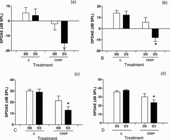
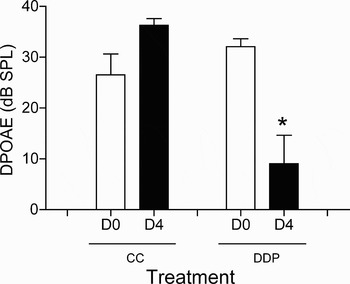
Distortion product evoked otoacoustic emission amplitudes decreased significantly in group one between baseline and D3 at the frequencies tested (3, 4, 6 and 8 kHz), compared with group two (the control group) (Figure 1). Mortality was high in group three on D4; at this time, only three of the eight animals treated with cisplatin were available for distortion product evoked otoacoustic emission testing. In two of these tests, emissions could not be detected due to a confirmed signal/noise difference below 6 dB SPL at 3 and 4 kHz. One animal had no measurable emissions at 6 kHz. Using a frequency of 8 kHz, distortion product evoked otoacoustic emission amplitudes were significantly lower on D4 than at baseline (Figure 2).
Distortion product evoked otoacoustic emission (DPOAE) average amplitudes (±standard error of the mean) in groups one and two, showing results at baseline (D0) and after three days (D3), for various test frequencies. Asterisks indicate statistically significant differences. (a) 3 kHz; *p = 0.0095, Student's t-test, comparing D0 and D3 for group one. (b) 4 kHz; *p = 0.0073, Student's t-test, comparing D0 and D3 for group one. (c) 6 kHz; *p = 0.0284, Student's t-test, comparing D0 and D3 for group one. (d) 8 kHz; *p = 0.0338, Student's t-test, comparing D0 and D3 for group one. C = control; CDDP = cisplatin

Distortion product evoked otoacoustic emission (DPOAE) average amplitudes (±standard error of the mean) at 8 kHz, in groups three and four, showing results at baseline (D0) and after four days (D4). Asterisks indicate statistically significant difference (*p = 0.0459, Student's t-test, comparing D0 and D4 for group three). C = control; CDDP = cisplatin

Morphological evaluation of ototoxicity
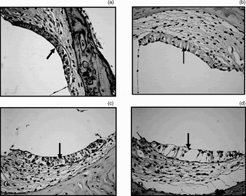
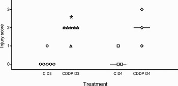
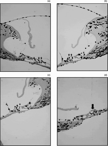
Some cochleae were lost during preparation, due either to fracture (three specimens from group one and one from group four) or malpositioning during paraffin embedding (one specimen from group one and one from group four). The changes observed were in the basal turn of the cochlear duct. A four-point scoring system was used to grade stria vascularis injury, with a score of zero indicating absence of shrinkage of the intermediate cells, and one, two and three indicating slight, moderate and severe shrinkage, respectively (Figure 3). On D3, the median score for stria vascularis injury was significantly greater in group one than group two (controls). Although a difference also existed between groups three and four, it was not statistically significant, possibly due to the small number of remaining cochleae (Figure 4).
Photomicrographs showing representative stria vascularis injury for each injury score grade, related to the degree of intermediate cell shrinkage (arrows). (a) Score of 0 = no shrinkage; (b) 1 = mild shrinkage; (c) 2 = moderate shrinkage; (d) 3 = severe shrinkage (H&E; ×400).

Stria vascularis injury scores for the four groups. Crossbars represent median values. p = 0.0047, Kruskal–Wallis test, comparing the four groups. Asterisks indicate statistically significant differences. *p < 0.05, Dunn's multiple comparison test, comparing groups one and two after three days (D3):. C = control; CDDP = cisplatin. Circle plots: injury score values in group 2 (D3); triangle plots: injury score values in group 1 (D3); square plots: injury score values in group 4 (D3); diamond plots: injury score values in group 3 (D3).

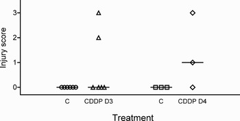
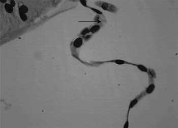
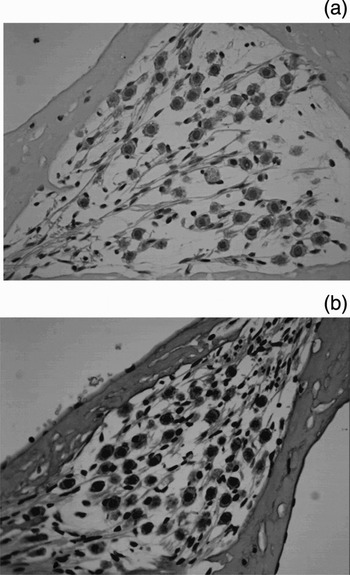
Injury to the external ciliated cells was also graded with a four-point scoring system, with a score of zero indicating the presence of three external ciliated cells with intact nuclei, and one, two and three indicating cochleae with injury to one, two or three external ciliated cells, respectively. Some animals scored as three displayed general changes to the architecture of the organ of Corti, including disappearance of the tunnel of Corti and the spaces of Nuel, which in some cases had been filled with supporting cells (Figure 5). Despite such changes, there were no statistically significant differences between the groups (Figure 6). In one animal from group three, the cells of Reissner's membrane showed signs of vacuolisation (Figure 7). Injury to the spiral ganglion was not observed in any specimen (Figure 8).
Photomicrographs showing representative organ of Corti injury for each injury score grade, related to the degree of outer hair cell injury. (a) Score of 0 = presence of three intact outer hair cells (OHCs) with well defined nuclei (arrow heads); (b) 1 = two intact OHCs (arrow heads); (c) 2 = one intact OHC (arrow head); the other OHC seen displays almost indiscernible, degenerated nuclei (asterisk); (d) 3 = absence of OHCs, with space filled with supporting cells and visible changes to the architecture of the organ of Corti (broad arrow) (H&E; ×400).

Outer hair cell injury scores for the four groups. Crossbars represent median values. There wasn't statistically significant difference between groups (p = 0.1231, Kruskal–Wallis test, comparing the four groups). C = control; CDDP = cisplatin. Circle plots: injury score values in group 2 (D3); triangle plots: injury score values in group 1 (D3); square plots: injury score values in group 4 (D3); diamond plots: injury score values in group 3 (D3).

Photomicrograph of Reissner's membrane of rat cochlea treated with 24 mg cisplatin/kg and evaluated after four days. Vacuolisation of epithelial cells is visible (arrow) (H&E; ×1000).

Photomicrographs of spiral ganglion of rat cochlea treated with 24 mg cisplatin/kg. (a) Baseline; (b) after four days (H&E; ×400).

Discussion
Morphological evaluation of rat cochleae is commonly performed in studies of cisplatin-induced ototoxicity, with or without functional evaluation. Many such studies have used scanning electron microscopy, due to the ease with which changes to cell cilia in the organ of Corti may be observed.Reference Campbell, Rybac and Meech5–Reference Fetoni, Quaranta, Marchese, Cadoni, Paludetti and Sergi10 However, light microscopy of longitudinal sections from the modiolus makes it possible to concomitantly identify injury to other structures, such as the stria vascularis, spiral ganglion and Reissner's membrane.Reference Cardinaal, De Groot, Huizing, Veldman and Smoorenburg15, Reference Wang, Faulconbridge, Fetoni, Guitton, Pujol and Puel20 Light microscopy is also less expensive and easier to use, since only routine histological procedures are required.
Few studies on cisplatin-induced ototoxicity in rats have used light microscopy.Reference Campbell, Meech, Rybak and Hughes11, Reference Lynch, Gu, Pierce and Kil19 The technique has been more frequently employed in studies involving guinea pigs, most of which have been published by a single group of researchers.Reference De Groot, Hamers, Gispen and Smoorenburg13–Reference Cardinaal, De Groot, Huizing, Veldman and Smoorenburg17, Reference Smoorenburg, De Groot, Hamers and Klis21–Reference Van Ruijven, De Groot, Klis and Smoorenburg26 These studies used a technique described by De Groot et al. Reference De Groot, Hamers, Gispen and Smoorenburg13 and based on intralabyrinthine perfusion of a fixative composed of 3 per cent glutaraldehyde, 2 per cent formaldehyde, 1 per cent acrolein and 2.5 per cent dimethylsulphoxide in 0.1 M cacodylate buffer at pH 7.4. Samples were also post-fixed after decalcification with 4 per cent osmium tetroxide and K4Ru(CN)6. Using guinea pig cochleae, Sergi et al. Reference Sergi, Ferraresi, Troiani, Paludetti and Fetoni18 fixed and post-fixed their samples with 2.5 per cent glutaraldehyde and 2 per cent paraformaldehyde, while Wang et al. Reference Wang, Faulconbridge, Fetoni, Guitton, Pujol and Puel20 used 4 per cent paraformaldehyde. In their study on rats, Campbell et al. Reference Campbell, Meech, Rybak and Hughes11 employed 2.5 per cent glutaraldehyde for fixation and osmium tetroxide for post-fixation. In contrast, Lynch et al. Reference Lynch, Gu, Pierce and Kil19 fixed their samples only once using 4 per cent paraformaldehyde. In the present study, immersion in 10 per cent buffered formaldehyde prior to decalcification, with no post-fixation, fixed tissues efficiently.
In the literature reviewed, cochlear decalcification was achieved by immersion in EDTA for either fourReference De Groot, Hamers, Gispen and Smoorenburg13–Reference Cardinaal, De Groot, Huizing, Veldman and Smoorenburg17, Reference Smoorenburg, De Groot, Hamers and Klis21–Reference Van Ruijven, De Groot, Klis and Smoorenburg26 or seven days.Reference Lynch, Gu, Pierce and Kil19 In our study, immersion in 10 per cent EDTA for one week decalcified and preserved samples adequately.
Among our references, only Lynch and colleaguesReference Lynch, Gu, Pierce and Kil19 used haematoxylin and eosin staining to evaluate morphological changes restricted to the stria vascularis. In the present study, this staining technique provided good definition of the structures of interest, with a clear distinction between nucleus and cytoplasm and easy visualisation of injury. Other studies have employed toluidine blueReference Campbell, Meech, Rybak and Hughes11, Reference Sergi, Ferraresi, Troiani, Paludetti and Fetoni18 or 1 per cent methylene blue with 1 per cent azur II in 1 per cent sodium tetraborate.Reference De Groot, Hamers, Gispen and Smoorenburg13–Reference Cardinaal, De Groot, Huizing, Veldman and Smoorenburg17, Reference Smoorenburg, De Groot, Hamers and Klis21–Reference Sluyter, Klis, De Groot and Smoorenburg23, Reference Van Ruijven, De Groot and Smoorenburg25, Reference Van Ruijven, De Groot, Klis and Smoorenburg26
Rats treated with cisplatin showed considerable changes to the basal cochlear turn, affecting mainly the external ciliated cells and the stria vascularis. In the former, the extent of injury ranged from one external ciliated cell to the entire architecture of the organ of Corti, including the disappearance of the tunnel of Corti and the spaces of Nuel and, in some cases, the filling of these spaces by supporting cells (Figure 4). In the stria vascularis, the most evident finding was shrinkage of the intermediate cells (Figure 1). Cochlear changes were confirmed by functional evaluation via distortion product evoked otoacoustic emission testing.
Most authors have reported cisplatin-induced injury to the external ciliated cells and the stria vascularis.Reference Cardinaal, De Groot, Huizing, Veldman and Smoorenburg15–Reference Cardinaal, De Groot, Huizing, Veldman and Smoorenburg17, Reference Lynch, Gu, Pierce and Kil19, Reference Smoorenburg, De Groot, Hamers and Klis21, Reference O'Leary, Klis, De Groot, Hamers and Smoorenburg22 Injury to the latter included shrinkage of the intermediate cells,Reference Cardinaal, De Groot, Huizing, Veldman and Smoorenburg15–Reference Cardinaal, De Groot, Huizing, Veldman and Smoorenburg17, Reference Lynch, Gu, Pierce and Kil19, Reference Smoorenburg, De Groot, Hamers and Klis21, Reference O'Leary, Klis, De Groot, Hamers and Smoorenburg22 blebbing of the marginal cells,Reference Cardinaal, De Groot, Huizing, Veldman and Smoorenburg15–Reference Cardinaal, De Groot, Huizing, Veldman and Smoorenburg17 protrusion of the marginal cells into the endolymphatic space,Reference Campbell, Meech, Rybak and Hughes11, Reference Smoorenburg, De Groot, Hamers and Klis21 oedemaReference Lynch, Gu, Pierce and Kil19 and atrophy.Reference Sluyter, Klis, De Groot and Smoorenburg23 After administering 2 mg/kg cisplatin for eight consecutive days, neither De Groot et al.,Reference De Groot, Hamers, Gispen and Smoorenburg13 O'Leary et al. Reference O'Leary, Klis, De Groot, Hamers and Smoorenburg22 nor Van Ruijven et al. Reference Van Ruijven, De Groot and Smoorenburg25 observed any changes to the stria vascularis.
• Cisplatin (cis-diamminedichloroplatinum) is commonly used in the treatment of a variety of neoplasms, especially those of the head and neck
• Most studies on rodent ototoxicity have been carried out with scanning electron microscopy, a reliable way to identify ciliated cell injury. However, this method is expensive and is available in only a few laboratories in our country
To our knowledge, all studies using guinea pigs and light microscopy have found cisplatin to cause injury to the external ciliated cells, predominantly to the basal cochlear turn and progressively less towards the apical turn.Reference De Groot, Hamers, Gispen and Smoorenburg13–Reference Sergi, Ferraresi, Troiani, Paludetti and Fetoni18, Reference Wang, Faulconbridge, Fetoni, Guitton, Pujol and Puel20, Reference O'Leary, Klis, De Groot, Hamers and Smoorenburg22, Reference Wolters, Klis, Hamers, De Groot and Smoorenburg24–Reference Van Ruijven, De Groot, Klis and Smoorenburg26 Campbell et al. Reference Campbell, Meech, Rybak and Hughes11 limited their observations to changes in the stria vascularis of rats, and made no mention of the external ciliated cells. Although they used surface tissue extracts, Lynch et al. Reference Lynch, Gu, Pierce and Kil19 reported injury to external ciliated cells in rats similar to that observed in guinea pigs.
Thus, in the present study, the main target of injury described in the literature, the basal cochlear turn, was used for the purpose of evaluation. However, since the rat cochlea contains only two and a half turns, and the apical opening (used in preparation) may have caused secondary injury to the apical turn, the middle turn of some specimens may have been affected.
Reissner's membrane was generally left intact. Only one animal from group three had vacuolised epithelial cells. Similar findings were reported by Cardinaal et al. Reference Cardinaal, De Groot, Huizing, Veldman and Smoorenburg15 for two out of 18 animals. In contrast with the present study, some authors also observed distension of Reissner's membrane suggestive of endolymphatic hydrops.Reference De Groot, Hamers, Gispen and Smoorenburg13, Reference Cardinaal, De Groot, Huizing, Veldman and Smoorenburg15, Reference Cardinaal, De Groot, Huizing, Veldman and Smoorenburg17
No spiral ganglion injury was observed in this study. Injury to the spiral ganglion, such as cell vacuolisation, cell shrinkage and decreased cell numbers, has been reported by some authorsReference Cardinaal, De Groot, Huizing, Veldman and Smoorenburg15–Reference Cardinaal, De Groot, Huizing, Veldman and Smoorenburg17, Reference Smoorenburg, De Groot, Hamers and Klis21, Reference Van Ruijven, De Groot and Smoorenburg25, Reference Van Ruijven, De Groot, Klis and Smoorenburg26 but not others.Reference De Groot, Hamers, Gispen and Smoorenburg13, Reference Sergi, Ferraresi, Troiani, Paludetti and Fetoni18
The method described in this paper is simple to perform with routine histology equipment, and is appropriate for the study of acute, cisplatin-induced ototoxicity in rats.
Conclusion
The described, routine histology technique, using fixation with 10 per cent formaldehyde and haematoxylin and eosin staining, enabled visualisation of morphological changes linked to changes in hearing function, evaluated with distortion product evoked otoacoustic emission testing. This preparation technique is appropriate for the study of acute cisplatin-induced ototoxicity in rats.