I. INTRODUCTION
As a kind of new unsupported nanomaterials, nanoporous metals and alloys have attracted great attention in a wide range of applications, such as catalysis,Reference Christensen and Nørskov1–Reference Xu, Xu, Su and Ding5 energy storage,Reference Chen and Qiao6 optical properties,Reference Dixon, Daniel, Hieda, Smilgies, Chan and Allara7, Reference Jiao, Ryckman, Ciesielski, Escobar, Jennings and Weiss8 detection,Reference Kim, Kim, Joo, Ko and Yi9 medicineReference Jeon, Yang, Byun and Kim10 and printing.Reference Luechinger, Walt and Stark11 In the last few years, more and more concerns were focused on the nanoporous metals prepared by dealloying.Reference Hodge, Doucette, Biener, Biener, Cervantes and Hamza12–Reference Zhang, Wang, Qi, Somsen, Wang and Zhao14 Nanoporous noble metals, such as nanoporous Au,Reference Hakamada and Mabuchi15 PtReference Pugh, Dursun and Corcoran16 and Pd,Reference Hakamada and Mabuchi17 have been widely studied by dealloying of different precursory alloys, respectively. During dealloying, one or more elements in the precursory alloys are removed selectively, resulting in the formation of a nanoporous structure.Reference Erlebacher, Aziz, Karma, Dimitrov and Sieradzki18 Recently, nanoporous Ag has caused more interest due to its unique catalytic and optical properties.Reference Xu, Li, Tian and Ding19–Reference Morrish and Muscat21 Some alloy systems such as Ag-AlReference Wang, Qi, Zhao, Wang and Zhang22 and Ag-ZnReference Li, Li, Qin and Lu23 were used to prepare nanoporous Ag through chemical dealloying. Nanoporous Ag exhibited different length scales and morphology of ligament/channel, depending on the precursory alloys and dealloying solution. The studies suggest that the phase constitutions of precursory alloys have a significant influence on the formation of nanoporous Ag.
It has been proved that the peculiar optical and catalytic properties of nanoporous metals were closely related to their microstructures.Reference Ding and Chen24 For example, Chen et al.Reference Chen, Yu, Fujita and Chen25 reported that the surface enhanced Raman scattering (SERS) intensity of nanoporous Cu started to increase with the pore size from 15 to 34 nm and reached its maximum at a nanopore size of 34 nm. Thus, much research has been carried out to control nanoporous metals by adjusting the alloy composition and dealloying conditions. Yeh et al.Reference Yeh, Tai, Huang and Sun26 prepared nanoporous Ag with different morphology by dealloying Ag–Zn films. The precursory alloys were adjusted through manipulating the composition, deposition potential, deposition current and deposition stripping temperature. QianReference Qian and Chen27 tailored the pore size of nanoporous Au from 33 nm to 5 nm by dealloying of Ag-Au leaf at low temperature. It is generally recognized that the fast diffusion of noble atoms along alloy/electrolyte interfaces could inevitably cause the ligament coarsening.Reference Sieradzki, Corderman, Shukla and Newman28 Therefore, it is still a great challenge to control nanoporous metals by inhibiting the ligament coarsening under mild dealloying conditions. Recently, Snyder et al.Reference Snyder, Asanithi, Dalton and Erlebacher29 reported that the addition of small amounts of noble metals to precursory alloys could stabilize the morphology of nanoporous metals. Ji et al.Reference Ji, Wang, Zhao, Zhang, Xu and Zhang30 prepared the nanoporous Ag with pore size of about 5 nm by adding a certain amount of Pd into the Mg-Ag precursory alloys. However, to the best of our knowledge, the preparation and control of nanoporous Ag through adding the sacrificial base metals into precursory alloys has not been reported.
Our previous workReference Li, Song, Sun, Yang, Ding, Yang, Yang and Wang31 prepared nanoporous Ag by electrochemical corrosion of the melt-spun Cu-Ag alloys at a low voltage. The ligaments of nanoporous Ag evolved from the Ag-rich phase. However, the ligament/pore sizes were dependant on the morphology of the Ag-rich phase in the precursory alloys. As a result, it is difficult to control nanoporous Ag by adjusting the corrosion parameters. The present investigation reported a novel approach to control the morphology of nanoporous Ag through adding the third element Ce into the precursory alloys combined with applied potentials. The ultrafine nanoporous Ag ribbons with granular ligaments were successfully prepared by electrochemical dealloying of the melt-spun Cu-Ag-Ce alloys. Meanwhile, the dealloying mechanism of the Cu-Ag-Ce alloys with different phase constitutions was discussed.
II. EXPERIMENTAL
Cu80Ag20 and Cu80-XAg20CeX (X = 2, 4, 6, at.%) alloys with nominal compositions were prepared from pure Ag (99.99%), pure Cu (99.99%) and pure Ce (99.00%) in a vacuum arc furnace at an argon atmosphere. The starting materials with a mass of 6 g were melted by arc employing a nonconsumable tungsten electrode in a water-cooled copper crucible. For obtaining homogeneous samples, several remelting cycles were performed with the ingots turned upside down before each remelting. Subsequently, the ribbons were achieved by a single roller melt spinning at a speed of 43 m/s under a pressure of 0 5 × 10⁻⁵ Pa Ar gas. The width and thickness of the ribbons were approximately 2 mm and 30 μm, respectively. It was found that the brittleness of the melt-spun ribbons increased much with a further increase of Ce, resulting in a formation of ribbon segments. Therefore, the maximum Ce contents in the precursory alloys was 6 at.% in the present experiment. The phase structures of the as-quenched ribbons were analyzed with the use of a Bruker D8 advanced x-ray diffraction (XRD; Bruker AXS GmbH, Karlsruhe, Germany). The microstructures of the ribbons were analyzed by a JEM-2100 transmission electron microscope (TEM; JEOL Ltd., Tokyo, Japan) and a JSM-7000F scanning electron microscope (SEM; JEOL Ltd., Tokyo, Japan) equipped with a backscattered electron detector.
The melt-spun ribbons were used to prepare nanoporous Ag by electrochemical dealloying in the 0.5 M CuSO4 aqueous solution. The dealloying was carried out by a CHI 1130A potentiostat (Xian Honghua electronic equipment factory, Xi'an, China), and the setting potential value was 0.05 V. The melt-spun ribbons served as the anode and two Ag rods as the cathode. The Cu-Ag-Ce ribbons with a surface area about 0.2 cm2 were dealloyed at room temperature. The dealloying duration ranged from 3 h to 5 h. For investigating the effect of the applied potentials, the Cu78Ag20Ce2 and Cu76Ag20Ce4 alloys were dealloyed in the 0.5 M CuSO4 aqueous solution at the potentials of 0.02 V, 0.1 V and 0.2 V, respectively.
The dealloyed alloys were cleaned in diluted hydrochloric acid and alcohol, respectively, and dried in air. The morphology characteristics of the as-dealloyed ribbons were analyzed with the help of the SEM. The electrochemical activities of the melt-spun Cu–Ag–Ce ribbons were measured by a VersaSTAT MC instrument (AMETEK, Mahwah, NJ) in the 0.5 M CuSO4 aqueous solution at a scanning speed of 1 mV/s. The experiments were carried out in a standard three-electrode electrochemical cell with a Pt plate electrode as the counter electrode, the ribbons as the working electrode and a saturated Ag/AgCl electrode as the reference electrode. The electrode potentials of Cu5Ce, Cu6Ce and Ag/Ce compounds were tested by a M273A electrochemical analyzer (Arbin, College Station, TX) in the 0.5 M CuSO4 solution. Graphite was used as an auxiliary electrode and the applied current was 0.0003A.
The SERS effects of nanoporous Ag were characterized by a Laboratory Ram JY-HR800 spectrometer (HORIBA Jobin Yvon, Paris, France). The spectra obtained by immersing the as-dealloyed Cu-Ag-Ce alloys in the 0.1 mM Rhodamine 6G solution for 20 min were measured at the output laser power of 17 mw/100 (D2 filter), the exposure time of 1 s and the operating wavelength of 632.8 nm.
III. RESULTS AND DISCUSSION
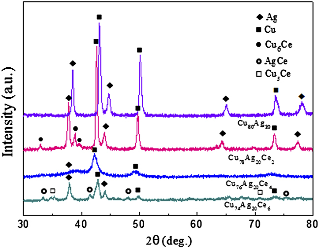
Figure 1 shows the XRD patterns of the melt-spun Cu80Ag20 and Cu-Ag-Ce alloys. The Cu80Ag20 alloy consisted of face-centered-cubic (fcc)-Cu and fcc-Ag, while the Cu78Ag20Ce2 alloy was composed of fcc-Cu, fcc-Ag, and Cu6Ce. It is noticed that the peaks of fcc-Cu and fcc-Ag shifted to left and the relative intensity of Ag peaks weakened due to the addition of 2% Ce, indicating that more Ag atoms dissolved in fcc-Cu. With the increase of Ce contents from 2% to 4%, the diffraction peaks of Ag almost disappeared. However, the Cu74Ag20Ce6 alloy contained fcc-Cu, fcc-Ag, Ag/Ce and Cu5Ce. It is obvious that the Ce contents had significant influence on the phase constitutions of the melt-spun Cu-Ag-Ce alloys.
XRD patterns of the melt-spun Cu80Ag20 and Cu80-XAg20CeX (x = 2, 4, 6) alloys.
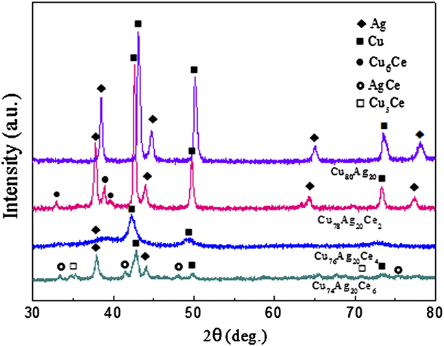
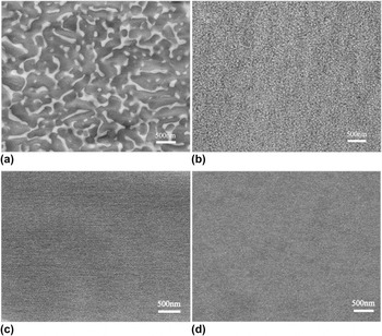
Figure 2 shows the backscattered electron images of the melt-spun Cu80Ag20 and Cu-Ag-Ce alloys. A typical divorced eutectic microstructure was observed in the Cu80Ag20 alloy. Where, the Ag-rich phase with a long rod-like shape distributed along the Cu-rich grain boundaries, seen in Fig. 2(a). For the Cu78Ag20Ce2 alloy, the divorced eutectic microstructure disappeared and a large number of spherical particles emerged in the Cu-rich matrix, as shown in Fig. 2(b). The particles might be the Ag-rich phase or Cu/Ce compounds according to the XRD patterns (Fig. 1). Although the types of the particles could not well be distinguished, it was very clear that the particles were obviously refined and homogeneously distributed. As the contents of Ce were 4% and 6%, the particles were not observed clearly in the backscattered electron images, as shown in Figs. 2(c) and 2(d).
Backscattered electron images of the melt-spun Cu80Ag20 (a), Cu78Ag20Ce2 (b) Cu76Ag20Ce4 (c) and Cu74Ag20Ce6 (d) alloys.

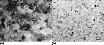
Figure 3 shows the TEM images of the melt-spun Cu78Ag20Ce2 and Cu76Ag20Ce4 alloys. It can be seen from Fig. 3(a) that the refined crystalline grains with a particle-like shape in the Cu78Ag20Ce2 alloy had an average size of about 100 nm. With the increase of Ce contents, the grain size of the Cu76Ag20Ce4 alloy was decreased to approximately 10 nm and a small quantity of compounds (darker particles) were homogeneously dispersed in the fcc-Cu matrix, as shown in Fig. 3(b).
TEM images of the melt-spun Cu78Ag20Ce2 (a) and Cu76Ag20Ce4 (b) alloys.

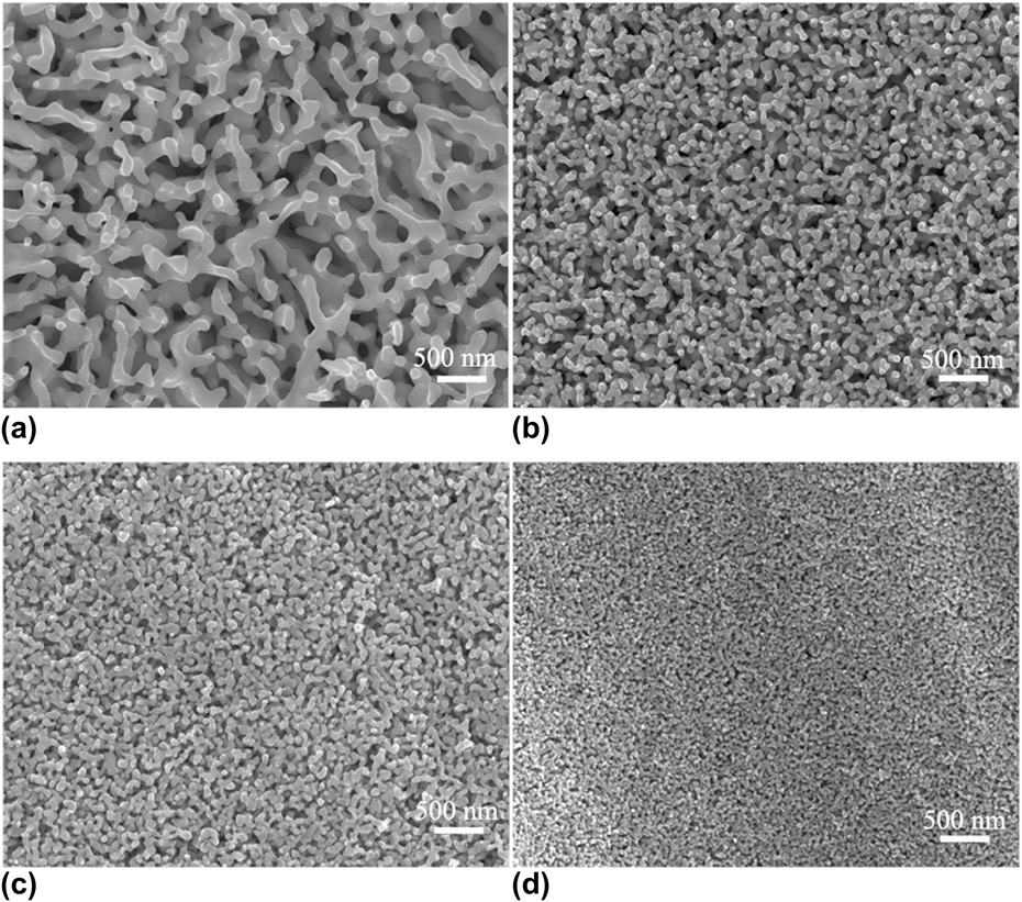
Figure 4 shows the plan-view of the ribbons prepared by electrochemical dealloying of the melt-spun Cu80Ag20 alloy for 12 h and Cu80-XAg20CeX (X = 2, 4, 6) for 3–5 h. The energy-dispersive x-ray spectrometry (EDS) showed that the dealloyed Cu-Ag-Ce alloys were close to pure Ag. It is obvious that all the dealloyed ribbons exhibited the bicontinuous and interpenetrating porous structure. As shown in Fig. 4(a), the pore channels with an average size of ∼200 nm were observed from the dealloyed Cu80Ag20 alloy. The ligaments comprised of long rod-like and short rod-like shapes that were similar to those of the Ag-rich phase in the precursory alloys [Fig. 2(a)]. As 2% Ce was added into the CuAg20 alloy, the Ag ligaments exhibited a particle-like shape, and the pore channels were about 60 nm. The distribution of the pores and ligaments was very uniform, as seen in Fig. 4(b). With the increase of Ce in precursory alloys, the pore and ligament sizes of the nanoporous Ag prepared from the Cu76Ag20Ce4 alloy, as shown in Fig. 4(c), further decreased. As the Ce contents increased to 6% in precursory alloys, the average pore size was about 15 nm, as shown in Fig. 4(d).
Plan-view SEM images of nanoporous Ag prepared by electrochemical dealloying of the melt-spun Cu80Ag20 (a), Cu78Ag20Ce2 (b), Cu76Ag20Ce4 (c) and Cu74Ag20Ce6 (d) alloys at the potential of 0.05 V in the 0.5 M CuSO4.
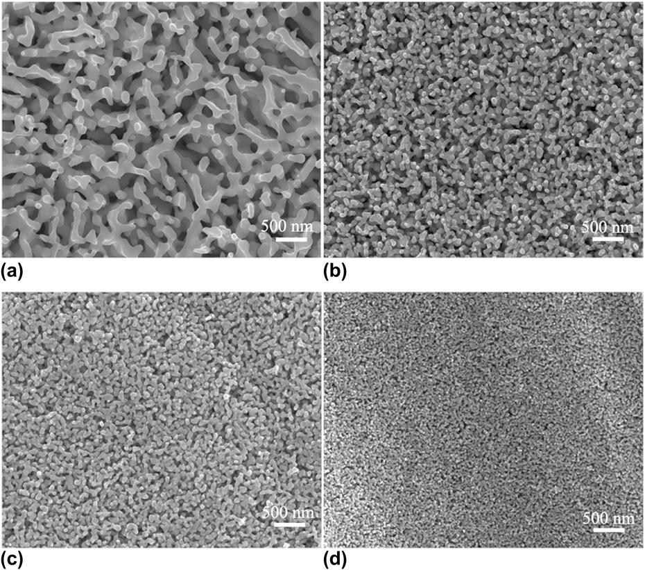
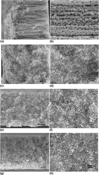
Figure 5 illustrates the cross-sectional view of the nanoporous Ag ribbons prepared from the Cu80Ag20 and Cu-Ag-Ce alloys. The pores in the dealloyed Cu80Ag20 alloy, as shown in Figs. 5(a) and 5(b), distributed across the entire ribbons. The morphology of the Ag ligaments was a long rod-like shape and perpendicular to the ribbon plane. From Fig. 5(b), it can be observed that the oriented straight Ag stocks with a channel size of about 160 nm were interconnected. As the Cu-Ag alloys contained 2% Ce (Cu78Ag20Ce2), the ligaments in nanoporous Ag exhibited a particle-like shape. The average size of the particles was about 80 nm, and the channels were about 50 nm, seen in Figs. 5(c) and 5(d). With the increase of Ce in precursory alloys, the ligaments of nanoporous Ag, as shown in Figs. 5(e) and 5(f), had similar shape to that from the Cu78Ag20Ce2 alloy, but the ligament/pore sizes reduced obviously. The ligament and pore sizes of nanoporous Ag prepared from the Cu74Ag20Ce6 alloy had length scales of about 40 nm and 15 nm, respectively, seen in Fig. 5(h). These results clearly indicated that the addition of Ce into the Cu80Ag20 alloy caused an evident refinement of nanoporous Ag, and the Ce contents in the precursory alloys had significant effects on the pore/ligament sizes.
Section-view of nanoporous Ag prepared by electrochemical dealloying of the melt-spun Cu80Ag20 (a)–(b), Cu78Ag20Ce2 (c)–(d), Cu76Ag20Ce4 (e)–(f) and Cu74Ag20Ce6 (g)–(h) alloys at the potential of 0.05v in the 0.5 M CuSO4 solution.

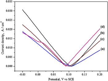
Figure 6 shows the typical Tafel curves of the melt-spun Cu80Ag20 and Cu-Ag-Ce ribbons. The critical potentials of the Cu80Ag20, Cu78Ag20Ce2, Cu76Ag20Ce4 and Cu74Ag20Ce6 alloys were 108 mV, 106 mV, 100 mV and 89 mV, respectively. These results confirmed that the addition of Ce had a significant influence on the electrochemical activities of the melt-spun Cu-Ag-Ce ribbons, indicating that the preparation of nanoporous Ag would be much easier with the increase of Ce contents in the precursory alloys.
Tafel polarization curves of the melt-spun Cu80Ag20 (a), Cu78Ag20Ce2 (b), Cu76Ag20Ce4 (c) and Cu74Ag20Ce6 (d) ribbons measured in the 0.5 M CuSO4 solution at room temperature.

The investigationsReference Liu, Meng and Zhang32, Reference Inoue, Park and Masumoto33 found that the rare earth (RE) elements in the Cu-Ag alloys could not only restrain the nucleation and growth of primary Cu phases but also decrease the eutectic temperature, resulting in the formation of the supersaturated solid solutions of Ag (Ce) in fcc-Cu and Cu (Ce) in fcc-Ag. During rapid solidification of the Cu78Ag20Ce2 alloy, the directional growth of the primary Cu was restrained and the number of crystal nuclei was increased due to larger supercooling. Therefore, the coexistent Cu (Ag, Ce) and Ag (Cu, Ce) solid solutions with a refined particle-like shape were formed [Fig. 3(a)]. As Ce contents further increased (Cu76Ag20Ce4), Ag almost dissolved in fcc-Cu (Fig. 1) and the grain sizes were dramatically reduced [Fig. 3(b)] due to the increase in the difficulty of nucleation and growth. However, for the Cu74Ag20Ce6 alloy, the fine supersaturated Cu (Ag, Ce), Ag (Cu, Ce) solid solutions and Ag/Ce compound were formed [Figs. 1 and 2(d)]. In other hand, more grain boundaries appeared in Cu-Ag-Ce alloys due to the refinement of Cu (Ag) matrix (Fig. 3).
During dealloying of the Cu-Ag alloys, the primary Ag phase evolved into the ligaments of nanoporous Ag after the Cu phase was removed, and the pore and ligament sizes were dependent on the morphology of primary Ag phase according to our previous work.Reference Li, Song, Sun, Yang, Ding, Yang, Yang and Wang31 For the Cu78Ag20Ce2 alloy, the crystalline grains were evidently refined due to the addition of 2% Ce [Fig. 2(b)] and the short rod-shaped Ag changed into nanoparticles. After the supersaturated Cu (Ag, Ce) solid solution and Cu6Ce compound were decomposed, the Ag-rich nanoparticles evolved into the ligaments, and then the pores with a small size were formed. The Ag atoms released from the Cu solution assembled attaching the primary Ag, which resulted in a slight growth of the Ag ligaments [seen Fig. 2(b)] and [Fig. 4(b)]. On the other hand, after the Cu and Ce atoms dissolved in the fcc-Ag were removed, the ligaments were directly formed, which would not be needed for the rearrangement of Ag atoms. In addition, the grain boundaries were corroded preferentiallyReference Sugawara and Ebiko34 and the existence of the active Ce in the fcc-Ag and fcc-Cu reduced the critical potentials of the Cu-Ag alloys (Fig. 6). Therefore, with the addition of Ce, the Cu-Ag alloys could be dealloyed easily.
The dealloying process of Ag-Au alloys with a single solid solution involved the dissolution of the Ag atoms and the rearrangement of the Au atoms.Reference Erlebacher, Aziz, Karma, Dimitrov and Sieradzki18, Reference Snyder, Livi and Erlebacher35 The structure coarsening caused by surface diffusion had an important influence on the length scales of pore/ligament in nanoporous metals.Reference Hakamada and Mabuchi36, Reference Dursun, Pugh and Corcoran37 Here, the melt-spun Cu76Ag20Ce4 alloy mainly consisted of a single supersaturated Cu (Ag, Ce) solid solution (Fig. 1). The released Ag atoms reorganize along alloy/electrolyte interfaces and formed a certain amount of islands during dealloying. According to the Ostwald ripening growth mechanism,Reference Sun, Mayers, Herricks and Xia38, Reference Xia, Xiong, Lim and Skrabalak39 the larger islands could grow at the expense of the smaller ones with the removal of Cu and Ce atoms. The pore and ligament sizes of nanoporous Ag depended on the coarsening rate of the islands. The refining grain could not only increase the density of diffusion path for the dissolution of the less noble atoms but also decrease the rearrangement distance of the noble atoms.Reference Lu, Li and Wang40 The grain size of the Cu76Ag20Ce4 alloy decreased to about 10 nm [Fig. 3(b)]. As a result, the coarsening of the Ag ligaments was inhibited after the Cu-based solid solution was decomposed. The pore size of the nanoporous Ag prepared from the Cu76Ag20Ce4 alloy was smaller than that from Cu78Ag20Ce2 [Figs. 5(d)–5(f)].
As the Ce contents increased to 6%, the alloys consisted of the refined Cu (Ag, Ce), Ag (Cu, Ce) grains, Ag/Ce and Cu5Ce compounds. It is very difficult to control nanoporous metals by dealloying the multiphase alloys with different intermetallic compounds because the homogeneity of ligament/channel was affected by the disparities in crystal structures between coexistent phases.Reference Dursun, Pugh and Corcoran41, Reference Zhang, Wang, Qi, Wang and Zhang42 For investigating the formation mechanism of nanoporous Ag, the electrode potentials of Ag/Ce, Cu6Ce and Cu5Ce compounds in the 0.5 M CuSO4 solution were tested. The results showed that their potentials were about 0.08 V, 0.18 V and 0.25 V, respectively, which indicated that Ag/Ce would decompose preferentially during dealloying. The released Ag atoms rearranged themselves into a great number of Ag islands. The islands would grow into the fine Ag particles at the expense of the Ag atoms released from the decomposed Cu-based solid solution. The Ag particles and the fine primary Ag evolved into the ligaments after Cu and Ce elements were removed. Because the number of the Ag atoms released from fcc-Cu was limited, the coarsening of the Ag ligaments was slight. As a result, the nanoporous Ag prepared from the Cu74Ag20Ce6 alloy had the finest pores and ligaments in the present work [Fig. 5(h)].
For investigating the control of nanoporous Ag further, the melt-spun Cu–Ag–Ce alloys were dealloyed at different potentials, where the concentration of the electrolyte was constant (0.5 M CuSO4). Figure 7 shows the plan-view images of the nanoporous Ag prepared from the Cu78Ag20Ce2 and Cu76Ag20Ce4 alloys at the potentials of 0.02 V, 0.10 V and 0.20 V, respectively. The EDS showed that all the dealloyed alloys were close to pure Ag. The ligaments and pores of the nanoporous Ag from Cu78Ag20Ce2, as shown in Fig. 7(a), were relatively coarser. As the applied potential was increased from 0.02 V to 0.10 V, the ligament and pore sizes of the nanoporous Ag decreased to ∼80 nm [Fig. 7(b)]. When the potential was 0.20 V, though the ligaments size changed little, a lot of cracks could be observed [Fig. 7(c)]. In contrast, the ligaments and pores of the nanoporous Ag prepared from the Cu76Ag20Ce4 alloy at the potential of 0.02 V, as shown in Fig. 7(d), were very fine and homogeneous. With the increase of applied potentials, they varied from very fine to relatively coarser ligaments and pores [Figs. 7(e)–7(f)], which was in contrast to that from the Cu78Ag20Ce2 alloy.
Plan-view SEM images of nanoporous Ag prepared by electrochemical dealloying of the melt-spun Cu78Ag20Ce2 alloy (a) 0.02 V for 12 h, (b) 0.1 V for 2 h, (c) 0.2 V for 0.5 h and Cu76Ag20Ce4 alloy (d) 0.02 V for 12 h, (e) 0.1 V for 2 h, (f) 0.2 V for 0.5 h in the 0.5 M CuSO4 solution.

It has been widely recognized that the surface diffusion of the more noble elements during dealloying has a significant influence on the ligament and pore sizes of nanoporous metals.Reference Zhang, Wang, Wang, Wang, Qi, Ji and Zhao43 The migration rate of Ag atoms in the electrolyte will be faster with the increase of applied potentials. The Cu78Ag20Ce2 alloy consisted of the Cu-rich phase, as also the Ag-rich phase. During dealloying at a potential of 0.02 V, the primary Ag would grow and aggregate at the expense of the Ag atoms released from the Cu-based solid solution, and thus nanoporous Ag had the larger ligaments and pores, seen in Fig. 7(a). With the increase of applied potentials, the faster diffusion promoted the rearrangement of the released Ag atoms, which played an important role in the refinement of Ag ligaments, as shown in Fig. 7(b). However, the rapid dealloying had caused inconsistent volume shrinkage among different layers in the precursory ribbons, resulting in some cracks on the surface of nanoporous Ag [Fig. 7(c)]. In contrast, the Cu76Ag20Ce4 alloy was mainly composed of the Cu-based solid solution. During dealloying at low potentials, the Ag atoms released from the solid solution formed a large numbers of Ag islands, and thus the nanoporous Ag with fine ligaments and pores could be obtained, as seen in Fig. 7(d). With the increase of applied potentials, the faster diffusion of Ag atoms caused coarsening and merging of the islands, which is consistent with the formation of nanoporous Au.Reference Sieradzki, Corderman, Shukla and Newman28 As a result, the coarser and nonuniform ligaments appeared in the nanoporous Ag prepared from the Cu76Ag20Ce4 alloy at higher applied potentials [Fig. 7(f)].
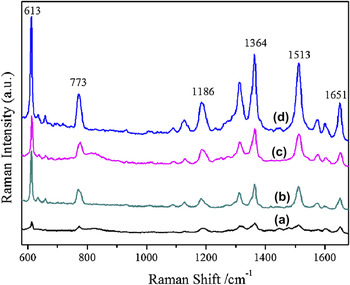
Figure 8 shows the Raman spectra of the as-dealloyed melt-spun Cu80Ag20 and Cu80-XAg20CeX (x = 2, 4, 6) alloys put in the 10−4 M R6G aqueous solution for 20 min. It is clear that the nanoporous Ag ribbons prepared from the Cu80-XAg20CeX alloys had the stronger surface-enhanced Raman signal intensities of R6G than those from the Cu80Ag20 alloy at the bands of 613, 773, 1186, 1364, 1515 and 1651 cm−1, and the SERS effects were enhanced with the increase of Ce contents in the precursory alloys. The researchReference Tiwari, Oleg, Darbha, Hardy, Singh and Ray44 suggested that the intensity of SERS signals for nanoparticles was stronger than that for nanorods. The ligament morphology of the nanoporous Ag prepared from the Cu78Ag20Ce2 alloy was particle-like, while that of the as-dealloyed Cu80Ag20 alloy was rod-like (Figs. 4 and 5). Therefore, the nanoporous Ag prepared from the Cu78Ag20Ce2 alloy had remarkable SERS effect than that from the Cu80Ag20 alloy. Furthermore, the junction between two Ag nanocrystals could yield single molecule SERS signals,Reference Michaels, Jiang and Brus45 and more hot spots could be obtained in the refined nanoporous Ag according to the hot spot phenomenon.Reference Li, Camargo, Lu and Xia46, Reference Nie and Emory47 For this reason, nanoporous Ag exhibited the stronger enhancement of the SERS effects with the increase of Ce contents in precursory alloys.
SERS spectra of the dealloyed Cu80Ag20 and Cu-Ag-Ce alloys. a, b, c and d indicate the SERS effects of nonporous Ag prepared from the melt-spun Cu80Ag20, Cu78Ag20Ce2, Cu76Ag20Ce4 and Cu74Ag20Ce6 alloys, respectively.

IV. CONCLUSIONS
In conclusion, nanoporous Ag ribbons with different pore/ligament sizes were successfully achieved by electrochemical dealloying of the melt-spun Cu-Ag-Ce alloys. The length scales of ligament/pore in nanoporous Ag can be controlled from 200 nm to 15 nm by adjusting the contents of Ce in precursory alloys. The addition of Ce had a significant influence on the phase constitutions of the melt-spun Cu-Ag alloys. The nanoporous Ag prepared from the Cu78Ag20Ce2 alloy with a refined Ag-rich phase had an average pore/ligament size of about 60 nm. With the increase of Ce contents, the Cu-Ag-Ce alloy contained a single solid solution with ultrafine crystal grains. The Cu-based solid solution could be dealloyed, and the pore and ligament sizes of nanoporous Ag further decreased. The pore size of ∼15 nm was achieved in the nanoporous Ag from the Cu74Ag20Ce6 alloy. The existence of Ag/Ce and Cu/Ce compounds in the precursory alloys was helpful to promote the dealloying. The pore/ligament sizes of nanoporous Ag could also be controlled by changing the applied potentials according to the phase constitution of the precursory alloys. Nanoporous Ag exhibited the stronger enhancement of the SERS effects with the increase of the Ce contents in precursory alloys. The present work could provide a flexible and simple method to control the morphology and ligament/pore sizes of nanoporous metals by the addition of RE elements into precursory alloys.
ACKNOWLEDGMENTS
This work is sponsored by the Natural Science Foundation of China (50871081, 51071116, 50901056, 50871080) and the National 863 Program Projects of China (2009AA03Z320).