Implications
In the present study, we investigated for the first time the effects of dietary arginine deficiency on feed intake and hypothalamic protein profiles in ducks using the isobaric tags for relative and absolute quantification technique. The results of the present investigation showed that dietary arginine deficiency inhibited appetite and decreased the expression of proteins involved in energy production and substance transport in the hypothalamus of ducks.
Introduction
Arginine is an essential amino acid for poultry and a conditioned essential amino acid for mammals. Most adult mammals can synthesize arginine, and therefore arginine is needed to be supplemented only to young and stressed mammals. However, in poultry, arginine cannot be synthesized and must be provided through diet to meet the need for their growth and production. Dietary arginine supplementation has been known to stimulate protein synthesis, enhance immunity, improve reproductive performance, and reduce fat deposition in pigs and poultry (Kwak et al., Reference Kwak, Austic and Dietert1999; Jobgen et al., Reference Jobgen, Fried, Fu, Meininger and Wu2006; Tan et al., Reference Tan, Y, Liu, Li, Xu, Kong, R, Tang, Shinzato, Smith and Wu2009; Wu et al., Reference Wu, Bazer, Davis, Kim, Li, Rhoads, Satterfield, Smith, Spencer and Yin2009). Furthermore, our previous study (Wang et al., Reference Wang, Xie, Huang, Yu and Hou2013) as well as another experiment (Kwak et al., Reference Kwak, Austic and Dietert1999) have shown that dietary arginine deficiency inhibits feed intake and thus reduces the growth of chickens and ducks.
The hypothalamus is a classic appestat, which integrates signals involved in feed intake originated from the peripheral tissues and organs, and finally makes the decision on satiety perception and food-seeking behaviour. Feed intake regulation is a complex process involved in a lot of peptides, including ghrelin, neuropeptide Y, agouti-related peptide, leptin, proopiomelanocortin and insulin, etc. and neurons, in which those peptides taking their functions gather in the hypothalamus (Kalra et al., Reference Kalra, Dube, Sahu, Phelpsf and Kalra1991; Cheung et al., Reference Cheung, Clifton and Steiner1997; Baskin et al., Reference Baskin, Figlewicz Lattemann, Seeley, Woods, Porte and Schwartz1999; Kamegai et al., Reference Kamegai, Tamura, Shimizu, Ishii, Sugihara and Wakabayashi2001; Gropp et al., Reference Gropp, Shanabrough, Borok, Xu, Janoschek, Buch, Plum, Balthasar, Hampel and Waisman2005). A different subnucleus of the hypothalamus is involved in an aspect of feeding behaviour. Lesions in the ventromedial hypothalamus led to hyperphagia, whereas anorexia occurred with lateral hypothalamus lesions (Anand and Brobeck, Reference Anand and Brobeck1951). Previous studies have shown that the ventromedial, dorsomedial and lateral hypothalamus were important functional sites to regulate appetite for amino acids (Leung and Rogers, Reference Leung and Rogers1980; Blevins et al., Reference Blevins, Dixon, Hernandez, Barrett and Gietzen2000). It is reported that arginine takes part in appetite regulation via agmatine and nitric oxide in the hypothalamus. The orexigenic effect of agmatine is elicited by stimulating α 2-adrenoceptors within the paraventricular hypothalamus to increase neuropeptide Y activity (Taksande et al., Reference Taksande, Kotagale, Nakhate, Mali, Kokare, Hirani, Subhedar, Chopde and Ugale2011). Other neuropeptides, such as leptin (Yang and Denbow, Reference Yang and Denbow2007), orexin-A (Farr et al., Reference Farr, Banks, Kumar and Morley2005) and ghrelin (Gaskin et al., Reference Gaskin, Farr, Banks, Kumar and Morley2003), can either inhibit or promote feed intake by modulating nitric oxide production in the hypothalamus of chicken and mouse.
Up to now, it has not reported yet whether arginine affects feeding behaviour through the changing expression of regulatory proteins in the hypothalamus of birds. Proteomic analysis is a high-throughput technique that can provide some valuable cues for understanding complicated biological processes. The technique has been successfully used to investigate the molecular mechanism of nutrient metabolism, antioxidation and steatosis development in ducks (Bax et al., Reference Bax, Chambon, Marty-Gasset, Remignon, Fernandez and Molette2012; Zheng et al., Reference Zheng, Liu, Zhang, Hou, Chang, Zhang, Cai and Chen2012). Therefore, the present study was conducted to evaluate hypothalamic protein profile change in ducks fed with arginine-deficient and -sufficient diet to provide more insights into the underlying mechanism of arginine on feed intake regulation in the ducks.
Material and methods
The present research was approved by the animal care and welfare committee of the Institute of Animal Sciences in the Chinese Academy of Agricultural Sciences.
Animal and feeding
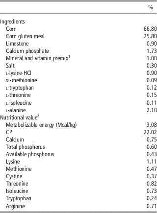
One hundred and twenty six 1-day-old male White Pekin ducks (Anas platyrhynchos domestica), obtained from Pekin duck breeding centre in Chinese Academy of Agricultural Sciences, were randomly divided into one of the two dietary treatments containing 0.71% (deficient) or 1.27% (sufficient) arginine, respectively. Ingredients and nutritional compositions of the arginine-deficient diet are shown in Table 1. The arginine-sufficient diet was prepared by adding l-arginine·HCl to arginine-deficient diet at the expense of corn. l-alanine was added to arginine-deficient diet to keep isonitrogenous level between the two experimental diets. Arginine content of the diets was analysed by ion-exchange chromatography with an L-8900 amino acid analyzer (Hitachi, Tokyo, Japan). Except for the arginine content in the arginine-deficient diet, all nutrients meet the requirements of White Pekin ducks in National Research Council (1994). All birds were kept in plastic-wire floor pens with 24 h lighting, and feed and water were provided ad libitum until 21 days of age. Room temperature was maintained at 28°C from 1 to 3 days of age, at 26°C from 4 to 7 days of age and at 25°C from 8 to 14 days of age. The feed intake was monitored throughout the experimental period.
Ingredients and chemical compositions of the arginine-deficient diet
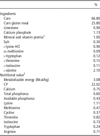
1 Premix supplied per kilogram diet: 10 mg of Cu, 60 mg of Fe, 60 mg of Zn, 80 mg of Mn, 0.3 mg of Se, 0.2 mg of I, 10 000 IU of vitamin A, 3000 IU of vitamin D3, 20 IU of vitamin E, 2 mg of vitamin K3, 2 mg of thiamin, 8 mg of riboflavin, 4 mg of pyridoxine hydrochloride, 0.06 mg of cobalamin, 20 mg of pantothenic acid, 50 mg of nicotinic acid, 1 mg of folic acid and 0.2 mg of biotin,1000 mg of choline chloride.
2 Values of CP and amino acid were analyzed.
Sample collection and preparation
At 21 days of age, live BW per pen was recorded after 12 h fasting. Five birds with the average BW of each treatment were slaughtered by manual exsanguinations, the hypothalamus was immediately removed, snap-frozen in liquid nitrogen and then stored at −80°C for proteomic analysis.
Proteomic analysis
Each hypothalamus was homogenized in liquid nitrogen, transferred into a 2 ml centrifuge tube and reconstituted in cold trichloroacetic acid–acetone. After votex-mixing for 15 s and standing at −20°C for 2 h, the mixture was then centrifuged at 30 000×g for 30 min at 4°C. Precipitate was collected and washed with 20 μl of acetone, left for 30 min and again centrifuged at 20 000×g for 30 min at 4°C. The procedure was repeated until the precipitate became white. The cleaned pellet was then dissolved in a resolubilization buffer (containing 8 M urea, 4% 3-[(3-cholamidopropyl)-dimethylammonio]-1-propane sulphonate, 30 mM 4-(2-hydroxyerhyl)piperazine-1-erhanesulfonic acid, 1 mM phenylmethanesulfonyl fluoride, 2 mM ethylene diamine tetraacetic acid, 10 mM 1,4-dithiothreitol (DTT) and was sonicated for 5 min to assist the reconstitution. After centrifugation for 25 min at 20 000×g, the supernatant was collected; DTT was added to the supernatant with final concentrate of 10 mM. After incubating for 1 h at 56°C, iodoacetamide was added to the solution to a final concentrate of 55 mM, and the mixture was kept in a dark room for 1 h. Four volumes of ice-cold acetone were added to sample, kept at 4°C for 3 h and then centrifuged at 20 000×g for 20 min at 4°C. The precipitate was collected, dissolved in 300 μl of resolubilization buffer and sonicated for 3 min to assist reconstitution of the precipitate. The protein concentration was determined using Bradford Method, and the quality and accuracy of protein quantification were inspected by SDS PAGE.
An equal amount of sample protein was pooled for each treatment and was adjusted to the same volume with TEAB containing 0.1% SDS (Sigma, St. Louis, MO, USA). Trypsin digestion of the protein was carried out at 37°C with 3.3 μg of trypsin per 100 μg of sample protein. After 24 h digestion, 1 μg of trypsin per 100 μg of sample protein was added to ensure the sufficient enzyme for the complete digestion. The total time of trypsin digestion was 36 h. Samples were subsequently dried using freeze dryer and were sequentially resolubilized in 50 μl of 50% TEAB containing 0.1% SDS. Digestion efficiency was checked by Ultraflex MALDI Tof/Tof mass spectrometer (Bruker Daltonics, Leipzig, Germany). Digested samples were mixed and then labelled with the iTRAQ® Kit (Applied Biosystems, Foster City, CA, USA), according to the manufacturer’s instructions. A volume of 70 μl isopropanol was added to iTRAQ Labeling reagent per tube, the mixture was vortexed for 1 min. Each sample was labelled with one of the labelling reagents. After incubation for 2 h at room temperature, equal volume of labelled samples from each treatment were mixed and loaded onto the strong cation exchange (SCX) liquid chromatography, using a Phenomenex Luna SCX-100A and eluted by buffer A (25% acetonitrile (ACN), 10 mM KH2PO4, pH=3.0) and buffer B (25% ACN, 2M KCl, 10 mM KH2PO4, pH=3.0). A total of 20 fractions containing different peptides were collected. Desalination was carried out by C18 reversed-phase chromatography using strata-X C18 (Phenomenex, Torrance, CA, USA). Pooled fractions were dried by vacuum centrifugation at 4°C, and then resolutilized in 20 μl of 0.1% formic acid. Labelling efficiency of peptides was determined by signals using Ultraflex MALDI Tof/Tof instrument (Bruker Daltonics) and fractions with similar wave shape in spectrogram were pooled together.
MS analysis was carried out on a Prominence Nano 2D system (Shimazhu, Tokyo, Japan) coupled with a micro-Tof-Q11 (Bruker Daltonics). MS/MS spectra were processed using Bruker Data Analysis software and screened against the full NCBInr database (release data, 13 Septemper 2012) using mascot search engine (version 2.3.01, Matrix Science, London, UK). Trypsin was selected as digestion enzyme with a maximum of one missed cleavage. Protein identification was accepted if they could be established at >90% probability, as specified by the Protein Prophet algorithm. When a protein was identified, the number of unique peptides was more than two for each protein.
Data analysis
Data on final live BW and cumulative feed intake were analysed using t-test in SAS software (SAS Institute Inc., 2003). The relative expression of identified proteins was based on the ratio of the reporter ions of the peptides in arginine-deficient group to arginine-sufficient group. Proteins above 1.5-fold change in expression (i.e. the ratio of the reporter ions ⩾1.50 or ⩽0.67) were regarded as differentially expressed proteins.
Results and discussion
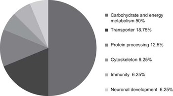
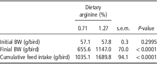
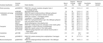
As shown in Table 2, the birds fed with low-arginine (0.71%) diet had a lower final live BW and cumulative feed intake (P<0.01) than those fed with arginine-sufficient diet (1.27%). Because lysine competes with arginine for entry into the cells, and excess lysine increases renal arginase activity and degradation of arginine to ornithine and urea (Austic and Nesheim, Reference Austic and Nesheim1970; Wu and Morris, Reference Wu and Morris1998), there is a classic antagonism between lysine and arginine. The observation in chickens fed diet containing normal level of arginine and high lysine indicates that a portion of the antagonism is because of a primary effect of lysine on the regulation of food intake (Austic and Scott, Reference Austic and Scott1975). It remains to be determined whether the low appetite in birds fed diet with low arginine and normal lysine is associated with dietary lysine concentration in the present study. Hypothalamic protein expression was compared between arginine-sufficient and -deficient treatments. A total of 16 identified differentially expressed proteins were found in the hypothalamus. Information (protein name, accession number, molecular mass expected, protein score, sequence coverage, quantitative ratio and expression variation) about these proteins was listed in Table 3. Among those proteins, the expression of nine proteins was upregulated and seven proteins downregulated by arginine deficiency. The differentially expressed proteins were grouped into six functional classes (Figure 1) related to carbohydrate metabolism and energy production, protein processing, transporter, cytoskeleton, immunity and neuronal development. Proteins associated with carbohydrate metabolism and energy production were the major protein families that were differentially expressed and constituted 50% of all the differentially expressed proteins identified.
Functional classification of the differentially expressed proteins identified from White Pekin duck hyperthalamus under dietary arginine deficiency.

Effect of dietary arginine on performance of White Pekin duck from 1 to 21 days of ageFootnote 1
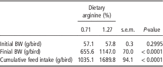
1 The value is the mean of seven repeats.
Effect of arginine on protein expression in the hypothalamus of White Pekin duck
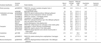
1 The relative expression of identified proteins was based on the ratio of the reporter ions of the peptides in arginine-deficient group to arginine-sufficient group. Only the proteins that changed more than 1.5-fold (i.e. ⩾1.50 or ⩽0.67) in expression were listed.
2 ‘↑’ and ‘↓’ denote upregulation and downregulation in protein expression, respectively.
It has been demonstrated that arginine can modulate glucose uptake as well as glucose and fatty-acid oxidation in the periphery tissues, such as the skeletal muscle, heart, liver and adipose tissue of mammals (Jobgen et al., Reference Jobgen, Fried, Fu, Meininger and Wu2006). Thus far, little is known about the involvement of arginine in regulating carbohydrate metabolism and energy production in the brain issue. In current studies, aldolase B fructose-bisphosphate and 6-phosphofructokinase type C-like, two enzymes involved in glycolysis were downregulated, suggesting dietary arginine deficiency may lead to the reduced glycolysis in the hypothalamus. Dietary arginine deficiency also decreased the expression of transaldolase, which would reduce the production of glyceraldehyde 3-phosphate and fructose 6-phosphate (two important substances for glycolysis) through non-oxidative phase from the pentose phosphate pathway. Aconitase was downregulated by dietary arginine deficiency, possibly leading to a disturbance of isocitrate production in tricarboxylic acid cycle. Guanidinoacetic acid is synthetized from arginine and glycine by glycine amidinotransferase, and transformed into creatine by methyl removing to participate in energy production (Bessman and Carpenter, Reference Bessman and Carpenter1985). The decreased expression of glycine amidinotransferase by dietary arginine deficiency possibly results in a low creatine biosynthesis. The results aforementioned showed that arginine not only took part in aerobic energy metabolism but also in anaerobic glycolysis in the brain tissue. As glycolysis and tricarboxylic acid cycle are the main ways by which the cells get energy, the decreased expression of enzymes associated with glycolysis and tricarboxylic acid cycle may inhibit carbohydrate metabolism and energy production.
Haemoglobin is a tetramer containing two α-subunits and two β-subunits, and functions to carry oxygen to the tissues of body and transport the resultant carbon dioxide back to the lungs. In the present study, haemoglobin α-subunit expression was decreased in the hypothalamus of low-arginine group, which would decrease oxygen supply to the hypothalamus and block carbon dioxide exportation from the tissue. Importin is a set of nuclear transport receptors and plays an important role in cell differentiation and apoptosis (Stewart, Reference Stewart2007). The decreased expression of importin subunit β -1-like caused by low arginine possibly inhibits nuclear protein transport and cell development in the hypothalamus. Voltage-dependent anion channel protein is a key porin allowing metabolite (ATP, ADP, K+, Na+, Cl− and Ca2+) flow across the mitochondrial outer membrane (Rostovtseva and Colombini, Reference Rostovtseva and Colombini1996; Tan and Colombini, Reference Tan and Colombini2007). Low arginine downregulated the expression of protein associated with energy production, compensatory expression of voltage-dependent anion channel protein presumably occurred to regulate ATP synthesis and material transport in low-arginine treatment.
Eukaryotic elongation factor 2 is an essential factor for peptide chain elongation, thus playing a central role in protein synthesis (Kaul et al., Reference Kaul, Pattan and Rafeequi2011). Myotrophin is required for stimulating protein synthesis through accumulating muscle structural proteins, actin and myosin (Shiraishi et al., Reference Shiraishi, Nakamura, Iwamoto, Haruno, Sato, Mori, Ikeuchi, Chikushi, Hayashi and Sato2006). α-actinin is a cytoskeletal actin-binding protein and a member of the spectrin superfamily. Its role ranges from bundling actin filaments to acting as a versatile protein interaction platform for structural, membrane and signalling proteins (Sjoeblom et al., Reference Sjoeblom, Salmazo and Djinovic-Carugo2008). Arginine is a necessary block for body protein and its deficiency would limit protein synthesis. Therefore, expression upregulation of the three proteins presumably is a compensatory reaction induced by dietary arginine deficiency in the present study.
Members of the dihydropyrimidinase-related proteins family are involved in the axonal outgrowth and pathfinding through the transmission and modulation of extracellular signals (Minturn et al., Reference Minturn, Fryer, Geschwind and Hockfield1995). Platelet-activating factor has many actions in addition to activation of the platelets: activation of polymorphonuclear leukocytes, monocytes and macrophages, increased vascular permeability, decreased cardiac output and others (Prescott et al., Reference Prescott, Zimmerman and McIntyre1990). Platelet-activating factor degradation to inactive products is catalyzed by platelet-activating factor acetylhydrolase. However, it is not clear why the platelet-activating factor acetylhydrolase and dihydropyrimidinase-related protein 1-like were increased under arginine deficiency in the present experiment.
In the hypothalamus, quantification of the regulated proteins associated with appetite is upregulated or downregulated with changes in feed intake (Kamegai et al., Reference Kamegai, Tamura, Shimizu, Ishii, Sugihara and Wakabayashi2001; Li et al., Reference Li, Yan, Zheng, Liu, Zhang and Wang2008). In the present study, the low-arginine diet led to a decreased feed intake; however, the expression of proteins in the hypothalamus known to be involved in feed intake had not changed. It indicated that arginine possibly does not regulate feed intake in ducks by changing the feeding protein expression in the hypothalamus. Nitric oxide is produced by arginine decarboxylation, and deficiency of nitric oxide would block the orexigenic effect of neuropeptide Y (Morley et al., Reference Morley, Farr, Sell, Hileman and Banks2011), ghrelin (Gaskin et al., Reference Gaskin, Farr, Banks, Kumar and Morley2003), orexin (Farr et al., Reference Farr, Banks, Kumar and Morley2005) and methylamine (Raimondi et al., Reference Raimondi, Alfarano, Pacini, Livi, Ghelardini, DeSiena and Pirisino2007). Therefore, it remains to be determined whether dietary arginine must be converted to nitric oxide as downstream signalling molecule of several feeding proteins to regulate feed intake in ducks. However, dietary arginine deficiency downregulated the expression of proteins associated with energy production and oxygen transport in the hypothalamus, and possibly resulted in a biological malfunction of hypothalamus, which presumably takes an adverse effect on appetite regulation of the hypothalamus.
Conclusion
Arginine could be a critical nutrient for stimulating appetite in ducks. Arginine deficiency inhibits feed intake in ducks through a mechanism that does not alter the expression of appetite-associated proteins in the hypothalamus. Dietary arginine deficiency downregulates the expression of proteins involved in energy production and oxygen transport.
Acknowledgement
This work was supported by the Earmarked fund for Modern Waterfowl Industry Technology System of China (CARS-43).