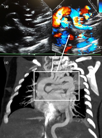
A 2-weeks-old boy was referred for systolic murmur. Cardiac ultrasound showed abnormal aortic arch with chicane shape isthmus (Fig 1 A and B) and tortuous pulmonary arteries although no intra-cardiac defect was detected. There were no evidence of aortic obstruction, pulmonary stenosis at any level, or secondary pulmonary hypertension.
( a and b ) Cardiac imaging of tortuous aortic arch with chicane shape isthmus ( a : echocardiography and b : echocardiography with colour Doppler). ( c ) Angioscanner of the entire tortuous aortic arch. The frame highlights the same vascular structures as in panels a and b .

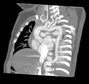
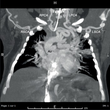
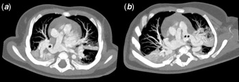
A thoracic angioscanner proved the tortuosity of aortic arch (Figs 1B and 2), great vessels (Fig 3), and pulmonary arteries (Fig 4). This observation suggested arterial tortuosity syndrome that was confirmed by genetic analysis.
Angioscanner image of the tortuous aortic arch and origin of the great vessels.

Angioscanner image of the tortuous great vessels. Ao = aorta, RSCA = right subclavian artery, RPCA = right primary carotid, LPCA = left primary carotid, LSCA = left subclavian artery.

Angioscanner images of the tortuous right and left pulmonary arteries. * = right pulmonary artery; ** = left pulmonary artery.

Arterial tortuosity syndrome is a rare autosomal recessive disorder involving SCL2A10 gene, responsible for abnormalities of connective tissue, causing elongation and tortuosity of the large and medium-sized arteries,Reference Callewaert, De Paepe, Coucke, Adam, Ardinger and Pagon1 with risks of aneurysms, dissections and stenosis of these vessels (sometimes requiring surgical repair, for example, peripheral pulmonary stenosis with secondary pulmonary hypertension).Reference Callewaert, De Paepe, Coucke, Adam, Ardinger and Pagon1-Reference Al-Khaldi, Momenah and Alsahari2 Dysmorphic facial features (elongated face, large ears, downslanted palpebral fissures, hypertelorism, high arched palate), hypotonia, hyperlaxity and cutis laxa, skeletal deformities (scoliosis, pectus abnormalities), ophthalmologic abnormalities (myopia, keratoconus), hernias (inguinal, umbilical, hiatal), and respiratory impairment (new-born respiratory distress syndrome, emphysema after severe bronchiolitis) have also been reported.Reference Callewaert, De Paepe, Coucke, Adam, Ardinger and Pagon1
Our patient presented inguinal hernias. Cutis laxa and dysmorphic features became evident during the first year of life. He also developed bronchial hyper reactivity requiring transient inhaled corticoids.
The concordance of both ultrasound and angioscanner images demonstrates that ultrasound is an efficient tool to study aortic arch. Abnormalities of great vessels do not necessarily imply cardiac malformation but may also be the sign of connective tissue disorders.
After 3 years of follow-up, the patient is asymptomatic except for subcutaneous spheroids on the legs. Echocardiography remains stable, without sign of aortic obstruction, pulmonary stenosis at any level, or secondary pulmonary hypertension. Total body angioMRI performed at the age of 2 years confirmed tortuosity of all the large and medium-sized arteries without aneurysm or stenosis.
The scheduled cardiovascular follow-up consists of echocardiography every 3 months and angioMRI every 5 years.
Financial support
This research received no specific grant from any funding agency, commercial, or not-for-profit sectors.
Conflicts of interest
None.
Ethical standards
The authors assert that all work reported complies with the ethical standards of the Helsinky convention, and consent for publication has been granted by the patient’s family.