Introduction
Larvae of the New World screwworm fly, Cochliomyia hominivorax, cause myiasis in animals and humans, constituting a public health problem in tropical and subtropical areas of South America (Silva et al., Reference Silva, Moya-Borja and Queiroz2011; Lacourse et al., Reference Lacourse, Martinez, Spach and Fang2015). This parasite affects cattle productivity and incurs economic losses, with an estimated cost of U$ 0.34 billion per year spent on curative and preventive treatments (Grisi et al., Reference Grisi, Leite, Martins, Barros, Andreotti, Cançado, Leon, Pereira and Vilella2014).
Topical and systemic antiparasitic agents are used to control C. hominivorax larvae in bovines (Lopes et al., Reference Lopes, Teixeira, Felipelli, Cruz, Maciel, Matos, Pereira, Buzulini, Soares, Oliveira and Costa2013). Approximately 160 products are currently registered in Brazil for the control of this parasite (SINDAN, 2018). According to the Brazilian Ministry of Agriculture and Livestock and the World Association for the Advancement of Veterinary Parasitology, 10 to 12 animals are recommended for use in registration tests (Brasil, 1997; Holdsworth et al., Reference Holdsworth, Vercruysse, Rehbein, Peter, Bruin, Lettonja and Green2006). Thus, at least 1600 to 1920 bovines were used to validate the efficacy of these formulations for sale against C. hominivorax larvae. As such studies are costly and involve an experimental protocol that is both painful and cruel, alternative methods are desirable.
The in vitro design has several advantages, such as a fast execution, relatively low cost and predictable hazards or risks (Nijhof and Tyson, Reference Nijhof, Tyson, Meng and Sluder2018). Moreover, this method is animal friendly, as it is not necessary to induce wounds in cattle to evaluate larval infestation and product efficacy. Thus, the aim of the present study was to create an in vitro method for the evaluation of the efficacy of topical products for the control of C. hominivorax larvae. An in vivo test on bovines was also performed to verify the consistency of the results found during the in vitro test.
Materials and methods
Cochliomyia hominivorax colony
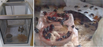
To obtain different larval stages of C. hominivorax for the in vitro test, a colony was developed using a method adapted from Oliveira et al. (Reference Oliveira, Brito, Giglioti and Chagas2009). A substrate denominated ‘oviposition mixture’ was prepared with 10 g of minced bovine meat and two test tubes containing blood (±10 mL) + anticoagulant (sodium citrate), which were refrigerated for 10 days. To obtain the egg masses, ~3 g of the oviposition mixture were deposited on the edge of a round plastic lid in a cage containing C. hominivorax adults. The same procedure was performed on the opposite edge of the lid and egg masses were extracted after 4 h (Fig. 1A and B).
(A) Cage to obtain C. hominivorax egg masses. (B) Cochliomyia hominivorax females realizing egg posture on an artificial substrate made principally with raw bovine meat.

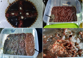
To obtain C. hominivorax L1, 30 g of minced meat (1 cm wide and 1 cm high) were placed cross-shaped in a round non-toxic polyethylene pot measuring 9.5 cm in width and 3.5 cm in height with a capacity of 150 mL. In the empty spaces between the ‘cross’ of meat, a liquid diet composed of blood powder (70 g) + egg powder (40 g) + formaldehyde (1 mL) + water (800 mL) was added. The egg masses were distributed over the minced meat, with ~0.5 cm of egg mass in each part of the ‘cross’. The pots were then sealed (with aeration) and placed in a biological oxygen demand (BOD) chamber with controlled temperature (35 to 37°C) and relative humidity (⩾70%). L1 larvae were obtained after 36 h.
Forty-eight hours after the preparation of the mixture described above, part of the L1 larvae was transported to a plastic vessel containing 150 g of minced bovine meat with 90 mL of the above-mentioned liquid diet. Cochliomyia hominivorax L2 larvae were obtained after 60 h. Next, the part of the L2 larvae was placed in a new plastic vessel with 50 g of minced bovine meat and 30 mL of the same liquid diet were added to the right corner of the bowl. Eighty-four hours after the initial preparation, L3 larvae were obtained and extracted (Fig. 2A–D). Throughout the period, the larvae were kept in BOD chambers with controlled temperature and relative humidity.
Cochliomyia hominivorax larvae (L) diets based on minced beef and liquid diet based on blood powder. (A) L1 recommended diet. (B) L2 recommended diet larvae, note how larvae were transferred from container A to the lower left corner of the container (arrow). (C) Diet for L3 diet, note that new meat has been added to the right side of the container (arrow). (D) Galleries formed in the diet by L3.

In vitro test
The in vitro test was performed from July to December 2017 at the Pathology and Parasitology Laboratories of UNIFRAN, Brazil. Six treatments were performed, five with commercial antiparasitic agents and one control. The following products were tested: T1 – coumaphos 3% + propoxur 2% (Tanidil®, Bayer Animal Health, Brazil); T2 – chlorfenvinphos 0.52% + dichlorvos 0.83% (Matabicheiras Forte SV®, Zoetis, Brazil); T3 – cypermethrin 0.5% + DDVP 1.8% + citronellal 4.64% + Ricinus oil 17.2% + metallic aluminium 5% (Couro Limpo Prata V18®, Noxon Animal Health, Brazil); T4 – cyhalothrin 1% + propoxur 1% (Cidental®, Bimeda, Brazil); T5 – cypermethrin 0.4% + DDVP 1.6% + silver sulphate 0.1% + aluminium 5% (Bactrovet Prata AM®, Konig, Brazil) and T6 – saline solution (control).
The C. hominivorax larvae used were from the colony described in Item 2.1. L1, L2 and L3 specimens with no anomalies and no visible mutilations were selected for the test. Nine containers identified with the product code, larval stage and date of treatment were destined for each tested product – three for each larval stage (triplicate). Two hundred larvae of the same stage were placed in each container. Thus, each product was tested on 1800 larvae (600 from each stage). The same procedure was performed for the control group.
The products were applied to the larvae following the manufacturer's recommendations. The volume was enough to cover the surface of the larvae with the respective product. The control group received the same amount of saline solution. All recipients were then stored a BOD chamber at 37°C and relative air humidity ⩾70% throughout the experiment. The samples were removed from the BOD chamber only for evaluation and then returned until the final reading. The viability of the larvae in each recipient of each group was evaluated after 5 and 30 min as well as 1, 2, 6, 12, 24, 48, 60 and 72 h post-treatment. The efficacy percentages obtained for each formulation at the different evaluation times were calculated using the following formula:

in which M is the mean of active larvae, CG is the control group and TG is the treatment group.
In vivo test
For the in vivo test, 30 crossbred male bovines (Bos taurus taurus × Bos taurus indicus) 20 months of age were obtained from Santos Reis ranch in the municipality of Itirapuã, São Paulo, Brazil. All animals had no contact with antiparasitic drugs for a minimum of 120 days before the onset of the trials and were identified by numbered earrings. During the experiment, the animals received pasture, water and mineral supplementation ad libitum.
We used a method adapted from Moya-Borja et al. (Reference Moya-Borja, Muniz, Umehara, Goncalves, Silva and McKenzie1997) and Lopes et al. (Reference Lopes, Teixeira, Felippelli, Cruz, Maciel, Matos, Pereira, Buzulini, Soares, Dos Santos, Oliveira and Costa2009). To verify the efficacy of the formulations tested, castration of the cattle was performed using the crude method. As the local anesthetic, each animal received 10 mL of 2% xylocaine (Anestésico Vansil®, Vansil Saúde Animal, Brazil) subcutaneously in the distal region of the scrotum (‘lid’). After an incision in the scrotum, the animals were then released and kept in the pasture area for natural infestation by C. hominivorax larvae (myiasis).
The cattle were examined daily to verify the presence of C. hominivorax eggs and/or larvae. After visualization of C. hominivorax larval infestation in the wounds, allocation of the animals was performed using a complete block randomization model based on individual weight. The animals in each group were kept in separate pastures. Six groups of five bovines each were formed: T1 – coumaphos 3% + propoxur 2% (Tanidil®, Bayer Animal Health, Brazil); T2 – chlorfenvinphos 0.52% + dichlorvos 0.83% (Matabicheiras Forte SV®, Zoetis, Brazil); T3 – cypermethrin 0.5% + DDVP 1.8% + citronellal 4.64% + Ricinus oil 17.2% + metallic aluminium 5% (Couro Limpo Prata V18®, Noxon Animal Health, Brazil); T4 – cyhalothrin 1% + propoxur 1% (Cidental®, Bimeda, Brazil); T5 –cypermethrin 0.4% + DDVP 1.6% + silver sulphate 0.1% + aluminium 5% (Bactrovet Prata AM®, Konig, Brazil) and T6 – saline solution (control).
The treatment was performed on Day Zero in all groups as recommended by the manufacturers. The control group (non-medicated) remained untreated until 72 h post-treatment. To evaluate the curative efficacy of the formulations, the scrotal lesions of the cattle were carefully examined at 12, 24, 36, 48, 60 and 72 h post-treatment. Cochliomyia hominivorax larvae in the scrotum incisions were classified as active (at least one live C. hominivorax larvae/lesion), which was identified by the number one (1), or non-active (absence of C. hominivorax larvae/lesion), which was identified by the number zero (0), as described by Lopes et al. (Reference Lopes, Teixeira, Felippelli, Cruz, Maciel, Matos, Pereira, Buzulini, Soares, Dos Santos, Oliveira and Costa2009). The percentages of efficacy obtained for each drug were calculated using the following formula:

in which N is the mean of active larvae, CG is the control group and TG is the treatment group.
Data analysis
The results regarding the presence or absence of viable C.hominivorax larvae in the in vitro and in vivo tests were analysed with the aid of the SAS V9.1 program (2002), using Fisher's non-parametric test with the significance level set to 5% (P ⩽ 0.05).
Results
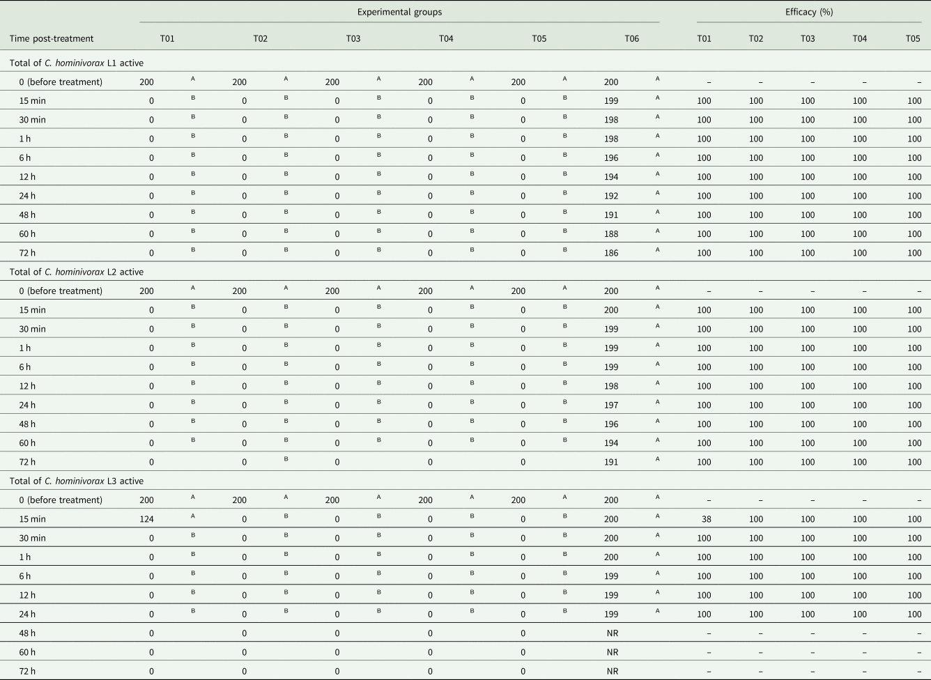
The efficacy results obtained for each formulation in the in vitro test are described in Table 1. All formulations demonstrated 100% efficacy against the L1 and L2 stages C. hominivorax at all times. For the L3 stage of this parasite, all formulations were 100% effective, except T1, for which viable L3 larvae were found 15 min after contact with the product (38% efficacy). However, this combination also demonstrated 100% efficacy at the subsequent evaluation (Table 1). Viable L1, L2 and L3 counts were higher in the control group than the treated groups at all post-treatment evaluations (P ⩽ 0.05), with the exception of the number of viable L3 larvae in T1 15 min post-treatment, which did not differ significantly from the number in the control group (P > 0.05). As L3 larvae in the control group molted to pupae after the 48-h evaluation, it was impossible to infer the efficacy and statistically analyse the formulations in question.
Number of C. hominivorax larvae stages and efficacy of the formulations used in the in vitro test

Means followed by the same letter in the line do not differ from each other (P > 0.05)
T01: coumafos 3% + propoxur 2% (Tanidil® – Bayer Saúde Animal, Brazil); T02: chlorfenvinphos 0.52% + dichlorvos 0.83% (Matabicheiras Forte SV® – Zoetis, Brazil); T03: cypermethrin 0.5% + DDVP 1.8% + citronelal 4.64% + Ricinus oil 17.2% + metallic aluminium 5% (Couro Limpo Prata V18® – Noxon Saúde Animal, Brazil); T04: cyhalothrin 1% + propoxur 1% (Cidental® – Bimeda, Brazil); T05: cypermethrin 0.4% + DDVP 1.6% + silver sulphate 0.1% + aluminium 5% (Bactrovet Prata AM® – Konig, Brazil) and T06 control (saline solution). NR: not realized, larvae molting to pupae.
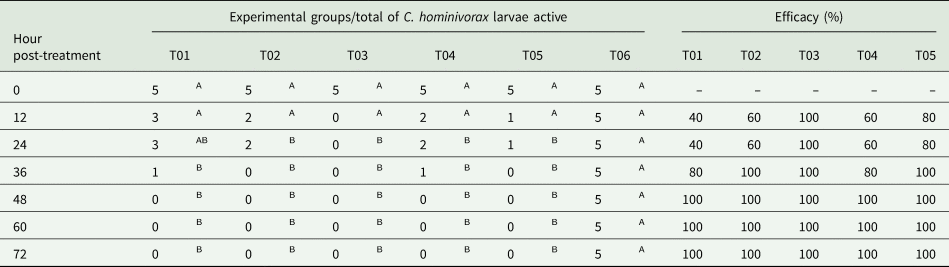
The results of the in vivo test revealed that treatments T2 and T5 achieved maximum efficacy (100%) against C. hominivorax larvae 36 h post-treatment, whereas treatments T1 and T4 achieved 100% efficacy beginning at 48 h post-treatment. Treatment T3 was the only formulation to achieve 100% efficacy at all post-treatment evaluation times (Table 2). The number of active larvae was significantly lower in groups T2, T3, T4 and T5 than the number found in the control group beginning at 24 h post-treatment through the final evaluation (P ⩽ 0.05). T1 differed significantly from the control group (P ⩽ 0.05) beginning at 36 h post treatment.
Number of active C. hominivorax larvae and therapeutic efficacy of the formulations used in the treatment of scrotal myiasis in cattle naturally infested with C. hominivorax larvae

Means followed by the same letter in the line do not differ from each other (P > 0.05)
T01: coumafos 3% + propoxur 2% (Tanidil® – Bayer Saúde Animal, Brazil); T02: chlorfenvinphos 0.52% + dichlorvos 0.83% (Matabicheiras Forte SV® – Zoetis, Brazil); T03: cypermethrin 0.5% + DDVP 1.8% + citronelal 4.64% + Ricinus oil 17.2% + metallic aluminium 5% (Couro Limpo Prata V18® – Noxon Saúde Animal, Brazil); T04: cyhalothrin 1% + propoxur 1% (Cidental® – Bimeda, Brazil); T05: cypermethrin 0.4% + DDVP 1.6% + silver sulphate 0.1% + aluminium 5% (Bactrovet Prata AM® – Konig, Brazil) and T06 control (saline solution).
Discussion
The results of the present study demonstrate that the method adopted in the in vitro test could be an alternative for the evaluation of the efficacy of topical products for the control of C. hominivorax larvae, as the results were similar to those obtained during the in vivo test.
Among all formulations evaluated during the in vitro experiment, coumaphos + propoxur combination achieved 100% efficacy against the L1 and L2 stages of C. hominivorax at all evaluation times. However, this product was the only one that did not achieve 100% efficacy after 15 min of contact with the L3 stage. All other formulations were 100% effective against all larval stages at all evaluation times. A drawback found in the in vitro experiment was the fact that L3 larvae pupated 48 h after treatment. This demonstrates the importance of considering the efficacy against this larval stage up to 24 h or perhaps the 84 h used to obtain this larval stage was too long. These issues should be investigated in future studies.
Analysing the results of the in vivo experiment, the formulations that took the longest time to demonstrate an effect against the ectoparasite in question were coumaphos + propoxur and cyclorin + propoxur, which achieved 100% efficacy beginning at 48 h after treatment. In the in vivo experiment, the formulations chlorfenvinphos + dichlorvos and cypermethrin + DDVP + silver sulphate + aluminium demonstrated 100% efficacy against C. hominivorax larvae beginning at 36 h post-treatment.
The superior efficacy of some formulations in the in vitro test when compared to the in vivo test may be explained by the methods employed. It is possible that the in vitro method enabled greater penetration of the chemical compounds into the cuticle of larvae, resulting in better absorption of the product by the ectoparasite and, consequently, greater efficacy.
The results found by Correa et al. (Reference Correa, Lopes, Teixeira, Cruz, Gomes, Felippelli, Maciel, Favero, Buzullini, Bichuette, Soares and Costa2015) in ticks may help understand the results obtained herein. The authors compared three methods for evaluating the effectiveness of topical formulations against the cattle tick Rhipicephalus microplus: adult immersion test (AIT), natural field infestation and stable test. The effectiveness of the topical formulations was higher in the AIT (100%) compared to the natural field infestation test (94.3 to 95.1%) and stable test (93.4 to 98.7%). It is important to mention that the AIT is a bioassay that has been recognized and accepted by the Food and Agriculture Organization of the United Nations (FAO, 2004). The search for in vitro methods to replace animal testing is also underway for pour-on and injectable products used on cattle against the tick R. microplus, as described by Klafke et al. (Reference Klafke, Sabatini, de Albuquerque, Martins, Kemp, Miller and Schumaker2006, Reference Klafke, Castro-Janer, Mendes, Namindome and Schumaker2012), Castro-Janer et al. (Reference Castro-Janer, Rifran, Gonzalez, Piaggio, Gill and Schumaker2010, Reference Castro-Janer, Rifran, Gonzalez, Niell, Piaggio, Gill and Schumaker2011) and Reck et al. (Reference Reck, Klafke, Webster, Dall'Angol, Scheffer, Souza, Corassini, Vargas, Santos and Martins2014).
Other aspects that may explain the difference in efficacy found in this study between the in vitro and in vivo tests are related to the fact that the wounds were open in the in vivo experiments, with the presence of animal tissue, organic matter and the proteolytic activity of the excretory/secretory products of the C. hominivorax larvae (Giglioti et al., Reference Giglioti, Guimarães, Oliveira-Sequeira, David, Brito, Huacca, Chagas and Oliveira2016), which may interfere with the action of the topical product. Furthermore, blood may flow in these open wounds as a result of C. hominivorax parasitism, which could cause the loss of part of the product, and some animals were able to lick the wound after treatment, which could diminish the amount of medicine at the wound site (Toutain et al., Reference Toutain, Modric, Bousquet-Mélou, Sallovitz and Lanusse2012). Although rainfall did not occur during the in vivo study, the presence of heat and dew may also interfere with the amount of product at the wound site, which could reduce the effectiveness of a formulation applied under in vivo conditions compared to in vitro conditions, as verified by Correa et al. (Reference Correa, Lopes, Teixeira, Cruz, Gomes, Felippelli, Maciel, Favero, Buzullini, Bichuette, Soares and Costa2015) and Silva et al. (Reference Silva, Prette, Lopes, Sakamoto, Buzullini, Santos, Cruz, Teixeira, Felippelli, Carvalho, Maciel and Costa2015) in studies involving bovine parasites.
The present results demonstrate that the in vitro method adopted in this study is a promising tool for the evaluation of the efficacy of topical products for the control of C. hominivorax larvae. However, further studies comparing both the in vitro and in vivo methods should be performed to confirm these findings.
Conclusion
The in vitro method used in this study to evaluate the efficacy of topical products for the control of C. hominivorax larvae proved to be valid, as the results were similar to those obtained during the in vivo test.
Financial support
This study was financed in part by the Coordenação de Aperfeiçoamento de Pessoal de Nível Superior – Brasil (CAPES) – Finance Code 001.
Conflict of interest
There were no conflicts of interest that may have biased the work reported in this paper.
Ethical standards
This study received approval from the animal welfare committee of Universidade de Franca (UNIFRAN, Franca, Brazil) under the protocol number 5746041116 and was conducted in accordance with the animal handling recommendations of the National Council of Animal Research.