Implications
The meat content of pig carcasses is a very important trait in pig production. Its improvement is one of the main breeding goals, and breeding animals have to be selected for their body composition, aiming for high meat content. Therefore, this study evaluated the imaging technique magnetic resonance imaging (MRI), regarding its potential to predict carcass composition in live pigs. It was concluded that area measurement of the loin eye region and its back fat layer on the basis of MRI images is a very promising technique to predict carcass composition in live pigs.
Introduction
One of the main breeding goals in pig production is the improvement of body composition and therefore carcass quality. Carcass dissection and chemical analysis are the gold standards for body composition analysis, but these are expensive and time-consuming methods and also fatal for the animal. In order to reduce costs and save valuable breeding animals, estimating body composition in vivo is of major concern in the field of animal breeding. An accurate evaluation of body composition in vivo is given by several techniques, which were recently discussed and compared by Scholz and Baulain (Reference Scholz and Baulain2009). The authors recommend preselecting breeding animals by ultrasound. Subsequently, as ultrasound is subject to biases associated with different problems (Houghton and Turlington, Reference Houghton and Turlington1992; Müller and Polten, Reference Müller and Polten2004), they recommend the use of a more exact method such as magnetic resonance imaging (MRI) or computed tomography (CT) to classify potential breeding animals more accurately. MRI and CT are both techniques belonging to the field of non-invasive imaging. Both provide similar images of nearly every part of the body, which offer the options of calculation of two-dimensional areas or three-dimensional reconstruction and volume rendering of distinct muscles, fat or bone tissue of particular regions – although the methods of image acquisition are completely different. CT provides X-ray-based cross-sectional images, whereas the MRI technique is based on a magnetic field combined with the application of radiofrequency waves. Therefore, MRI offers a non-invasive in vivo imaging technique completely free from any radiation exposure, which is of major concern for humans and also for the animal. Formerly, because of its costs, the use of MRI has been restricted to research facilities only (Baulain, Reference Baulain1997). At present, there is a market for older second-hand low-field MRI devices from human medical facilities. Compared with basic CT prices between 300.000 and 600.000 Euros (Kongsro et al. Reference Kongsro, Røe, Kvaal, Aastveit and Egelandsdal2009), these devices are very reasonably priced (<100.000 Euro) and go along with low operating costs, as they are chilled by water instead of helium or liquid nitrogen. These facts open new technical opportunities for performance testing even for smaller livestock research and industry facilities. Consequently, the objective of this study was to analyze whether the use of MRI as an imaging technique of highest accuracy regarding soft tissue imaging on the loin eye area in live pigs can provide sufficient information to predict the in vivo body composition. Dual energy X-ray absorptiometry (DXA) was used as a reference method for determining the body composition of the whole pigs in vivo, as it offers repeatable and accurate results in predicting the body composition of live pigs. DXA is a quantitative body composition analysis method based on the tissue-specific attenuation of X-rays (Pietrobelli et al., Reference Pietrobelli, Formica, Wang and Heymsfield1996). The method has often been proved to accurately predict body composition in live pigs compared with chemical analysis (Mitchell et al., Reference Mitchell, Scholz and Conway1998a; Scholz, Reference Scholz2002; Suster et al., Reference Suster, Leury, Ostrowska, Butler, Kerton, Wark and Dunschea2003) or dissection (Scholz and Förster, Reference Scholz and Förster2006). In the latter study using the same DXA device as in the present one, DXA variables explained a high proportion of variation (R 2 > 0.8; √MSE = 70 to 784 g) in the tissue masses of the reference parts from dissection (fat, muscle, bone) in two BW groups (I: 30 to 50 kg; II: 70 to 90 kg). Almost similar relationships resulted for tissue percentages as was shown on pig carcasses by Scholz et al. (Reference Scholz, Soffner, Littmann, Peschke and Foerster2002) with R 2 = 0.89, √MSE =1.30% for dissection fat% v. DXA traits and R 2 = 0.81; √MSE = 1.57% for dissection lean meat% v. DXA traits.
Material and methods
The study was conducted at the Livestock Center Oberschleissheim of the Veterinary Faculty of the University of Munich. The protocol of the study was approved by the Government of Upper Bavaria with the tracking number 55.2-1-54-2531.2-60-07.
Animals
In this study – similar to the related study by Kremer et al. (Reference Kremer, Fernández-Fígares, Förster and Scholz2012), 77 pigs (seven boars, 33 barrows and 37 gilts) were examined for their in vivo body composition by using MRI and DXA. The pigs originated from different extensive or conventional breeds or crossbreds: Cerdo Iberico (n = 5), Duroc ×Cerdo Iberico (n = 15), German Landrace and German Large White = White Sow Lines (n = 8), Hampshire and Piétrain ×Hampshire and Piétrain × Piétrain–Hampshire (n = 6), Piétrain × Duroc and Piétrain × Piétrain–Duroc (n = 24) and Piétrain or Piétrain–Duroc × White Sow Lines (n = 19). The pigs ranged from 45 to 102 kg in BW and from 122 to 189 days in age. Arithmetic means and standard deviations for traits measured by DXA and MRI are summarized in Table 1.
Descriptive statistics for traits measured by MRI and DXA

MRI = magnetic resonance imaging; DXA = dual energy X-ray absorptiometry; DXA-LM = DXA soft lean tissue mass; DXA-%LM = DXA soft lean tissue percentage; DXA-FM = DXA fat tissue mass; DXA-%FM = DXA fat tissue percentage; MRI-FA = MRI fat area above the corresponding MRI-LA loin eye area of both body sides at 13th/14th vertebra.
MRI scans
A 0.2 T Magnetom Open combined with a large body coil (Siemens, Erlangen, Germany) was used for MRI scanning. The MRI protocol consisted of a T1 weighted spin echo sequence (time of repetition (TR) 380 ms, time between echoes (TE) 15 ms, flip angle 90°, 10 slices), a field of view of 461 × 461 mm with an image matrix of 256 × 256 pixels and a transversal slice thickness of 15 mm with a distance factor of 0.25. The acquisition time for one sequence with four repetitions was 6 min 32 s. The MRI scans were performed to compare loin eye area (MRI-LA) and its above fat area (MRI-FA) in the region of the 14th vertebra of both body sides – according to the site of the standard performance testing procedure of German pig breeders. Therefore, after performing a localizer scan, the slices of the specified protocol were positioned on the coronary plane with the first slice in the region of the 13th vertebra, in order to see the region of interest on the second or third slice. The muscle and fat areas were calculated using the image analysis software Dicom Works 1.3.5 (Inviweb, Lyon, France). The corresponding regions of interest for both body sides were defined manually after visually distinguishing between muscle and fat by qualified personnel as described in Kremer et al. (Reference Kremer, Fernández-Fígares, Förster and Scholz2012).
DXA analysis
Single DXA scans were performed by using a GE Lunar DPX-IQ pencil beam scanner (Lunar software; version 4.7e, GE Healthcare, Chalfont St. Giles, UK). The whole body ‘adult normal’ scan mode was used on all animals. Every 2nd day a calibration scan of a phantom was performed to ensure the maintenance of accurate body composition results. Measurements taken by DXA included soft lean tissue mass (DXA-LM), soft lean tissue percentage (DXA-%LM), fat tissue mass (DXA-FM), fat percentage (DXA-%FM) and total tissue mass as BW.
Animal preparation and scanning
Both examinations of the pigs were conducted in one session. Before the scans, the pigs were fasted for ∼16 h. Examinations started with weighing the pigs in order to calculate anesthesia. The animals were sedated by an intramuscular injection of Azaperone (1.2 mg/kg BW; Stresnil®; Janssen Pharmaceutica, Beerse, Belgium), followed by intramuscularly administered Ketamine (40 mg/kg BW; Ursotamin®; Serumwerk Bernburg AG, Bernburg, Germany). After the effect occurred, an intravenous (i.v.) catheter was inserted into an ear vein and fixed with tape, enabling i.v. follow-up dosing of Ketamine if necessary. Primarily, the pigs were placed on the MRI scanner in a prone position with fore and hind legs extended. Subsequently, the DXA scan was performed. The pigs were also fixed in prone position with hind legs extended and slightly tied together, but the front legs were positioned caudally separated from the body by two foam plastic wedges. After both scans, the pigs were moved into a separate pen to enable a gentle recovery phase after anesthesia.
Statistical analysis
The SAS software package (version 9.2; SAS Institute Inc., Cary, NC, USA) provided the tools for data analysis.
A linear single regression analysis using the ‘Proc Reg’ module was performed to quantify the linear relationships between DXA-LM and MRI-LA, DXA-%LM and MRI-LA, DXA-FM and MRI-FA and DXA-%FM and MRI-FA. In addition, a stepwise regression procedure with a significance level of P < 0.05 for the explanatory MRI variables and BW entering and staying in the equations were performed to calculate the best-fitting (multiple) regression equations with the DXA traits as response variables. The results of the multiple regression analyses are given as coefficient of determination (R 2), root mean squares error (√MSE), √MSE divided by the standard deviation (s.d.) of the response variable, intercept and slopes. In addition, after performing a PLS (partial least squares) analysis for the above calculated ‘multiple’ relationships, we provide the minimum root mean PRESS value for the leave-one-out cross-validation test using the standard iterative NIPALS algorithm.
Results
The detailed results and statistical differences of the DXA and MRI measurements especially for the different genotype groups are provided and discussed in a study by Kremer et al. (Reference Kremer, Fernández-Fígares, Förster and Scholz2012).
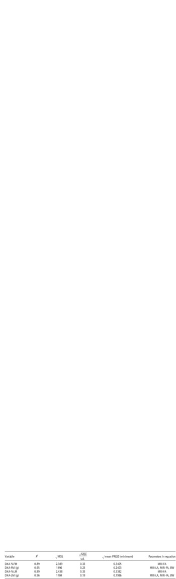
During the current study, the calculated areas of the loin eye muscle and the above back fat of all pigs were evaluated for predicting the response variables DXA-%LM and DXA-LM, DXA-%FM and DXA-FM, respectively, using regression analysis (Tables 2 and 3). As a result, MRI-FA predicts DXA-%FM and DXA-FM with a high accuracy. Similarly, MRI-LA shows a high relationship with DXA-LM. However, MRI-LA shows no relationship with DXA-%LM. The following prediction equations resulted from the multiple regression analysis:
Relationship between DXA-%LM (g) and MRI-LA (cm2)

DXA = dual energy X-ray absorptiometry; LM = lean tissue mass; MRI = magnetic resonance imaging; MSE = mean squares error; DXA-%LM =DXA soft lean tissue percentage; DXA-LM = DXA soft lean tissue mass.
Relationship between DXA-FM (g) and MRI-FA (cm2)

DXA = dual energy X-ray absorptiometry; FM = fat mass; MRI = magnetic resonance imaging; MRI-FA = fat area above the loin muscle, calculated from MRI images; MSE = mean squares error; DXA-%FM = DXA fat tissue percentage; DXA-FM = DXA fat tissue mass.
 $${ {\rm{DXA {\hbox -} LM(g)}}\: {\rm{ = }}\:{\rm{11501}}\:{\rm{ + }}\:{\rm{(83}}{\rm{.34}}\:\times \:{\rm{MRI {\hbox -} LA}}\,({\rm{cm^2))}} \cr \quad- {\rm{(360}}{\rm{.40}}\:\times \:{\rm{MRI {\hbox -} FA}}\,({\rm{cm^2))}}\:\cr \quad{\rm{ + }}\:{\rm{(644}}{\rm{.42}}\:\times \:{\rm{BW}}\,({\rm{kg))}} $$
 $$ {\rm{DXA {\hbox -} \% FM}}\:{\rm{ = }}\:{\rm{1}}{\rm{.787}}\:{\rm{ + }}\:{\rm{(0}}{\rm{.599}}\:\times \:{\rm{MRI {\hbox -} FA}}\,({\rm{cm^2))}} $$
 $$ {\rm{DXA {\hbox -} \% LM}}\:{\rm{ = }}\:{\rm{95}}{\rm{.72}} \,{ -} \,{\rm{(0}}{\rm{.62}}\:\times \:{\rm{MRI {\hbox -} FA}}\,({\rm{cm^2))}} $$
 $${ {\rm{DXA {\hbox -} FM}}\,{\rm{(g)}}\:{\rm{ = }}\: \minus {\rm{10025}} \,{ -}\, {\rm{(53}}{\rm{.87}}\:\times \:{\rm{MRI {\hbox -} LA}}\,({\rm{cm^2))}}\:\cr \quad{\rm{ + }}\:{\rm{(315}}{\rm{.62}}\:\times \:{\rm{MRI {\hbox -} FA}}\,({\rm{cm^2))}}\:\cr \quad{\rm{ + }}\:{\rm{(270}}{\rm{.94}}\:\times \:{\rm{BW}}\,({\rm{kg))}} $$
The accuracies of the prediction equations are described by the coefficient of determination (R 2) and the corresponding root mean squares errors √MSE or prediction errors (√mean PRESS) as shown in Table 4.
Models selected for prediction of DXA values of in vivo body composition of pigs using MRI

DXA = dual energy X-ray absorptiometry; MRI = magnetic resonance imaging; MSE = mean squares error; PRESS = sum of squares of predicted residual errors; DXA-%FM = DXA fat tissue percentage; MRI-FA = fat area above the loin muscle, calculated from MRI images; DXA-FM = DXA fat tissue mass; MRI-LA = loin area, calculated from MRI images; DXA-%LM = DXA soft lean tissue percentage; DXA-LM = DXA soft lean tissue mass.
Discussion
In connection with the still increasing demand for pork and especially lean meat content in slaughter pigs, it is of major concern to establish methods of accurate in vivo body composition analysis and estimation of breeding values to select healthy and valuable breeding animals. Currently, selection of animals is based on progeny testing. Ultrasound – mainly by using A-mode devices – is used routinely for performance testing to estimate the lean meat content. The ultrasound method has several advantages, namely mobility, the fact that no anesthesia is required and it is a time-saving method. Compared with other body composition analysis methods, however, the accuracy of ultrasound measurements depends on many factors. It can be affected by the examining person, site of measurement, the device used and the pig's live weight, breed and gender (Mueller and Polten, Reference Müller and Polten2004). Progeny testing is an expensive and time-consuming method to estimate meat yield, and an indirect method to evaluate breeding animals. Therefore, as recommended by Scholz and Baulain (Reference Scholz and Baulain2009), it could be reasonable and economically effective to preselect animals by the ultrasound technique, and later, choose the animals carefully out of the preselected pool by more sophisticated techniques. Because performance and progeny testing not only provide data about lean meat and fat tissue content, but also about distinct regions of the carcass, for example, loin eye area, back fat thickness, proportion of valuable lean cuts and proportion of ham (Kernerová et al., Reference Kernerová, Václavovský, Matoušek and Hanyková2006), a method is needed, which provides reliable information about in vivo body composition and distinct regions of the body in live pigs. As mentioned above, the recommended imaging techniques by Scholz and Baulain (Reference Scholz and Baulain2009) are MRI and CT.
Both methods are relatively expensive, need a high level of expertise and offer cross-sectional images of nearly every part of the body (Szabo et al., Reference Szabo, Babinszky, Verstegen, Vangen, Jansman and Kanis1999; Scholz and Baulain, Reference Scholz and Baulain2009). CT creates X-ray-based images of the body, and therefore the application of ionizing radiation is an important difference between both techniques. CT exposes animals to high whole-body radiation and underlies strict government regulations. In contrast, MRI is based on a magnetic field combined with the application of radiofrequency waves. No ionizing radiation may cause damage to the human operator or to the livestock, which is of crucial importance when dealing with breeding animals. Furthermore, MRI is favored to differentiate soft tissues (Moon et al., Reference Moon, Genant, Helms, Chafetz, Crooks and Kaufman1983). High acquisition, operating costs and the requirement for high technical expertise restricted the use of MRI for livestock breeding, basically to research during the last few years (Baulain, Reference Baulain1997; Scholz and Baulain, Reference Scholz and Baulain2009). The availability of MRI devices with lower magnetic field strength (0.2 to 0.5 T) offers the opportunity for a more efficient livestock imaging. These permanent-magnet systems are very reasonably priced. The operating costs are low, because the cooling system runs with water instead of helium and liquid nitrogen. Monziols et al. (Reference Monziols, Collewet, Bonneau, Mariette, Davenel and Kouba2006) used a low-field MRI device in order to predict tissue composition of pig carcasses and primary cuts. Compared with full dissection, the authors achieved very high relationships regarding muscle weights (R 2 ranging from 0.970 to 0.997 with very low residual s.d. between 81 and 129 g) and total fat weight (R 2 ranging from 0.877 to 0.981 with very low residual s.d. (RSD) between 143 and 292 g). The subcutaneous fat weight at the loin was even estimated with an R 2 = 0.99 (RSD = 105 g). However, MRI has also been used for in vivo body composition analysis in several species, for example, in pigs (Scholz et al., Reference Scholz, Baulain and Kallweit1993; Baulain, Reference Baulain1997; Mitchell et al., Reference Mitchell, Scholz, Wang and Song2001), in sheep (Streitz et al., Reference Streitz, Baulain and Kallweit1995) and in chicken (Davenel et al., Reference Davenel, Seigneurin, Collewet and Rémignon2000). MRI analysis offers a wide range of area and volume calculations of distinct regions, organs or muscles of live animals. Mitchell et al. (Reference Mitchell, Scholz, Wang and Song2001) verified MRI-derived volume calculations of the brain, the heart, left and right kidney and the liver in 28 freshly killed pigs by dissection and found corresponding relationships (R 2) of 0.67, 0.64, 0.84, 0.86 and 0.90 combined with error estimates (SEE) of 8, 65, 17, 15 and 125 g, respectively. The weight of shoulder, longissimus dorsi and ham muscle showed in the average for both body sides also have high coefficients of determination (R 2) of 0.85 (SEE = 241.5 g), 0.93 (SEE = 117.5 g) and 0.965 (SEE = 209 g), respectively. During the same study, they also evaluated different MRI-derived muscle and fat volumes of another 61 pigs (Experiments 3 and 4) by stepwise regression for the prediction of total body fat percentage and total body lean percentage. They also found high relationships (R 2) between the MRI tissue volumes (fat + muscle) and chemical carcass composition of 0.91 (fat%) and 0.89 (lean%), respectively. The corresponding error estimates (SEE) were 1.761% and 1.984%. These results document the very promising potential offered by MRI for the prediction of different carcass and body composition traits in vivo.
For establishing new methods, a reliable reference method is needed. As mentioned before, chemical analysis is the gold standard for body composition analysis, but fatal for the animal. On the contrary, DXA offers a non-invasive method of quantitative analysis and is an approved method to measure in vivo body composition of pigs. It accurately provides information about fat, lean tissue and bone mineral, regarding mass and percentage distribution of the whole body – though it does not provide three-dimensional information about the distribution of the tissues of interest, such as fat, soft lean or bone mineral. DXA has been used successfully to predict body composition in live pigs (Mitchell et al., Reference Mitchell, Conway and Potts1996 and Reference Mitchell, Scholz and Conway1998a; Scholz, Reference Scholz2002; Suster et al., Reference Suster, Leury, Ostrowska, Butler, Kerton, Wark and Dunschea2003; Scholz and Foerster, Reference Scholz and Förster2006) and carcasses (Mitchell et al., Reference Mitchell, Scholz and Conway1998, Scholz et al., Reference Scholz, Soffner, Littmann, Peschke and Foerster2002; Marcoux et al., Reference Marcoux, Faucitano and Pomar2005) and has been proven for several BW categories (Mitchell et al., Reference Mitchell, Scholz and Conway1998a and Reference Mitchell, Scholz and Pursel2000; Lukaski et al., Reference Lukaski, Marchello, Hall, Schafer and Siders1999; Scholz Reference Scholz2002) compared with chemical analysis. Above all, Suster et al. (Reference Suster, Leury, Ostrowska, Butler, Kerton, Wark and Dunschea2003) found that in vivo DXA-derived values for fat tissue mass were more strongly related with values of chemical analysis-derived lipid than P2 back fat measurements in carcasses. That means DXA is not only an alternative technique for progeny testing, but also superior in accuracy and represents a reference technique, which reliably offers repeatability and measurement precision – though results among different manufacturers or technologies are not directly comparable without cross-calibration (Ruge, Reference Ruge2006; Lösel et al., Reference Lösel, Kremer, Albrecht and Scholz2010).
Therefore, by using only one device and software mode, the present study used DXA as quantitative in vivo analysis reference technique to evaluate the potential of MRI as in vivo imaging technique for predicting body composition in live pigs. The pigs originated from different extensive or conventional breeds and their crosses, such as Cerdo Iberico or German Landrace, German Large White, Hampshire, Duroc and Piétrain, representing a wide range of body composition as described in a study by Kremer et al. (Reference Kremer, Fernández-Fígares, Förster and Scholz2012). First of all, the coefficients of determination and error estimates between the MRI-derived muscle or fat areas and the DXA-derived tissue masses or proportions were evaluated by linear regression and an additional PLS analysis. The measurement values of both devices showed – with exception of the relation between MRI-LA and DXA-%LM – high R 2 and reasonable error terms. The MRI-derived FA was highly related with DXA-%FM and with DXA-FM, whereas the regression coefficient between MRI-LA and DXA-LM was R 2 = 0.82 (√MSE = 4018 g). Alone, as expected for our material, MRI-LA and DXA-%LM were not related at all because the DXA lean percentage is higher related to the MRI-derived subcutaneous fat layer, which shows a higher variation than does the loin eye area in pigs.
Thereupon, a stepwise regression analysis was performed to predict DXA-%FM, DXA-FM, DXA-%LM and DXA-LM with MRI-derived traits and BW (Table 4). Both regression equations for the dependent variables DXA-%FM and DXA-%LM contained the independent variable MRI-FA alone. DXA-FM and DXA-LM were both predicted with a significance level of P < 0.05 for entry and stay by the independent variables MRI-LA, MRI-FA and BW. The relatively high R 2 (R 2 > 0.89) combined with reasonable root mean squares error (and prediction errors = √mean PRESS × s.d. of response variable) of <2.5% for DXA%FM or DXA%LM and of <1852 g for DXA-FM or DXA-LM of the generated prediction equations (Table 4) prove expectedly the prediction potential of the chosen MRI-derived areas. Scholz (Reference Scholz2002) found in comparison with the above results an R 2 of 0.91 and an RMSE of 1801 g for the relationship between DXA-FM and the MR fat volume above the loin eye region within a 10 cm wide body region starting at 14th thoracic vertebra in cranial direction. From a practical point of view, simple area measurements of the loin eye area and its above back fat can be used favorably, because they provide almost as exact prediction equations as those by Mitchell et al. (Reference Mitchell, Scholz, Wang and Song2001), using MRI-derived tissue volumes to predict tissue weights of pigs after dissection. These authors reported coefficients of determination of R 2 = 0.99 for a multiple regression equation predicting lean mass after dissection by MRI-derived volumes of back fat, shoulder muscle, longissimus muscle and psoas muscle as independent variables, and of R 2 = 0.97 for predicting fat mass using the MRI-derived volumes of jowl fat, back fat and shoulder muscle as independent variables. The relationship between ham muscle volume (by MRI) and total lean mass from chemical analysis yielded an R 2 of 0.982 with an RMSE = 1511 g, whereas the relationship between MRI back fat volume and total fat mass resulted in an R 2 of 0.952 with an RMSE = 803 g. In consideration of the number of examined body regions and the fact that area calculations are quicker and easier to perform than volume renderings, the method of the current paper seemed to be very suitable for an accurate in vivo evaluation of breeding pigs.
Conclusions
MRI offers a non-invasive in vivo imaging technique without the use of any ionizing radiation. MRI-derived images of the loin region of live pigs offer the possibility to exactly calculate loin eye area and the above back fat area. These parameters – combined with BW – not only provide important information about common ‘carcass-traits’ in live pigs, but can also be used to predict the in vivo body composition of pigs with high accuracy. The results of this study, therefore, strongly support the potential of MRI for the in vivo evaluation of breeding pigs.