INTRODUCTION
Morphogenetic studies of ciliates using silver impregnation methods are widely considered to provide critical evidence to reconstruct phylogenies of ciliates in general and scuticociliates in particular (Corliss, Reference Corliss1968, Reference Corliss1979). The pioneering studies of Evans & Corliss (Reference Evans and Corliss1964) and Small (Reference Small1967) were among the first to demonstrate the taxonomic value of morphogenetic data for the identification of scuticociliates. These authors emphasized the necessity of a comparative approach in order to reveal taxonomic and phylogenetic relationships.
Currently, the genus Philasterides comprises two species: the type species P. armata and the recently described P. armatalis (Song, Reference Song2000). Morphogenetic studies have so far been carried out on P. armata only (Grolière, Reference Grolière1980). The present study provides a description of stomatogenesis and a redescription of the morphology of P. armatalis, in order to gain a better understanding of morphogenetic patterns in the genus Philasterides.
MATERIALS AND METHODS
Cells were found with low abundances in offshore mollusc-culturing waters near Qingdao (Tsingtao, 36°08′N 120°43′E), China during the period of June to October, 2000. Water temperature 23–27°C, pH 7.8–8.2, salinity 28–37‰, and dissolved oxygen concentration 8.8–10.6 mg l–1. After isolation, specimens were maintained in the laboratory for 7–10 days either as pure or raw cultures in Petri dishes with rice grains for enriching bacterial food.
Observations of living cells were made using a differential interference contrast microscope. Cells in different morphogenetic stages were selected for protargol impregnation in order to reveal the infraciliature.
All measurements were made under oil immersion (×1250). Drawings were performed with the help of a camera lucida. Terminology is mostly according to Morado & Small (Reference Morado and Small1994).
RESULTS
Philasterides armatalis Song, 2000 (Table 1; Figures 1–4)
Since our population corresponds well with the original population, our redescription based on the current observations will concentrate on those features that differ from the original description, or are more clearly illustrated from new preparations.
MORPHOLOGICAL REDESCRIPTION
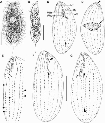
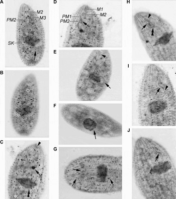
Body shape, pellicle, cytoplasm, ciliary pattern, nuclear apparatus and locomotion are very similar between the current and the original populations and are not re-described here, although morphometric data are given in Table 1, and some features are illustrated. Cells approximately 60–90 × 20–35 µm in vivo (Figure 1A). Buccal field approximately 25–35% of cell length, with shallow depression. Extrusomes ~1.5 µm long in vivo and ~3 µm long when ejected after fixation (arrows in Figure 3G). After several days in culture, cells are more slender than those in their natural state (Figure 1B).
(A & B) Morphology and stomatogenesis of Philasterides armatalis in vivo and (C–G) after protargol impregnation. (A) Typical body shape, right lateral view; (B) slender body shape, left lateral view; (C & D) infraciliature on ventral (C) and dorsal (D) sides, arrow in C to show the scutica, while in D to indicate the irregularly shaped macronucleus, arrowhead marks the apical plate; (E) ventral view, arrow to show the kinetosomal proliferation in the scutica; arrowheads to show the number of basal bodies increasing in the somatic kineties; and (F & G) ventral views, arrows to show the posterior part of the paroral membrane splitting longitudinally, arrowhead in F to show the dedifferentiation of membranelle 2, arrowheads in G mark the proliferation of basal bodies in the scutica. M1, 2, 3, membranelles 1, 2 and 3; PM1, 2, anterior and posterior parts of the paroral membrane. Scale bars: 20 µm.

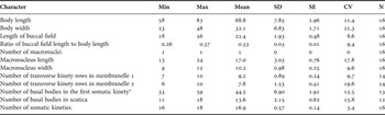
Morphometric characterization of Philasterides armatalis. All data based on protargol-impregnated specimens.

CV, coefficient of variation in %; Max, maximum; Min, minimum; SD, standard deviation; SE, standard error of the mean; N, sample size. Measurements in μm. *, Each basal body pair counted as one.
Infraciliature and nuclear apparatus as shown in Figures 1C, D, 2A & 3A–H. Somatic kineties (SK) 16–18 (mean 17) in number, mainly composed of monokinetids throughout except the anteriormost end of each kinety (Figure 3A & B), although in larger (possibly pre-dividing?) cells there is usually a mixture of dikinetids and monokinetids (Figures 1C, D & 2A).
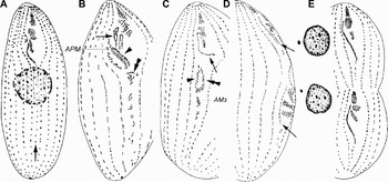
Protargol-impregnated specimens of Philasterides armatilis showing (A) its morphology and (B–E) stomatogenesis. Arrow in A to show the scutica, arrows in B & C to show the anlage of the paroral membrane in the proter, while in D to indicate the single-rowed anlage of the paroral membrane accompanied by a short row of kinetosomes at its posterior portion; arrowhead in B and double-arrowhead in C to show the anlagen of membranelles 1 and 2, arrowhead in C to mark the anlage of the paroral membrane in the opisthe, double-arrowhead in B to show the anlage of membranelle 3; and (E) infraciliature at late stage: inset shows macronucleus and micronucleus after division. APM, anlage of paroral membrane; AM3, adoral membranelle 3.

Photomicrographs showing the morphology and stomatogenesis in Philasterides armatalis. (A & B) Ventral (A) and dorsal (B) views of the same cell, arrow to show the scutica; (C) dorsal view, arrowhead to show the apical plate, arrow to show the micronucleus, double-arrowhead to mark the irregularly shaped macronucleus; (D, E & H) ventral view, depicting the buccal apparatus and somatic kineties, arrow in D to show membranelle 3, arrow in E to indicate a somatic kinety composed of monokinetids, in H arrow shows the anterior part of the paroral membrane, double-arrowhead indicates the posterior part of the paroral membrane, arrowhead marks membranelle 2; (F) spherical macronucleus (arrow) and somatic kineties; (G) somatic kineties and extrusomes (arrows); and (I & J) ventral views at early stages of stomatogenesis to show the PM2 splitting longitudinally (arrow), arrowhead to indicate the kinetosomal proliferation in the scutica. M1, 2, 3, membranelles 1, 2 and 3; PM1, 2, the anterior and posterior parts of the paroral membrane; SK, somatic kineties.

Buccal apparatus consisting of paroral membrane and three well-formed membranelles (Figures 1C, 2A & 3A, D, H): membranelle 1 and 2 (M1, 2) consisting of 7–10 and 6–10 transverse rows of kinetosomes, respectively; membranelle 3 (M3) generally double-rowed but its posterior portion is accompanied by another one or two basal bodies; paroral membrane (PM) conspicuously bipartite with the anterior part double-rowed and the posterior part (PM2) in a zig-zag-formation. Scutica (Sc) with 11–18 basal bodies (arrows in Figures 1C, 2A & 3A).
STOMATOGENESIS DURING BINARY FISSION
Stomatogenesis commences with the proliferation of kinetosomes in the scutica below the cytostome forming the primary field (PF; arrow in Figure 1E; arrowhead in Figure 3I). Meanwhile, the kinetosomes in the somatic kineties begin to increase in number (arrowheads in Figures 1E & 3I).
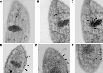
Soon, the zig-zag structure of the PM2 begins to split longitudinally (arrow in Figures 3I, J & 4A). Shortly afterwards, PM2 splits into two lines (arrow in Figure 1F). At almost the same time the kinetosomes in membranelle 2 became dedifferentiated (arrowhead in Figure 1F). Thereafter, both PM1 and PM2 lengthen and the two lines from PM2 begin to curve inwards and are designated the secondary field (SF; arrow in Figure 1G); the number of basal bodies increases in PF (arrowheads in Figure 1G). The kinetosomes in SF and PF continue to proliferate, the proliferation on the interior side of the SF being faster than that on the exterior side.
Photomicrographs showing stomatogenesis in Philasterides armatalis. (A) Ventral view at early stage, arrow to show the posterior part of the paroral membrane splitting longitudinally; (B & C) ventral view of the same cell, arrow in B to show the anlage of the paroral membrane in the proter, which is composed of one row of basal bodies; arrow in C to show the organization of membranelle 3 in the opisthe, arrowhead to indicate the anlage of the paroral membrane in the opisthe, double-arrowhead to mark the anlagen of membranelles 1 and 2 in the opisthe; (D) middle-late stage, arrow to show the point where the anlage curves, arrowhead to indicate further development of the anlagen of membranelles 1 and 2 in the opisthe, double-arrowhead to mark the reorganization of membranelle 3 in the opisthe; and (E & F) ventral views of the proter (E) and opisthe (F), arrows to show the hook-like structure in the posterior part of the anlage of the paroral membrane, arrowheads to indicate remnant kinetosomes from the anlage of the paroral membrane.

During the next stage the SF divides into two parts which migrate posteriad. As shown in Figure 2B, the interior part comprises many short rows of kinetosomes regularly arranged in an inverted U-shape. By contrast, the exterior part develops more slowly and contains several loosely arranged kinetosomes. These two parts form the anlagen of membranelles 1 and 2 & (AM1, 2; arrowhead in Figure 2B), and the paroral membrane (APM; Figure 2B), respectively. The PM1 gradually develops into a curved and single-rowed structure (arrow in Figure 2B), which is the anlage of paroral membrane (APM) in the proter. By this time, the anterior portion of the PF has formed a group of irregularly arranged kinetosomes, which is the anlage of membranelle 3 (AM3; double-arrowhead in Figure 2B).
Next, the anlagen of the opisthe move further downward. The AM1, 2 reorganize and gradually form a bow-shaped structure (double-arrowhead in Figures 2C & 4C; arrowhead in Figure 4D), and the kinetosomes in APM begin to reorganize (arrowhead in Figures 2C & 4D). Meanwhile the kinetosomes in AM3 begin to arrange in order from anterior to posterior, each row with 2–4 kinetosomes (Figure 2C; arrow in Figure 4C; double-arrowhead in Figure 4D). The kinetosomes below AM3 gradually decrease in number because some of those in the anterior portion join in the formation of AM3 and the posterior kinetosomes are possibly resorbed. In the proter, the APM stretches and becomes an arc (arrows in Figures 2C & 4B).
Subsequently, AM1, 2 divides at the middle into two parts and AM3 begins to move to a transverse position. The kinetosomes in the parental scutica continue to decrease in number. In both the proter and the opisthe, the APM further lengthens to become a long structure with the anterior part single-rowed and the posterior portion two-rowed (arrows in Figures 2D & 4E, F). At the same time, a few basal body pairs are always present posterior to the APM (arrowheads in Figure 4E & F).
At the late stage of stomatogenesis, the differentiation and orientation of the oral apparatus become clear. The reorganization of the paroral membrane in the dividers is almost complete with its migration posteriad to the right of membranelle 2. At this time a small portion at the anterior end of PM is single-rowed but will eventually become double-rowed, while the posteriormost portion is zig-zag. A few basal bodies below the paroral membrane will subsequently proliferate to form the scutica of the daughter cells. At this time the three parental membranelles complete their reorganization and become the new oral structure of the proter. Meanwhile, the division of the nuclear apparatus is completed and one macronucleus, with one accompanying micronucleus, is present in each divider (Figure 2E, insets).
DISCUSSION
Considering the body shape and the structure of the oral apparatus, the current population corresponds well with the original population described by Song (Reference Song2000) (Table 1).
Compared with Philasterides armata Kahl, 1926, the type species of the genus Philasterides, P. armatalis is distinguished by the possession of 16–18 rows of somatic kineties (vs 26–32 rows), the terminally positioned contractile vacuole (vs just below the equatorial region) and its marine (vs freshwater) biotope (Grolière, Reference Grolière1980; Song & Wilbert, Reference Song and Wilbert1989; Song, Reference Song2000).
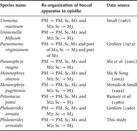
Stomatogenesis in the genus Philasterides has been only studied in P. armata (Grolière, Reference Grolière1980). That investigation revealed the dual origin of the buccal apparatus in the opisthe developing from two fields: one derived from the scutica and the other from the paroral membrane. This kind of stomatogenetic mode, which was described by Grolière (Reference Grolière1980) as the ‘Philasterides’ subtype, is also found in several other genera in Philasterina, i.e. Uronema (Small, Reference Small1967), Paranophrys (Ma et al., Reference Ma, Song and Hu2001), Metanophrys (Ma & Song, Reference Ma and Song2003), Mesanophrys (Morado & Small, Reference Morado and Small1994), Potomacus (Ramsey et al., Reference Ramsey, Brownlee and Small1980), Uronemella (Ma et al., Reference Ma, Song and Ma2002) and Paruronema (Grolière, Reference Grolière1974) (Table 2). Based on our observations of its ontogenesis, specifically with respect to the paroral membrane, membranelles 1 and 2, and the scutica in the opisthe (which originates from the parental paroral membrane whereas membranelle 3 generates from the parental scutica), Philasterides armatalis undoubtedly belongs to this subtype. Philasterides armata and P. armatalis, however, differ at least in two respects: (1) in the proter, the paroral membrane and scutica are generated from the remnant of the whole parental paroral membrane involving PM1 and PM2 in P. armata (vs from PM1 only in P. armatalis); and (2) in the opisthe, PM2 gives rise to the APM, AM1, 2 and the scutica in P. armata (vs PM2 gives rise to AM1, 2, while the APM and scutica derive from PM1 in P. armatalis).
Morphogenetic comparison of the buccal apparatus of the opisthe in some scuticociliates.

M1, membranelle 1; M2, membranelle 2; M3, membranelle 3; PM, paroral membrane; Sc, scutica.
Small (Reference Small1967) erected the order Scuticociliatida on the basis of its stomatogenetic process, that is ‘scuticobuccokinetal’. In our opinion, however, it is more reasonable to reconstruct ciliate phylogeny at the family level according to the subtypes of morphogenesis within this order, while simultaneously paying attention to morphological characters, than basing it only on similarity in microstructure (e.g. infraciliature) and/or ultrastructure. Unfortunately, this point was not taken into account in the classification systems of Corliss (Reference Corliss1979), Small & Lynn (Reference Small, Lynn and Lee1985) and Lynn & Small (Reference Lynn, Small and Lee2002). Small & Lynn (Reference Small, Lynn and Lee1985), for example, placed Philasterides in Philasteridae, Potomacus and Paruronema in Parauronematidae, and Paranophrys, Metanophrys and Mesanophrys in Paranophryidae, although these latter three were subsequently transferred to the Orchitophryidae (Lynn & Small, Reference Lynn, Small and Lee2002). Based on their infraciliature and patterns of stomatogenesis, however, we suggest that all these genera should be assigned to one family, Uronematidae Thompson, 1964.
ACKNOWLEDGEMENTS
This work was supported by the Natural Science Foundation of China (Project Nos.: 30570236, 30430090), The Royal Society, UK Short Visit Programme, the Programme for New Century Excellent Talents in Universities (NCET 05-0595) and the Darwin Initiative Programme (Project no. 14-105) which is funded by the UK Department for Environment, Food and Rural Affairs.