Introduction
Lichenicolous fungi encompass a wide range of ecological and taxonomic groups of fungi associated with lichens, including specialized saprobes, parasymbionts and pathogens belonging to the Ascomycota as well as Basidiomycota (Lawrey & Diederich Reference Lawrey and Diederich2012). Lichenicolous fungi are worldwide in distribution, occur on all kinds of taxonomic groups of lichens and use all types of lichen thalli as well as fruit bodies as ecological niches. The exploration of lichen-inhabiting fungi was strongly stimulated by the comprehensive treatments of hyphomycetes and coelomycetes of this ecological group in Hawksworth (Reference Hawksworth1979, Reference Hawksworth1981) and increased rapidly. Nevertheless, the inventory of lichenicolous fungi is far from complete, and many taxa are still unnamed and undescribed.
In the course of examinations of numerous specimens of anamorphic lichenicolous fungi found in the Holarctic (mainly in Russia), several interesting species have been identified. Three of the taxa concerned are treated here, including a new hyphomycete genus, a new species of Phoma and an additional species described and discussed in comparison with the latter species.
Material and Methods
The material was examined and photographed using a Zeiss Stemi 2000-CS microscope and Axio Imager A1 equipped with Nomarski differential interference contrast optics. Microscopic examination was carried out with material mounted in water, 10% KOH (K), Lugol's iodine, directly (I) or after a KOH pre-treatment (K/I), phloxin or erythrosin. The length, breadth and length/breadth ratio (l/b) of conidia are given as: (min–){
 $\bar x$
–SD}–{
$\bar x$
+SD}(–max), where min and max are the extreme values,
$\bar x$
the arithmetic mean, and SD the corresponding standard deviation. The material examined is housed in the herbarium of V. L. Komarov Botanical Institute in St.-Petersburg, Russia (LE) and the herbarium of the Martin Luther University in Halle (Saale), Germany (HAL).
Ameroconium U. Braun & Zhurb. gen. nov.
MycoBank No.: MB 803390
Lichenicolous dematiaceous hyphomycetes. Superficially similar to Heteroconium Petr., but distinguished by forming aseptate (amerosporous) conidia in simple acropetal chains, conidiogenesis holoblastic, conidiogenous loci and hila of the conidia truncate and relatively broad, proliferation of the conidiogenous cells enteroblastically percurrent, with several coarse, flaring annellations.
Type species: Ameroconium cladoniae U. Braun & Zhurb.
Etymology. Composed of ‘amero’ (referring to amerosporous conidia) and ‘conium’ (referring to the superficially similar genus Heteroconium).
Ameroconium cladoniae U. Braun & Zhurb. sp. nov.
MycoBank No.: MB 803391
Conidiophores subcylindrical, 15–50×3·5–6·0 µm, septate, conidiogenous cells integrated, terminal, percurrently proliferating, with several coarse, flaring annellations. Conidia catenate, doliiform to lemon-shaped, (3·8–)4·7–7·3(–11·0)×(2·7–)3·3–4·5(–5·1) µm, aseptate, pigmented.
Type: Russia, Siberia, Irkutsk Region, near Erbogachen, 60°59′15·40″N, 108°28′55·30″E, on Cladonia rangiferina, 10 September 2008, K. E. Vershinin (LE 260838—holotype).
(Fig. 1)
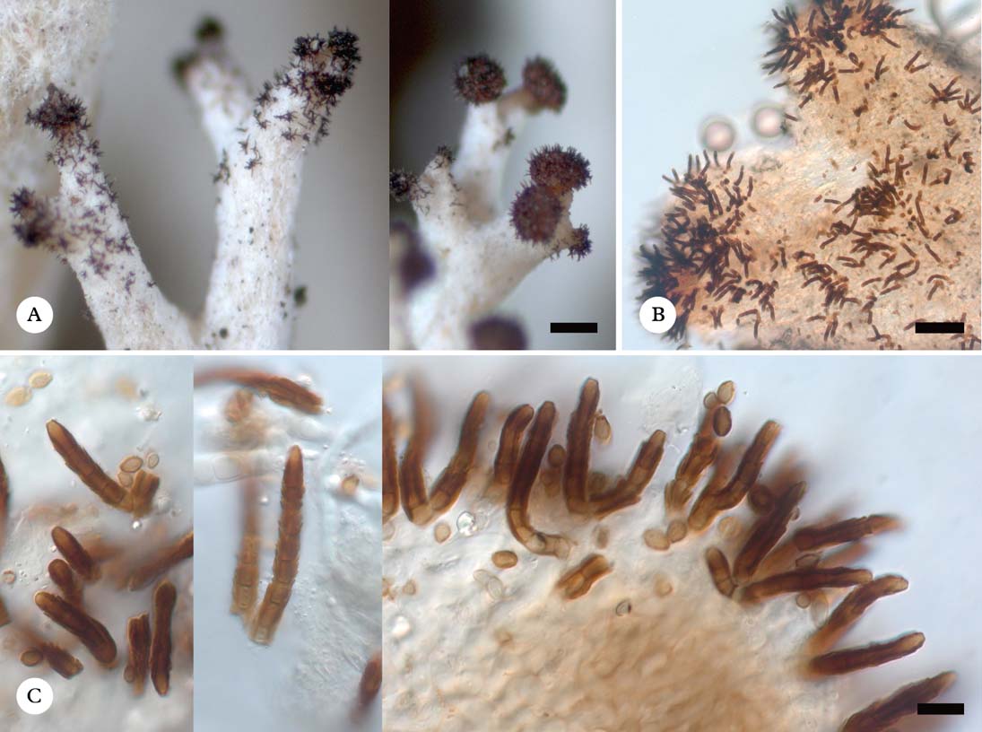
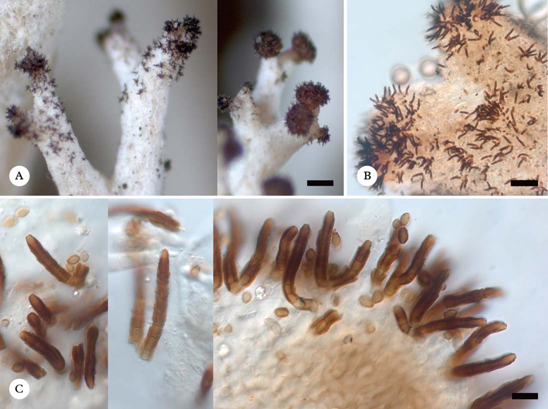
Ameroconium cladoniae (holotype). A, colonies on host thalli (habitus); B, colonies (squashed preparation in water); C, conidiophores and conidia (in K). Scales: A=200 µm; B=50 µm; C=10 µm.
Colonies on thalli of Cladonia species, short caespitose, aggregated or effuse, blackish. Vegetative hyphae not observed.
Conidiophores solitary or in small aggregations, scattered, arising from internal hyphae, erumpent, erect, straight to curved, subcylindrical, septate, medium to dark brown (basal cells pale brown to subhyaline), unbranched, but sometimes a few conidiophores arising from a single basal cell, 15–50 µm long (including conidiogenous cells) and 3·5–6·0 µm wide, wall 1·0–1·5 µm thick, K−, smooth. Conidiogenous cells integrated, terminal, about 5–8 µm long, medium to dark brown, unilocal, monopodial, proliferation enteroblastically percurrent with several conspicuous, coarse, flaring annellations, conidiogenesis holoblastic, conidiogenous loci truncate, neither thickened nor darkened, 1·5–3·0 µm diam. Conidia formed in unbranched acropetal chains, doliiform to lemon-shaped, primary conidia with rounded apex and truncate to short obconically truncate base, subsequent (secondary) conidia usually with distinctly truncate apex and base, (3·8–)4·7–7·3(–11·0) × (2·7–)3·3–4·5(–5·1) µm, l/b=(1·1–)1·2–1·8(–2·8) (n=95, in water or K), aseptate, wall thin, pale or medium brown to olivaceous brown, dry, smooth (but wall evidently delicate and thus easily cracking in microscopic preparations), with small guttules seen in K, K−, hila (1·0–)1·5–2·0 µm diam., base occasionally slightly concave and then with minute frill, but conidial secession schizolytic.
Notes
In dematiaceous hyphomycetes, including lichenicolous ones, it is usually relatively easy to identify new species, but the proper assignment to existing genera is often difficult due to intricate morphological characters, intermediate between certain genera. The present, obviously undescribed, new lichenicolous hyphomycete is a striking example.
The new species on Cladonia is a lichenicolous dematiaceous hyphomycete, well characterized by having distinctly percurrently proliferating conidiogenous cells with several coarse, flaring annellations, holoblastic conidiogenesis, and amerosporous (aseptate) conidia formed in unbranched acropetal chains.
Hitherto known lichenicolous hyphomycete genera and other hyphomycete genera encompassing at least some lichenicolous species have been compared, but attempts to find an appropriate genus failed (Seifert et al. Reference Seifert, Morgan-Jones, Gams and Kendrick2011). The lichen-inhabiting genus Ampullifera Deighton is somewhat similar, but differs in forming hyphopodia and unilocal, determinate (non-percurrent) conidiogenous cells (Hawksworth Reference Hawksworth1979; Seifert et al. Reference Seifert, Morgan-Jones, Gams and Kendrick2011). The coarse and flaring annellations in the new species resemble those of the lichenicolous genus Refractohilum D. Hawksw. (Hawksworth Reference Hawksworth1979; Seifert et al. Reference Seifert, Morgan-Jones, Gams and Kendrick2011), but the latter genus is mucedinaceous (non-pigmented) and the amero- to phragmosporous conidia are formed singly. Among other non-lichenicolous hyphomycetes, there are some morphologically similar genera. Fusicladium Bonord. emend. Schubert et al. (Schubert et al. Reference Schubert, Ritschel and Braun2003) [nom. cons.] comprises several foliicolous, plant-pathogenic species with unilocal, annellate conidiogenous cells, but in all of them the conidia are consistently formed singly (catenate conidia are only connected with taxa having multilocal, sympodial conidiogenous cells), and the annellations are finer and laterally appressed (i.e. non-flaring). Heteroconium and allied genera, which are also characterized by having pigmented conidiophores and conidia, unilocal, determinate to percurrently proliferating conidiogenous cells, and conidia formed in simple acropetal chains (Ellis Reference Ellis1971; Morgan-Jones Reference Morgan-Jones1976; Castañeda et al. Reference Castañeda, Saikawa and Guarro1999, Reference Castañeda, Iturrinaga, Abarca, Minter, Gené, Stadler, Saikawa and Silvera-Simón2008; Hughes Reference Hughes2007), are comparable with the new lichenicolous hyphomycete. However, the conidia of H. citharexyli Petr., the type species of Heteroconium, and most other species assigned to this genus are phragmosporous and annellations are lacking or, if present, they are quite distinct by being rather inconspicuous, fine, with an appressed, non-flaring margin. Only two currently recognized species of Heteroconium, H. indicum Varghese & V.G. Rao and H. ponapense Matsush., have 1-septate conidia (Castañeda et al. Reference Castañeda, Iturrinaga, Abarca, Minter, Gené, Stadler, Saikawa and Silvera-Simón2008). The phylogenetic position of the type species of Heteroconium is still unknown, but the whole genus is undoubtedly polyphyletic and morphologically heterogeneous (Hughes Reference Hughes2007; Castañeda et al. Reference Castañeda, Iturrinaga, Abarca, Minter, Gené, Stadler, Saikawa and Silvera-Simón2008). Several species previously included in Heteroconium have been reallocated to other genera, such as H. chaetospira (Grove) M. B. Ellis to Cladophialophora Borelli (Crous et al. Reference Crous, Schubert, Braun, de Hoog, Hocking, Shin and Groenewald2007) and H. solanicola (Sacc. & P. Syd.) M. B. Ellis to the new genus Pirozynskiella S. Hughes (Hughes Reference Hughes2007). The reassessment of H. tetracoilum (Corda) M. B. Ellis as a species of Lylea Morgan-Jones (Holubová-Jechová Reference Holubová-Jechová1978) was questioned by Hughes (Reference Hughes2007). A survey of Heteroconium species, including key, was published by Castañeda et al. (Reference Castañeda, Iturrinaga, Abarca, Minter, Gené, Stadler, Saikawa and Silvera-Simón2008). Inclusion of the new species in Heteroconium would demand a further unacceptable, unnatural widening of the concept of the already heterogeneous genus Heteroconium. Therefore, we prefer to introduce a new genus for this lichenicolous hyphomycete, which is characterized by a peculiar combination of characters, although in general one should be very cautious with new genera in such assemblages of phylogenetically untreated or little examined anamorph groups.
Some other Heteroconium-like genera or segregations of Heteroconium are easily distinguishable from Ameroconium and not qualified as candidates to accommodate the new lichenicolous species. Species of Lylea are characterized by having distoseptate conidia, and in Xenoheteroconium Bhat et al. (Bhat & Kendrick Reference Bhat and Kendrick1993) the basal cells of the conidiophores are radially lobed, the conidiogenous cells are terminal as well as usually intercalary with terminal and lateral conidial formation, or even with conidia laterally arising from other conidia. Pirozynskiella S. Hughes was introduced for fungicolous Heteroconium-like species characterized by centrifugal septation in conidia, in contrast to basifugal formation in true Heterosporium species (Hughes Reference Hughes2007). The phylogenetic and taxonomic meaning of differences in the formation of eusepta in conidia, either with the first septum developed at the base or in the middle, is still unclear. The type species of Pirozynskiella was classified and recognized as a Heteroconium species and keyed out in Castañeda et al. (Reference Castañeda, Iturrinaga, Abarca, Minter, Gené, Stadler, Saikawa and Silvera-Simón2008), whereas Pirozynskiella was treated as separate genus in Seifert et al. (Reference Seifert, Morgan-Jones, Gams and Kendrick2011).
Distribution and host
Known from the boreal zone of Asia and North America. The species grows on branches (mainly on their tips), apothecia and pycnidia of Cladonia arbuscula and C. rangiferina. Symptoms of pathogenicity not observed.
Additional specimen examined. Canada: North West Territories: 40 km S of Inuvik, 100 m W of Dempster Highway, 68°06′39·3″N, 133°28′38·2″W, lichen-dwarf shrub-Picea mariana woodland, on Cladonia arbuscula ssp. arbuscula, 14 vii 2004, D. Walker (LE 260870a).
Phoma lobariae Diederich & Etayo
(Fig. 2)
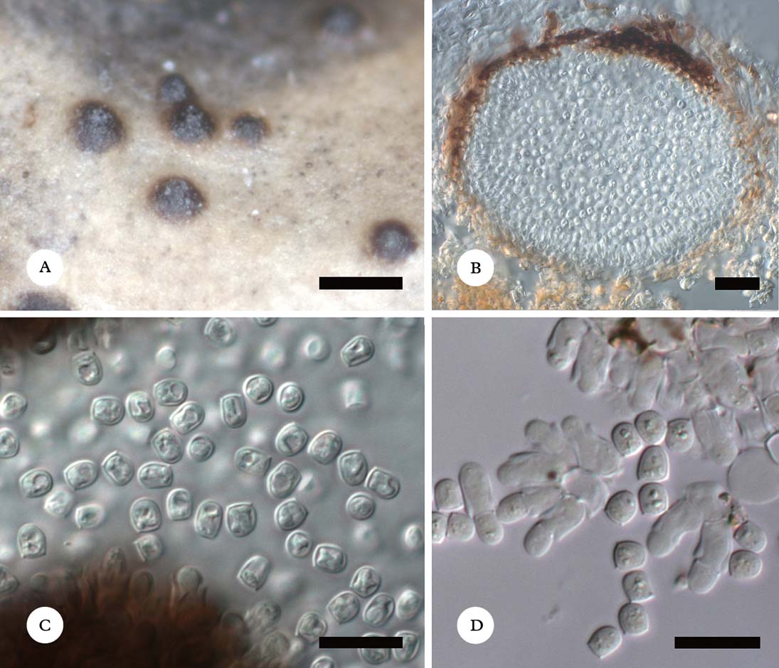
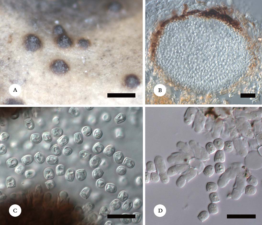
Phoma lobariae (LE 260668). A, habitus of conidiomata; B, conidioma in cross-section (in water); C & D, conidia and conidiogenous cells (C, in water; D, in K then I). Scales: A=200 µm; B=20 µm; C & D=10 µm.
Vegetative hyphae not observed.
Conidiomata pycnidial, brownish black, glossy, subglobose, almost immersed to 1/2 erumpent, (50–)100–160 µm diam., ostiole central, occasionally with tiny indistinct papilla, circular, 5–20 µm diam., sometimes showing white interior, dispersed to occasionally contiguous; wall strongly differentiated in structure and pigmentation, dark brown and 10–20 µm thick above, pale brown to almost hyaline and c. 10 µm thick below, in surface view resembling textura angularis, K+ causing distinct olive tint; gel I−, K/I−. Conidiophores absent. Conidiogenous cells enteroblastic, phialidic, collarette-like ends conspicuous, periclinal wall about 1 µm thick, hyaline, doliiform, elongate-campanulate, broadly lageniform or short cylindrical, entirely lining the pycnidial cavity, aseptate, conidiogenous cells (4·5–)5·4–7·0(–8·0) × (2·7–)3·2–4·0(–4·7) µm (n=65, in water, K, K/I or K then phloxin), smooth-walled. Conidia solitary, hyaline, broadly ellipsoid to almost campanulate, sometimes subglobose, cuboid or doliiform, apex rounded, base usually distinctly broadly truncate to slightly convex, often with tiny marginal frill, aseptate, (3·1–)3·7–4·5(–4·9)×(2·7–)3·1–3·5(–4·0) µm, l/b=(1·0–)1·1–1·3(–1·7) (n=88, in water or K), dry, wall thin (c. 0·3 µm thick), smooth, small lipid guttules occasionally seen only in K.
Notes
There are some discrepancies between the features of the specimen examined and the species protologue, in which Etayo & Diederich (Reference Etayo, Diederich, Daniels, Schulz and Peine1995) described conidiomata being 50–100 µm in diam., conidiogenous cells 4–6×1·7–3·5 µm, and conidia 3–4×2·5–3·0 µm, without any reference to marginal frills. However, we suggest that these differences reflect infraspecific variability, which is supported by the observations of Brackel (Reference von Brackel2011) and Lawrey et al. (Reference Lawrey, Diederich, Nelsen, Freebury, Van den Broeck, Sikaroodi and Ertz2012). The K+ olive reaction of the conidiomatal wall was not mentioned in the species protologue. The reaction is not typical for lichenicolous Phoma species and, incidentally, was one of the reasons for creating a separate genus Briancoppinsia Diederich et al. for Phoma cytospora (Vouaux) D. Hawksw., characterized by the same reaction of the conidiomatal wall (Diederich et al. Reference Diederich, Lawrey, Sikaroodi, van den Boom and Ertz2012). Phoma lobariae is quite similar and comparable to P. psoromatis described below, but both species are unusual among plant-inhabiting species of this genus due to an extraordinary structure of the pycnidial wall (see discussion under the latter species). The species is new to Russia, the Arctic and Asia in general, and Lobaria linita is a new host species. The infection causes bleaching of the host lobes.
Specimen examined. Russia: Chukotka: vicinity of Baran'e Lake, 66°54′N, 175°15′E, tundra on a mountain slope, on Lobaria linita (upper lobe surface), 31 vii 1980, I. Makarova (LE 260668).
Phoma psoromatis Zhurb. & U. Braun sp. nov.
MycoBank No.: MB 803393
Similar to Phoma lobariae, but with longer conidiogenous cells, larger conidia, (3·7–)4·8–6·4(–8·3) × (2·9–)3·6–4·4(–5·1) µm versus (3·1–)3·7–4·5(–4·9) × (2·7–)3·1–3·5(–4·0) µm, and K− wall of conidiomata.
Type: Russia, Central Siberia, Taimyr Peninsula, Enisey Gulf coast at Ragozinka River mouth, 72°48′N, 80°53′E, alt. 30 m, sedge-Dryas-moss tundra, on Psoroma hypnorum, 13 July 1990, M. Zhurbenko 90717 (LE 261532—holotype).
(Fig. 3)
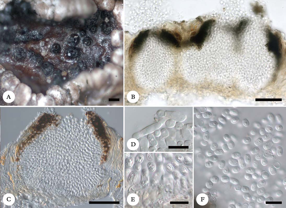
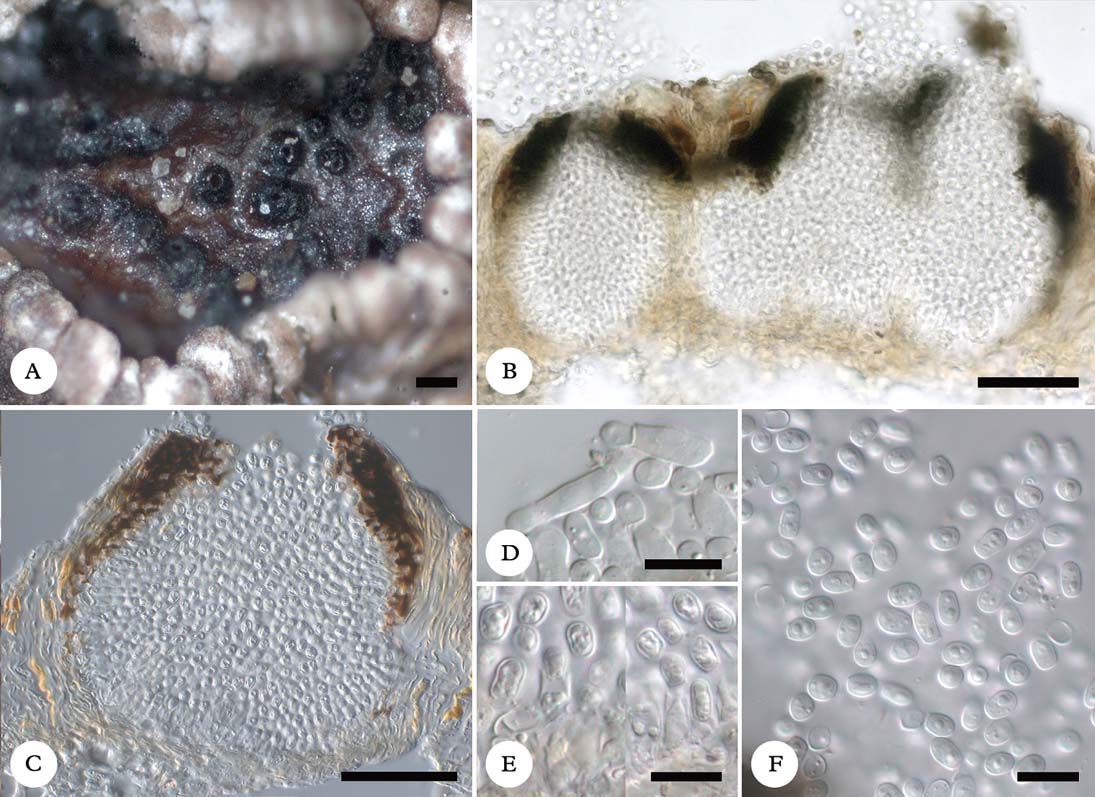
Phoma psoromatis (A, holotype; B–F, LE 261612). A, habitus of conidiomata; B & C, a single and fused conidiomata in cross-section (in water); D, conidiogenous cells (in K); E, conidiogenous cells with collarette (in water); F, conidia (in K). Scales: A=100 µm; B & C=50 µm; D–F=10 µm.
Vegetative hyphae not observed.
Conidiomata pycnidial, black, glossy, rough, subglobose to ampulliform or lageniform, often with more or less flat base, 1/5 to 2/3 erumpent, 80–180 µm diam., ostiole central, sometimes slightly papillate, circular or irregular in outline, at later stages sometimes gaping, 15–50 µm diam., occasionally showing white interior; pycnidia dispersed, solitary or sometimes contiguous or two to three pycnidia even fusing, then with several ostioles, resembling multilocular pseudopycnidia; wall strongly differentiated in structure and pigmentation, dark brown and 15–30 µm thick above, pale brown to almost hyaline and c. 10 µm thick below, in surface view resembling textura angularis, composed of pseudoparenchymatous cells 4–10 µm across, K−; gel I−, K/I−. Conidiophores absent. Conidiogenous cells lining the entire pycnidial cavity, enteroblastic, phialidic, with broad terminal opening, collarette-like ends only occasionally observed, periclinal thickening often indistinct, hyaline, lageniform, doliiform, almost short cylindrical or occasionally elongate-ampulliform, (6·7–)7·9–11·0(–13·0) × (3·0–)3·5–4·7(–5·3) µm (n=45, in water, erythrosin, I or K/I), aseptate, thin-walled, smooth. Conidia solitary, hyaline, cuneiform, broadly ovoid, ellipsoid, oblong and sometimes slightly constricted below to pyriform, occasionally subglobose or cuboid, apex rounded, base often distinctly broadly truncate to slightly convex, sometimes with tiny marginal frill, aseptate, (3·7–)4·8–6·4(–8·3)×(2·9–)3·6–4·4(–5·1) µm, l/b =(1·0–)1·2–1·6(–2·3) (n=206, in water, I, phloxin or erythrosin), dry, thin-walled, smooth, with 1–3 small lipid guttules clearly seen only in K.
Notes
The new species is provisionally placed in Phoma s. lat., which is undoubtedly polyphyletic. The lichenicolous habit of Phoma psoromatis, and some morphological characteristics of the ascomata and conidia, deviate from the type species of Phoma and common plant-inhabiting taxa. The wall of the conidiomata is strongly differentiated in width and pigmentation, the conidiogenous cells are often more or less subcylindrical, and the conidia are mostly truncate at the base with a tiny marginal frill and without distinct guttules (the pycnidial wall in Phoma is usually uniformly pigmented, conidiogenous cells are usually not cylindrical or subcylindrical, and conidia are rounded at the base; Sutton Reference Sutton1980).
About 20 lichenicolous Phoma species have been described, but only two of them have basally truncate conidia and similarly differentiated walls of conidiomata, viz. P. dubia (Linds.) Sacc. & Trotter, known from Usnea spp., and P. lobariae known from Lobaria spp. (Hawksworth Reference Hawksworth1981; Diederich et al. Reference Diederich, Lawrey, Sikaroodi, van den Boom and Ertz2012; Lawrey & Diederich Reference Lawrey and Diederich2012). The latter species differs from Phoma psoromatis in having much smaller pycnidia, 25–60 µm diam., and smaller conidia, 3·5–5·0×1·5–2·0 µm, that are narrowly ellipsoid to almost bacilliform only sometimes slightly truncate at the base. Phoma lobariae, described above in detail as comparable with P. psoromatis, is in general very similar to the new species, but has a K+ olive conidiomatal wall, shorter conidiogenous cells with more conspicuous collarettes and smaller conidia. Phoma lobariae and P. psoromatis may represent an undescribed genus, but final conclusions should be based on phylogenetic analyses.
With regard to other coelomycete genera with lichenicolous species lacking conidiophores and with hyaline, aseptate conidia, the fungus on Psoroma hypnorum has to be compared with species of Asterophoma D. Hawksw., Bachmanniomyces D. Hawksw., Briancoppinsia Diederich, Ertz et al., Diederichia D. Hawksw., Epicladonia D. Hawksw., Lawalreea Diederich, Minutophoma D. Hawksw., Pseudoseptoria Speg., Subhysteropycnis Wedin & Hafellner, and Vouauxiomyces Dyko & D. Hawksw. (Hawksworth & Dyko Reference Hawksworth and Dyko1979; Sutton Reference Sutton1980; Hawksworth Reference Hawksworth1981, Reference Hawksworth2003; Diederich Reference Diederich1990; Wedin Reference Wedin1994; Etayo & Diederich Reference Etayo and Diederich1996; Wedin & Hafellner Reference Wedin and Hafellner1998; Ihlen & Wedin Reference Ihlen and Wedin2005; Etayo & Sancho Reference Etayo and Sancho2008; Etayo Reference Etayo2010; Diederich et al. Reference Diederich, Lawrey, Sikaroodi, van den Boom and Ertz2012; Zhurbenko & Tugi Reference Zhurbenko and Tugi2013). Asterophoma can be distinguished from Phoma psoromatis by its conidioma wall composed of extremely attenuated cells projecting at the exterior, occasionally biphialidic conidiogenous cells, and subcylindrical conidia rounded at both ends; Bachmanniomyces is characterized by its holoblastic, annellate conidiogenous cells; Briancoppinsia has a K+ dark olivaceous pycnidial wall of textura intricata, I+ and K/I+ red pycnidial gel and short ampulliform conidiogenous cells; the conidiogenous cells in Diederichia are ampulliform, often indistinct, and the conidia are cylindrical, ellipsoid or irregularly shaped and obtuse at both ends; Epicladonia has holoblastic, annellate conidiogenous cells, its conidia are sometimes 1-septate and genuine conidiophores are sometimes present; Lawalreea forms conidiomata without ostioles and falciform conidia; Minutophoma is characterized by conidiogenous cells forming the lower part of the pycnidial wall, often pale brown at the base, and subcylindrical conidia rounded at both ends; Pseudoseptoria has holoblastic, annellate conidiogenous cells and falcate, fusiform or cymbiform conidia; Subhysteropycnis differs in having usually densely aggregated conidiomata with irregularly rounded, elongated or slit-like ostioles, K+ dark green wall and I+ red, K/I+ blue pycnidial gel, bacilliform conidia, and is a gall inducer; Vouauxiomyces is quite distinct in having holoblastic, percurrently proliferating conidiogenous cells and sometimes weakly echinulate and occasionally slightly pigmented conidia.
Acaroconium punctiforme Kocourk. & D. Hawksw., Microsphaeropsis lichenicola Etayo and species of Lichenoconium Petrak & Syd. (Hawksworth Reference Hawksworth1977; Etayo & Sancho Reference Etayo and Sancho2008; Kocourkova & Hawksworth Reference Kocourkova and Hawksworth2008) are some additional, superficially similar, lichenicolous species. Acaroconium punctiforme differs from the new species in having pycnidia with a distinctly thickened ostiolar collar and conidia which are at first colourless but later become very pale brown, rounded at both ends and lacking any basal scar or frill; Microsphaeropsis lichenicola has brown conidia with rounded ends; and species of Lichenoconium have percurrently proliferating conidiogenous cells and brown conidia with ornamentation.
As the conidiomata of the new species are sometimes fused and have strongly differentiated walls resembling multilocular pseudopycnidia, fungal genera with this type of conidioma also have to be compared. Among stromatic genera with pseudopycnidial conidiomata, the monotypic genus Ceuthodiplospora Died., based on the type species C. robiniae (Bubák) Died. growing on twigs of Robinia, is comparable and similar (Sutton Reference Sutton1980; Kiffer & Morelet Reference Kiffer and Morelet2000). However, the latter genus and its type species differ in having immersed conidiomata, ampulliform conidiogenous cells, and 0–1-septate conidia without a distinctly truncate base and without any marginal frill.
The taxonomic and phylogenetic significance of differentiated structures of the pycnidial wall with darker pigmentation near the ostiolar region and paler below is quite unclear within Phoma-like genera (Phoma complex), and can only be elucidated using molecular analyses including lichenicolous species belonging to this morphological type. The relevance of differences in the conidial shape in general, and of conidia with truncate base and at least partly minute frills in particular, is also unknown. We tentatively follow previous interpretations of Phoma in a broader sense (Etayo & Diederich Reference Etayo, Diederich, Daniels, Schulz and Peine1995; Diederich et al. Reference Diederich, Lawrey, Sikaroodi, van den Boom and Ertz2012; Lawrey & Diederich Reference Lawrey and Diederich2012) and assign the new species to this genus. The relation of some Phoma-like genera, such as Diederichia and Minutophoma, to Phoma s. str. is not yet clear and needs to be examined in detail, above all based on molecular approaches.
Distribution and host
Known from tundra biomes of the Russian Arctic. Formed in the hymenium of apothecia of Psoroma hypnorum, which is usually bleached or blackish caused by infection, suggesting pathogenicity of the fungus.
Additional specimens examined [all in dwarf shrub-moss-lichen tundra, growing in hymenium of apothecia of Psoroma hypnorum]. Russia: Nenetz Region: S of Yamboto Lake, 68°05′N, 61°15′E, 18 viii 1998, A. Kustysheva (LE 261602). Central Siberia: Taimyr Peninsula, Enisey Gulf coast at Ragozinka River mouth, 72°48′N, 80°53′E, alt. 30 m, 1990, M. Zhurbenko 90713 (LE 261612). Wrangel Island: Gusinaya River basin, 71°12′N, 179°01′E, alt. 200 m, 23 vii 1991, S. Kholod (LE 260879). Chukotka: E coast of Kolyuchin Inlet, Yuniveem River basin, 66°40′N, 173°54′W, 4 viii 1980, A. Katenin (HAL 2533 F).
We are grateful to Paul Diederich for valuable comments on the manuscript. We thank Konstantin Vershinin and Svetlana Vershinina for placing the collection of Ameroconium cladoniae at our disposal.