

Alcohol and pancreatitis
Acute pancreatitis may be triggered by diverse precipitants, however, alcohol and gallstones account for approximately 70–80% of cases reported.Reference Pandol, Saluja, Imrie and Banks4 Although it was recognised for many centuries that ‘excesses of lifestyle’, including elevated alcohol consumption, were linked to pancreatic disease,Reference Cawley5 the first concrete description of the direct correlation between alcohol and pancreatitis was made by Friedrich in 1878, who coined the term the ‘Drunkard’s Pancreas’.Reference Friedreich and Ziemssen6 However, at that time the underlying pathological mechanisms of the disease were unknown. An important development in the understanding of acute pancreatitis was the observation by the pathologist Hans Chiari in 1896 that patients who had died from pancreatitis appeared to have succumbed to the digestive properties of this organ.Reference Chiari7 The exocrine portion of the pancreas is responsible for the synthesis and secretion of important digestive enzymes that break down food once it reaches the duodenum. Under physiological conditions, stimulation of the pancreas initiates the release of proteolytic enzymes, such as trypsin, from stores (‘zymogen granules’) in the acinar cell as inactive precursor forms that are transported via the pancreatic duct and secreted to the intestinal tract where activation takes place. The exocrine pancreas may therefore be envisaged as a ticking time-bomb which, under certain pathological conditions, may be triggered inappropriately to release digestive enzymes within the acinar cell, thereby initiating autodigestion of the organ. The importance of premature trypsin activation to the aetiology of the disease is highlighted from observations of hereditary pancreatitis, which is known to occur via defects in the cationic trypsinogen gene (PRSS1) that cause premature activation and/or impaired inactivation of the digestive enzyme within the pancreas.Reference Whitcomb, Gorry, Preston, Furey, Sossenheimer, Ulrich, Martin, Gates, Amann, Toskes, Liddle, McGrath, Uomo, Post and Ehrlich8 Furthermore, mutations of the serine protease inhibitor Kazal type I (SPINK1) confer an increased risk of pancreatitis,Reference Witt, Luck, Hennies, Classen, Kage, Lass, Landt and Becker9 whilst overexpression of SPINK I ameliorates the severity of experimental acute pancreatitis.Reference Nathan, Romac, Peng, Peyton, Macdonald and Liddle10 It has been shown that long-term alcohol consumption augments the pancreatic content of digestive enzymes,Reference Apte, Wilson, McCaughan, Korsten, Haber, Norton and Pirola11 and that ethanol increases the fragility of these organelles.Reference Haber, Wilson, Apte, Korsten and Pirola12
Despite the recognised association between alcohol consumption and pancreatitis, quantitative information relating the risk of alcohol consumption to incidence of disease is sparse, with few case-control studies available. Recently, a large population-based cohort study in Copenhagen has shown that there was an increased risk of pancreatitis in both men and women who consumed more than 14 drinks per week, although no difference between the type of beverage consumed or the frequency of intake was detected.Reference Kristiansen, Gronbaek, Becker and Tolstrup13 However, although excessive alcohol consumption increases the risk of pancreatitis, it has been estimated that less than 10% of chronic alcoholics will develop the disease, suggesting a susceptibility of certain individuals to the detrimental effects of excessive drinking. Why this should be is at present unclear. Intriguingly, it is difficult to induce experimental pancreatitis in animals with ethanol feeding alone, and additional factors, such as hyperstimulation of the organ with cerulein, an analogue of the hormone cholecystokinin (CCK) or ductal obstruction, are required for the disease to develop. Over the past few decades many theories have been proposed to explain the toxic actions of alcohol on the pancreas, including sensitizing effects of alcohol on the above processes, formation of protein-rich plugs that obstruct the pancreatic duct and generation of oxidative stress, amongst others. However, it is beyond the scope of this article to provide detailed discussion of such mechanisms, and the interested reader is directed to a recent review of alcoholic pancreatitis.Reference Pandol, Saluja, Imrie and Banks4, Reference Pandol and Raraty14 Instead, attention will focus on recent evidence suggesting the importance of the generation of toxic alcohol metabolites to pancreatic acinar cell death. In order to understand the role that alcohol and its metabolites may play in pancreatic acinar cell death, it is necessary firstly to describe how calcium signalling, energy production and enzyme secretion are physiologically regulated in the acinar cell and how disruption of homeostasis may occur to induce pathological consequences.
Physiological Ca2+-dependant enzyme secretion
The exocrine portion of the pancreas responds to both nervous and hormonal stimuli to generate large quantities of digestive enzymes necessary to break down food ingested into the gastrointestinal tract. The basis of such a coordinated secretory response is the formation of tightly controlled calcium signals that are restricted to microdomains within pancreatic acinar cells.Reference Petersen, Sutton and Criddle15 Until recently it had been widely held that the hormone CCK only acted indirectly to stimulate pancreatic exocrine secretion via release of the neurotransmitter acetylcholine (ACh) from vagal parasympathetic nerves.Reference Owyang and Logsdon16 This tenet was largely based upon an inability to detect calcium signals in response to application of CCK to isolated human pancreatic acinar cells.Reference Ji, Bi, Simeone, Mortensen and Logsdon17 However, these findings were in contrast to the robust calcium elevations detected in rodent cell experiments in response to direct CCK stimulation, the basis of which has been extensively characterised by many research groups.Reference Petersen18 It was perhaps surprising that humans should differ so fundamentally from animals with respect to mechanisms of exocrine secretion. However, experimentation with human pancreatic tissue is technically difficult and many factors, including the presence of pancreatic ductal obstruction in patients from whom the sample is obtained, the time of transit from surgical removal to arrival at the laboratory and the tissue dispersal procedures utilised, may all influence final cell quality. Recently, in a study involving pancreatic specimens taken from patients at the Royal Liverpool University Hospital over a period of several years, we have shown that direct stimulation of isolated human pancreatic acinar cells by physiological (picomolar) CCK induces oscillatory cytosolic calcium ([Ca2+]c) signals that are coupled to digestive enzyme secretion (Figure 1).Reference Murphy, Criddle, Sherwood, Chvanov, Mukherjee, McLaughlin, Booth, Gerasimenko, Raraty, Ghaneh, Neoptolemos, Gerasimenko, Tepikin, Green, Reeve, Petersen and Sutton19 Any potential indirect nerve transmitter release was excluded in this study by use of atropine, which blocks muscarinic (M3) receptors activated by ACh, and tetrodotoxin, which blocks transmitter release from nerve terminals. In addition, our study also demonstrated that physiological (nanomolar) ACh induced oscillatory [Ca2+]c rises, that were blocked by atropine, in contrast to the high supramaximal (micromolar) concentrations required to elicit a signal in the previous report, suggesting that a difference in cell quality may underlie the previous difficulty in observing responses to physiological CCK.Reference Saluja, Logsdon and Garg20 Our recent study has therefore emphasised the similarity between rodent and human exocrine pancreatic physiology, confirming the importance of a wealth of prior observations from animal experiments within the field.
Direct effects of the hormone cholecystokinin (CCK) on cytosolic calcium ([Ca2+]C) and digestive enzyme secretion in human pancreatic acinar cells. (a) CCK at a low physiological concentration (10 pM) evoked oscillatory rises of [Ca2+]C in isolated human cells (inset: light-transmitted image of a cell cluster) in the presence of both atropine and tetrodotoxin to block potential indirect (nerve-mediated) effects. Caffeine completely and reversibly blocked these responses, indicating involvement of calcium release via the inositol trisphosphate (IP3) receptor. (b) Amylase secretion from superfused segments of human pancreatic tissue in response to CCK, monitored fluorometically in the flow cell effluent, and obtained in the presence of tetrodotoxin. The basal amylase output, observed when the tissue is online (arrow), and reversible stimulatory effect of physiological CCK are shown (*denotes air bubble through system). (c) CCK elicited exocytosis, seen as a fall of quinacrine fluorescence from the region of interest (inset: circle), in the presence of atropine and tetrodotoxin. A coordinated increase in NAD(P)H autofluorescence occurred at the start of CCK stimulation, indicative of increased ATP production to fuel secretion (adapted from Ref. Reference Murphy, Criddle, Sherwood, Chvanov, Mukherjee, McLaughlin, Booth, Gerasimenko, Raraty, Ghaneh, Neoptolemos, Gerasimenko, Tepikin, Green, Reeve, Petersen and Sutton19)

The signal transduction pathways utilised by the acinar cell in response to neurohormonal stimulation have been thoroughly elaborated over the past few decades.Reference Petersen and Tepikin21 The hormone CCK and neurotransmitter ACh, whose release from specialised I-cells and parasympathetic nerves, respectively, is triggered by the presence of food in the gut, are known to bind to their specific receptor proteins on the plasma membrane of the acinar cell in order to induce a physiological response. Activation of CCK1 and M3 receptors, respectively, by these agonists causes the release of specific intracellular second messenger molecules, inositol trisphosphate (IP3), cyclic adenosine dinucleotide phosphate ribose (cADPR) and nicotinic acid adenine dinucleotide phosphate (NAADP), which in turn triggers calcium release from internal stores to generate transient, oscillatory signals in the apical granular pole. The major calcium store in the pancreatic acinar cell is the endoplasmic reticulum (ER), which responds to second messenger stimulation through the opening of discrete calcium release channels, the IP3 receptor (subtypes 2 and 3) and the ryanodine receptor (RyR). The calcium signals induced by CCK in both human and rodent acinar cells appear to be mediated predominantly via the release of calcium from the ER store through caffeine-sensitive IP3 receptor channels (Figure 1(a)). However, recent evidence suggests that acidic organelles may also be important in contributing to the [Ca2+]c rises observed upon neurohumoural stimulation and complex interactions between second messengers and these two stores may underlie the concert of oscillatory [Ca2+]c signals observed in response to physiological neurohumoural simulation.Reference Gerasimenko, Sherwood, Tepikin, Petersen and Gerasimenko22, Reference Petersen23 Importantly, these precise, transient changes of [Ca2+]c provide the crucial trigger for the process of exocytosis, which allows fusion of the zymogen granular membrane with the apical plasmalemma to secrete inactive digestive enzyme precursors from the acinar cell, which are then flushed away from the pancreas into the duodenum via the pancreatic duct.
The importance of energy
An important requirement of the process of secretion is the provision of energy by specialised organelles, the mitochondria. It is estimated that actively respiring mitochondria, often referred to as the ‘powerhouses’ of the cell, synthesise approximately 40 kg of adenosine triphosphate (ATP) per day in adult humans. In pancreatic acinar cells the mitochondria display a distinct spatial distribution, being situated close to the zymogen granules in a perigranular belt, and they respond to transient elevations of [Ca2+]c by increased metabolism that ultimately generates the ATP necessary to fuel secretion. Thus, normal secretory function is dependent on the elaboration of finely-tuned spatiotemporal [Ca2+]c signals that are linked to energy formation within the cell. In addition to stoking the fires of secretion, ATP production by mitochondria is fundamental for cellular calcium homeostasis, maintaining [Ca2+]c levels within a narrow range. This is achieved through supply of ATP to energy-requiring calcium pumps present on the cell membrane, the plasmalemmal calcium ATPase (PMCA) which extrudes calcium from the cell against its electrochemical gradient, and on the endoplasmic reticulum, the sarco-endoplasmic reticulum ATPase (SERCA) which uptakes calcium back into the ER. Without the correct functioning of these pumps, rises of [Ca2+]c that occur by calcium release from stores and/or entry from the external medium in response to diverse stimuli may become sustained instead of transient, thereby initiating a disruption of cellular homeostasis. An inability to manage intracellular calcium levels poses significant problems for the cell.
Pathological [Ca2+]c signals and toxicity
It has been proposed that disruption of calcium homeostasis in pancreatic acinar cells, induced by a variety of precipitants, may constitute a common theme in acute pancreatitis that ultimately leads to necrotic cell death.Reference Ward, Sutton, Jenkins and Petersen24, Reference Sutton, Criddle, Raraty, Tepikin, Neoptolemos and Petersen25 Hyperstimulation of the pancreas with cerulein, an analogue of the gastrointestinal secretory hormone CCK, is a recognised model of experimental acute pancreatitis. At the cellular level, stimulation of both isolated human and murine pancreatic acinar cells with supra-physiological (nanomolar) CCK causes large sustained elevations of [Ca2+]c instead of the characteristic transient oscillatory responses generated by physiological (picomolar) CCK.Reference Murphy, Criddle, Sherwood, Chvanov, Mukherjee, McLaughlin, Booth, Gerasimenko, Raraty, Ghaneh, Neoptolemos, Gerasimenko, Tepikin, Green, Reeve, Petersen and Sutton19, Reference Raraty, Ward, Erdemli, Vaillant, Neoptolemos, Sutton and Petersen26 This type of prolonged signal is composed of both calcium release from the ER and sustained calcium entry into the cell via as yet undefined channels, and is associated with intracellular activation of trypsinogen to trypsin and vacuolisation in the apical pole of the cell, changes indicative of acute pancreatitis. Similarly, large, sustained calcium signals generated in response to bile acids,Reference Voronina, Longbottom, Sutton, Petersen and Tepikin27 recognised precipitants of acute pancreatitis, have been shown to induce necrosis of isolated pancreatic acinar cells.Reference Kim, Kim, Lee, Namkung, Sun, Ananthanarayanan, Suchy, Shin, Muallem and Lee28 According to such a unifying hypothesis it might also be predicted that alcohol could induce a similar disruption of calcium homeostasis to underlie its ability to cause acute pancreatitis.
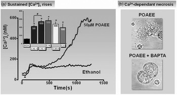
However, consistent with the inability of ethanol to cause experimental acute pancreatitis when administered alone to rodents in vivo, even very high supraphysiological (millimolar) concentrations of ethanol applied to isolated murine pancreatic acinar cells produced only modest elevations of [Ca2+]c in a majority (Figure 2).Reference Criddle, Raraty, Neoptolemos, Tepikin, Petersen and Sutton29 These rises of [Ca2+]c did not adversely affect [Ca2+]c signals produced by other agents, including the neurotransmitter ACh, suggesting that homeostasis was not adversely affected by ethanol alone. However, in a minority of cells (<30%) some larger, initial ‘spike’ elevations of [Ca2+]c were triggered that returned to lower resting levels, suggesting heterogeneity in the response to alcohol. On closer investigation of the mechanism of action, a slow, partial release of calcium from the ER, which occurred in a time-dependant manner, was observed in response to application of alcohol at these high (millimolar) concentrations. Such a release from internal stores was minor compared to that of thapsigargin, a pharmacological tool that completely depleted the ER calcium store via inhibition of the SERCA pump. Furthermore, this effect of thapsigargin was not prevented by ethanol. Overall, the results from experiments assessing the effects of ethanol on [Ca2+]c suggested little effect of ethanol per se to disrupt pancreatic acinar cell function. If alcohol itself was not responsible for significant cellular damage, could its metabolism provide a key to understanding toxicity?
Contrasting effects of ethanol and non-oxidative metabolites, fatty acid ethyl esters (FAEEs) on cytosolic calcium ([Ca2+]C) in isolated perfused pancreatic acinar cells. (a) Ethanol (850 mM) produced only small elevations of [Ca2+]C in a majority of cells, whereas palmitoleic acid ethyl ester (POAEE; 50 μM) caused large, global and sustained rises. Inset shows the large rises of [Ca2+]C induced by FAEEs of various acyl chain lengths and degree of saturation compared to the modest effect of ethanol. (b) Light-transmitted images showing the severe damage of pancreatic acinar cells exposed to POAEE (upper) and protective effects of BAPTA (lower) which prevented both POAEE-induced [Ca2+]C rises and consequent necrotic cell death (adapted from Ref. Reference Criddle, Raraty, Neoptolemos, Tepikin, Petersen and Sutton29)

Metabolism of ethanol: the role of fat
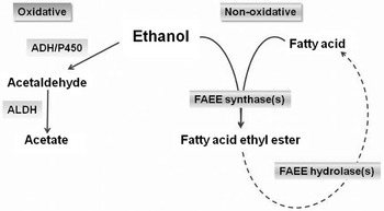
In the pancreas, as in many other organs, alcohol may be metabolised in two principal ways; the oxidative and non-oxidative pathways (Figure 3). Oxidative metabolism of ethanol in cells, perhaps the more familiar route, involves the action of alcohol dehydrogenase (ADH) and cytochrome P450 2E1 (CYP2E1) to convert ethanol to acetaldehyde and then via aldehyde dehydrogenase (ALDH) to acetate. Such an oxidative conversion of alcohol to acetaldehyde has been implicated in causing hepatic damage and liver failure, a process that may be linked to the formation of reactive oxygen species and subsequent damage to cellular membranes via lipid peroxidation.
Oxidative and non-oxidative metabolism of ethanol. In the oxidative pathway, ethanol is metabolised to acetaldehyde by alcohol dehydrogenase (ADH) and cytochrome P450 2E1 (CYP2E1) and then via aldehyde dehydrogenase (ALDH) to acetate. In the non-oxidative pathway, ethanol and fatty acids are converted to fatty ethyl esters (FAEEs) via synthases. Hydrolysis of the ester linkage also occurs via FAEE hydrolase (esterase) enzymes releasing fatty acids

In contrast, non-oxidative metabolism of ethanol occurs via a group of enzymes denominated fatty ethyl ester (FAEE) synthases that cause esterification of fatty acids to FAEEs. Interestingly, a study in rats has shown that whereas a high ethanol diet alone did not induce pancreatitis, a combination of alcohol and high fat caused pancreatic inflammation and necrosis.Reference Kono, Nakagami, Rusyn, Connor, Stefanovic, Brenner, Mason, Arteel and Thurman30 Pancreatic cholesterol esterase and synthase III enzyme expression are increased in pancreas and liver, but not brain or heart, in response to long-term ethanol feeding in rats.Reference Pfutzer, Tadic, Li, Thompson, Zhang, Ford, Eagon and Whitcomb31 Intriguingly, a gene polymorphism of one candidate FAEE synthase, carboxylester lipase, a digestive enzyme that is secreted by the exocrine pancreas, has been associated with the development of alcoholic pancreatitis;Reference Miyasaka, Ohta, Takano, Hayashi, Higuchi, Maruyama, Tando, Nakamura, Takata and Funakoshi32 an increase in the frequency of the L allele was detected in patients with alcoholic pancreatitis compared with those with non-alcohol induced forms, suggesting the potential importance of pancreatic non-oxidative metabolism to the disease. However, it should be noted that there are many enzymes that will perform non-oxidative metabolism of ethanol, including acyl-CoA: ethanol O-acyltransferase (AEAT), and non FAEE-specific enzymes, such as lipoprotein lipase and cholesterol esterase, indicating that the situation may be complex and, consequently, the potential development of specific therapeutic agents may be a significant challenge. Furthermore, FAEEs may possess broad substrate specificity. For example, carboxylester lipase performs many actions including the digestion of fat-soluble vitamin esters and cholesterol esters in the small intestine, and the hydrolysis of dietary mono-, di- and triacylglycerols and bile salts.
The potential role of FAEEs as mediators of pancreatic injury was first suggested from important findings of an autopsy study of individuals who died from acute alcohol intoxication.Reference Laposata and Lange33 The highest levels of FAEEs were found in the pancreas, compared with relatively low levels in other organs, e.g. the heart, that are also adversely affected by excessive alcohol, whilst the concentration of non-oxidative metabolites correlated directly with blood alcohol levels. The human pancreas has been shown to possess high levels of FAEE synthase activity, attaining levels in excess of 100 nmol/g of tissueReference Diczfalusy, Bjorkhem, Einarsson, Hillebrant and Alexson34 and elevated free fatty acids reaching this organ will be readily metabolised via the non-oxidative route. The association between hyperlipidaemia and pancreatitis, especially the form related to alcohol, has long been recognised and although the underlying pathological basis remains to be defined such a link would appear to support the potential danger of non-oxidative ethanol metabolism to pancreatic damage. Although increased levels of blood lipids linked to pancreatitis may be independent of or secondary to alcohol abuse, it may be that the combination of excess alcohol and free fatty acids is important to the development of acute pancreatitis. A study has shown that the carboxylester lipase gene polymorphism appears not to be increased in patients with hyperlipidaemia alone, whereas this occurs in alcoholic pancreatitis.Reference Miyasaka, Ohta, Takano, Hayashi, Higuchi, Maruyama, Tando, Nakamura, Takata and Funakoshi32
At the experimental level, acute i.v. administration of ethanol to anaesthetised rats increased levels of FAEEs, when oxidative metabolism was inhibited by 4-methylpyrazole, and induced changes indicative of acute pancreatitis, including pancreatic oedema, elevation of pancreatic trypsinogen activation peptide and subcellular vacuolization.Reference Werner, Laposata, Fernandez-del Castillo, Saghir, Iozzo, Lewandrowski and Warshaw35 Increased plasma FAEE levels have been reported in patients following inhibition of the oxidative metabolism of ethanol by 4-methylpyrazole application,Reference Best and Laposata36 although there are currently no data available evaluating potential damaging effects of oxidative metabolism inhibition on the pancreas. The detrimental actions of FAEEs on the pancreas were more directly demonstrated in an in vivo study in which i.v. administration of FAEEs to rats induced severe acute pancreatitis.Reference Werner, Saghir, Warshaw, Lewandrowski, Laposata, Iozzo, Carter, Schatz and Fernandez-del Castillo37 Taken together, these observations provide convincing evidence that non-oxidative metabolism of ethanol in the pancreas may be prejudicial for the organ under conditions of high alcohol intake. However, the mechanism of action of these metabolites was undefined in these studies. An intriguing question therefore was whether non-oxidative alcohol metabolites might in some way alter calcium homeostasis in isolated pancreatic acinar cells.
Toxic mechanisms of non-oxidative ethanol metabolites
Calcium overload
In contrast to the relatively weak effects of ethanol per se, FAEEs applied to the isolated acinar cell caused profound sustained global elevations of [Ca2+]c, that resulted in cell death (Figure 3).Reference Criddle, Raraty, Neoptolemos, Tepikin, Petersen and Sutton29 Interestingly, the oxidative metabolite of ethanol, acetaldehyde, induced no detectable changes of basal [Ca2+]c. In pancreatic acinar cells exposed to POAEE, the typical structural polarity was disrupted, cell membranes were damaged and the nuclei stained brightly with propidium iodide as necrotic cell death ensued. Importantly, if the pathological sustained rise of [Ca2+]c was prevented by pre-treatment of the cells with the Ca2+ chelator BAPTA-AM, then cells were protected from FAEE-induced necrotic cell death. The rises of [Ca2+]c induced by palmitoleic acid ethyl ester (POAEE) involved both the release of Ca2+ from the endoplasmic reticulum (ER) and entry of Ca2+ from the external medium. Using the technique of whole-cell patch-clamp, POAEE could be applied directly to the interior of the cell to instigate transient, oscillatory global elevations of [Ca2+]c that were detected as increases of Fluo-4 fluorescence and Ca2+-dependent chloride currents; such rises were mediated via the activation of caffeine-sensitive, IP3-dependant Ca2+-release channels.Reference Criddle, Murphy, Fistetto, Barrow, Tepikin, Neoptolemos, Sutton and Petersen38 Perfusion of the POAEE around the cell at a concentration of 100 μM, a level that may be attained in acute pancreatitis, resulted in the generation of a sustained global elevation of [Ca2+]c that was not inhibited by caffeine, suggesting an IP3-receptor-independent mechanism.
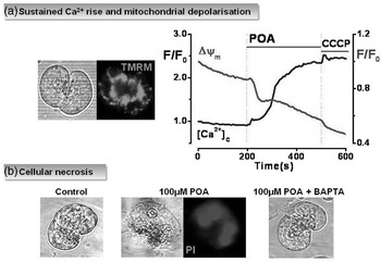
A previous study in human pancreas has demonstrated that not only is there high activity of FAEE synthase to produce FAEEs, but that there is substantial metabolism of FAEEs via hydrolase (esterase) enzymesReference Diczfalusy, Bjorkhem, Einarsson, Hillebrant and Alexson34 which de-esterify the non-oxidative ethanol metabolites back to their parent fatty acids (Figure 2). Such FAEE hydrolytic activity has been shown to be of the order of 1,100 nmol/min/g tissue in the human pancreas.Reference Diczfalusy, Bjorkhem, Einarsson, Hillebrant and Alexson34 A study in cardiac myocytes indicated that endogenously synthesized FAEEs partitioned to the mitochondria, where they underwent hydrolysis to release large quantities of free fatty acids.Reference Lange and Sobel39 Application of the unsaturated fatty acid palmitoleic acid (POA) alone to isolated murine pancreatic acinar cells caused a slow and sustained global elevation of [Ca2+]c that resulted in severe necrotic cell deathReference Criddle, Murphy, Fistetto, Barrow, Tepikin, Neoptolemos, Sutton and Petersen38 (Figure 4). The hydrolytic conversion of FAEEs back to fatty acids may be manipulated pharmacologically using the hydrolase inhibitor bis-4-nitrophenyl phosphate (BNPP), which has been shown previously to inhibit the activity of ethyl palmitate hydrolase in rat liver homogenates.Reference Diczfalusy, Bjorkhem, Einarsson, Hillebrant and Alexson34 In the absence of hydrolase inhibition POAEE induced a sustained elevation of [Ca2+]c and necrosis in murine pancreatic acinar cells, however, when BNPP was present the sustained component of the [Ca2+]c rise was greatly inhibited and cells were protected from Ca2+-dependent necrotic cell death.Reference Criddle, Murphy, Fistetto, Barrow, Tepikin, Neoptolemos, Sutton and Petersen38
Detrimental effects of fatty acids on cytosolic calcium ([Ca2+]C) and mitochondrial function to induce necrosis of isolated perfused pancreatic acinar cells. (a) Light-transmitted and TMRM fluorescence images of a doublet of pancreatic acinar cells (left) showing the typical perigranular mitochondrial belt, and recordings demonstrating the sustained increase of cytosolic calcium ([Ca2+]C) and depolarisation of mitochondrial membrane potential (ΔΨm) induced by palmitoleic acid (POA; 50 μM). (b) Calcium-dependent necrosis induced by POA. Light-transmitted images showing the damaging effects of POA on cellular morphology compared with untreated cells. POA induced calcium-dependent cellular necrosis, seen as bright nuclear staining with propidium iodide (PI) that was prevented by pretreatment with the calcium chelator BAPTA-AM (adapted from Ref. Reference Criddle, Murphy, Fistetto, Barrow, Tepikin, Neoptolemos, Sutton and Petersen38)

Inhibition of mitochondrial function
Application of fatty acids, such as POA, to isolated murine pancreatic acinar cells exerted an important inhibitory influence on the mitochondria.Reference Criddle, Murphy, Fistetto, Barrow, Tepikin, Neoptolemos, Sutton and Petersen38 The metabolic function of these organelles may be assessed experimentally in intact pancreatic acinar cells in a variety of ways, including autofluorescence measurements from the cell that detects NAD(P)H produced via the Krebs cycle; typically a bright perigranular belt of mitochondria is observed, with lesser perinuclear and sub-plasmalemmal regions also detectable. A similar distribution of mitochondria is apparent when cells are loaded with tetramethyl rhodamine methyl ester (TMRM), a fluorescent dye used to assess mitochondrial membrane potential (ΔΨm) (Figure 4). A marked depolarisation of mitochondria was observed in response to the perfusion of cells with POA, in parallel with the sustained rise of [Ca2+]c, suggesting that normal energy production would be compromised by overload of these organelles with fatty acids (Figure 4).Reference Criddle, Murphy, Fistetto, Barrow, Tepikin, Neoptolemos, Sutton and Petersen38 The inhibition of mitochondrial metabolism by excess fatty acids was also confirmed in separate experiments in which application of POA induced slow and sustained decreases of NAD(P)H levels. The damage to the mitochondria appeared largely to be due to the prolonged [Ca2+]c elevations, since the POA-induced depolarisation was prevented by the calcium chelator BAPTA-AM. However, a small Ca2+-independent metabolic inhibitory effect of fatty acids was also detected, suggesting a complex scenario.
Thus, the situation may be envisaged in which FAEEs are synthesised, via pancreatic FAEE synthases, in response to excessive alcohol consumption. A targeting to and metabolism of these extremely lipophilic substances by the mitochondria would lead to an overload of fatty acids in these organelles, causing profound inhibition of their function. Indeed, the earlier observation that 72% of intracellularly synthesised FAEEs were found to bind to mitochondria in cardiac myocytesReference Lange and Sobel39 supports this notion, whilst use of a novel fluorescent probe has indicated that conversion of FAEEs to fatty acids occurs in pancreatic acinar cell mitochondria.Reference Criddle40 As predicted from such a hypothesis, application of excess fatty acids to isolated pancreatic acinar cells caused a depletion of cellular ATP, measured indirectly using the fluorescent probe magnesium green.Reference Criddle, Murphy, Fistetto, Barrow, Tepikin, Neoptolemos, Sutton and Petersen38
The rundown of ATP production has very serious consequences for the cell since calcium homeostasis is greatly impaired. Inactivation of both the ATP-dependent PMCA and SERCA pumps would occur as energy reserves are depleted, resulting in an inhibition of calcium extrusion from the cell and uptake into the internal ER store, respectively. The calcium elevations induced by FAEEs cannot be adequately dealt with by the acinar cell in this compromised state and [Ca2+]c remains greatly elevated above the basal value. Furthermore, as the ER becomes depleted an important signal is generated that results in calcium entry into the cell, via store-operated channels, resulting in further calcium overload of the cytosol.Reference Criddle, Sutton and Petersen41 Thus, a vicious cycle is induced in which calcium levels rise unabated within the cell, further impairing mitochondrial ATP production, and necrotic cell death ensues.Reference Criddle, Gerasimenko, Baumgartner, Jaffar, Voronina, Sutton, Petersen and Gerasimenko3 It is this development of necrosis that is so damaging in acute pancreatitis since a full-blown inflammatory response may follow, potentially leading to multiple organ failure and death. Experiments were performed in which intracellular ATP was supplemented via a patch pipette in isolated perfused pancreatic acinar cells. Under these conditions, POA no longer produced the sustained elevation of [Ca2+]c, compared with rises observed in non-patched cells that received no ATP,Reference Criddle, Murphy, Fistetto, Barrow, Tepikin, Neoptolemos, Sutton and Petersen38 highlighting the crucial role of mitochondrial ATP production to fuel energy-dependent pumps that clear excess calcium from the cytosol.
Conclusions: Where Now?
Given the dramatic increase in incidence of alcoholic pancreatitis over the past few decades, especially in the younger population, it is important to understand more fully the basis of this severe disease in order to devise a specific therapy. There are many potential avenues for development of therapeutic agents derived from the evidence of the pathophysiological effects of alcohol metabolites, which ultimately induce necrotic cell death. Abrogation of the excessive [Ca2+]c rises induced by FAEEs may prevent damage to acinar cells. This might be performed in several different ways, including prevention of calcium release from internal calcium stores, using drugs such as caffeine, or calcium entry into the cell by store-operated calcium entry blockers. The latter option appears remote in the absence of clear molecular characterisation of the calcium entry channel in the pancreatic acinar cell. However, we have recently obtained preliminary data suggesting some protective effects of caffeine in a rodent model of acute pancreatitis, although further study is required. In accord, a previous clinical study has shown that coffee drinking was inversely related to the risk of alcohol-associated pancreatitis, but not other forms of the disease.Reference Morton, Klatsky and Udaltsova42 Alternatively, preservation of mitochondrial function, that would allow these organelles to maintain a supply of ATP to energy-requiring calcium pumps, could be beneficial to maintain calcium homeostasis in the face of an insult by ethanol metabolites.Reference Mukherjee, Criddle, Gukvoskaya, Pandol, Petersen and Sutton43 Finally, despite the multiplicity of enzymes that possess the ability to generate FAEEs from fat and alcohol and their rudimentary characterisation to date in this cell type, development of specific FAEE synthase inhibitors may be possible. It may also be feasible to prevent the hydrolysis of FAEEs to fatty acids, via inhibitors such as BNPP, thereby preventing sustained [Ca2+]c rises and mitochondrial dysfunction.Reference Criddle, Murphy, Fistetto, Barrow, Tepikin, Neoptolemos, Sutton and Petersen38 However, whatever the therapeutic potential for treatment of the disease might be, it is clear that a reduction of alcohol and fat consumption is desirable for the well-being of the patient. Furthermore, a greater dissemination of the dangers of excessive alcohol coupled with advice relating to nutrition is strongly indicated to reduce mortality from acute pancreatitis and to alleviate the associated burden on healthcare.
Acknowledgement
This paper is based on a lecture given by David N. Criddle at the 20th annual meeting of the Academia Europaea, held in Liverpool, UK, in September 2008.
David N. Criddle holds a PhD in Pharmacology from the University of Bath, UK. He is a senior Lecturer in Physiology, Department of Physiology, University of Liverpool. He has taught at the Universities of Bath and Manchester, and served as Visiting Professor, Department of Pharmacology, Rio de Janeiro State University, Brazil; Research Fellow of the National Council for Scientific and Technological Development (CNPq), Brazil; Senior Lecturer in Physiology and Director of International Relations Division, State University of Ceará, Brazil; and Research Associate, Departments of Pharmacology and Physiology, Federal University of Ceará, Brazil.