Introduction
Increasing evidence suggests that the onset of diseases in adults may originate in adverse events of fetal life, such as reduced nutrient supply and hypoxia.Reference Fowden, Giussani and Forhead 1 – Reference Luyckx and Brenner 3 Inadequate nutrition, particularly low protein, in the fetal period results in smaller birth size and may predispose humans and experimental animals to various health problems after maturation, including hypertension, type 2 diabetes, obesity, and cardiovascular and renal disorders.Reference Baum 4 – Reference Portha, Chavey and Movassat 8 Such developmental programming (developmental plasticity) is the ability of an organism to change its phenotype in response to changes in the environment.Reference Burdge and Lillycrop 9 , Reference Gluckman, Hanson, Buklijas, Low and Beedle 10 Furthermore, in advanced countries, the number of babies with low birth weight (LBW; 2500 g or lower) has been increasing since the 1980s.Reference Ikezumi, Suzuki and Karasawa 11 Also, our recent studyReference Ikezumi, Suzuki and Karasawa 11 shows that the incidence of focal segmental glomerulosclerosis (FSGS; diagnosed by renal biopsy) is increasing in children and is associated with enlarged glomeruli and a reduced number of nephrons. Causal relationships among these facts, however, are not completely understood.
Using a unique bird model, we tested the hypothesis that reduced nutrition during the critical time of kidney development retards nephrogenesis, resulting in a low number of nephrons. The remaining nephrons show compensatory enlargement. Increased glomerular pressure damages incompletely mature nephrons, leading to mesangial expansion and nephrosclerosis. Birds provide an ideal model for studying this hypothesis because avian embryos have a predetermined nutrient supply in the eggReference Prelipcean, Prelipcean and Teusan 12 and their nutrition is free from influence by daily maternal diet. Also, birds show a maturation-dependent increase in blood pressure. We therefore investigated whether Japanese quail in which the nutrient supply was reduced during development: (1) show LBW (low hatch weight) and a decreased number of renal glomeruli; and (2) show renal injury after maturation, particularly in the glomerular mesangium area, possibly leading to nephrosclerosis. Nutrition-reduced quail also provide a good model for studying the phenotype and mechanism of fetal programming and for examining whether ‘fetal programming’ evolved phylogenetically early.
Materials and methods
Animals, egg incubation and maintenance
Fertilized eggs of Coturnix japonica (Ebihara’s male/female differentiable strain) were purchased from a commercial hatchery (Ebihara Farm; Tochigi, Japan) and were incubated in a temperature (37.5°C) – and humidity (60%; increased to 80% 3 days before hatching) – controlled incubator with periodic rotation (Ehret Egg Incubator; AFOS, UK).Reference Miwa and Nishimura 13 – Reference Nishimura, Yang and Lau 15 Fertilized quail eggs were hatched on D17–D17.5. Chicks that were hatched after the 18th day were not included. Hatched birds were kept at 37°C for the first 3 days; the temperature was decreased by 2°C every 2 days until it reached 25°C. Thereafter, birds were kept in a temperature (25°C) – and humidity-controlled brooder (JQ-TECH; Toyohashi, Japan) for 3 weeks and then moved to a group pen at room temperature (22–24°C). Chick food (Ebihara Farm, Japan) containing protein (25% minimum), fiber (6%), crude minerals (13.7%), crude fat (3%), calcium (0.9%), phosphorus (0.7%) and crude carbohydrate (rest) was fed to the birds for 2 weeks. The chick food was gradually replaced with adult food containing 24% protein and 2.5–3.0% calcium (the other constituents are similar). Calcium in adult food is higher because females use a large amount of calcium for laying eggs. Drinking water containing multiple vitamins (GQF Manufacturing; Savannah, GA, USA) was given for the first 7 days, followed by tap water ad libitum. The photoperiod (12 h light–12 h dark cycle) was controlled. Birds were kept in groups separating control (CT) and experimental [egg white withdrawn (EwW)] quail. Animal protocols were reviewed and approved by the Institutional Animal Care and Use Committee of the Niigata University School of Medical and Dental Sciences, Niigata, Japan and University of Tennessee Health Science Center.
Withdrawal of partial egg white
Before the experiments, intact eggs were boiled to estimate approximate volume of egg white.Reference Nishimura, Yang and Lau 15 Egg whites and yolks were separated, and the average percentage of egg white over whole egg weight was calculated (57.8±0.6%; n=7). In all, 8–10% of estimated egg white was withdrawn before initiation of hatching (referred to as EwW). In preliminary studies, we found that EwW of <5% showed no effect, whereas EwW of >10% reduced the hatching rate from 75–80% to <5%. Fertilized eggs were gently cleaned with warm water. A sterile G19 blunted needle was inserted through a small hole in the steep end of the egg shell, and the egg white was withdrawn into a 1-ml syringe moistened with sterile phosphate-buffered saline (PBS). The punctured hole was completely sealed by covering it with a small piece of quail egg shell and non-organic adhesive gel. Eggs were discarded when egg white was contaminated with egg yolk, which contains vitamins, calcium, ions and other important materials for egg growth. Analysis of quail eggsReference Prelipcean, Prelipcean and Teusan 12 indicated that albumen (egg white) comprises 59.2% of the total egg weight.
Animal groups and experimental protocols
Quail incubation and hatches were repeated three times (Series A, B and C). Because of the low hatching rate of EwW groups and limitations in maintenance facilities and sample handling, it was not possible to process all examinations in the same group. We measured birth (hatch) weight in all series, and embryonic weight was measured in Series A and C. Body weight was measured to assess body growth every 2–3 days and then once a week in all groups. The specific aims of the study and protocol differed depending on the series.
Series A: the specific aim of Series A was to determine the morphological development and immunohistochemical properties of glomerular junction proteins in kidneys of embryonic and newborn quail. Kidney specimens were collected at embryonic day 10 (E10), E15, postnatal day 4 (D4) and D21 for examination by electron microscopy (EM) (n=2 each for CT and EwW), histology (n=3 each for CT and EwW), and immunohistochemistry (n=3 each). Trunk blood samples were collected by decapitation at D4 and D21. Series B: this group was used for studying time-dependent structural changes in epithelial cells and mesangium. Specimens for histology (n=3 each) and EM (n=2 each) and for trunk blood samples were collected at 5, 21 and 63 weeks. Series C: the specific aim of this series was to determine the mechanism of reduction of the number of glomeruli in the EwW group. Kidney specimens for measuring apoptosis in CT and EwW quail (n=3 each) were taken at E15, D4 and D16–D18. Histology and EM specimens were also examined (n=3 each) to confirm whether the morphological changes were similar to those in Series A and B. Trunk blood was collected by decapitation at D4 and D16–D18.
In all series examined, small EM specimens were collected from the caudal tip of the lower lobe of the left kidney, whereas the rest of the left lower lobe (largest lobe) was used for histochemical examination. For histology, right and left kidneys (embryonic and neonatal) or whole right kidneys were used. As tissue congestion often occurs due to anesthetic agents and affects a clear microscopic view, we did not use any chemical treatment before decapitation.
Tissue preparation for histology and EM
Kidneys from embryos and chicks were quickly excised and immersed in 4% paraformaldehyde (PFA; pH 7.2) for 3 days, dehydrated in ethanol, and embedded in paraffin. To maintain their gross structure, excised kidneys were placed on filter paper before fixation. Left and right kidneys were collected together from E10 and E15 embryos and postnatal birds. Tissues were sliced (longitudinal sections) to a thickness of 3–4 µm and stained with hematoxylin-eosin, periodic acid-Schiff (PAS), and periodic acid-methenamine-silver stain (PAM) for morphological examination. Histological examination was conducted with an Olympus Model BX50 microscope (Olympus America; New Hyde Park, NY, USA).
Tissue blocks from quail kidneys for EM examination were fixed in 2.5% glutaraldehyde in 0.1 m phosphate buffer (pH 7.4) at 4°C overnight and postfixed in 1% osmium tetroxide. After dehydration in a graded alcohol series, tissues were embedded in Epon 812 resin. Ultrathin sections were double-stained with uranium acetate and lead citrate for observation by EM (H600A; Hitachi, Tokyo, Japan).Reference Kihara, Yaoita and Kawasaki 16 For gross examination, thin sections (~2 µm) were stained with 0.1% of toluidine blue and viewed.
Morphometric analysis
Assessment of the number and size of glomeruli was conducted in longitudinally cut slices by (1) counting the number of glomeruli in a designated cortical (superficial) area (0.2 mm2, five areas per kidney slice, total 30 areas) and in a deeper zone (1 mm2, two areas per kidney slice, total 18 areas) and (2) determining the size of the glomerulus and mesangium areas using a Keyence HS All-in-One Fluorescence Microscope BZ-9000 (Keyence Co.; Osaka, Japan) and Dynamic Cell Count BZ-H1C kit (Keyence). We selected 6–60 glomeruli per kidney slice (depending on protocols; exact numbers are shown in the relevant figures), representing various parts of the kidney from three birds per group; glomeruli containing a vascular pole or a Bowman’s capsule transition to a proximal tubule were selected. In avian kidneys, the zonation between cortex and medulla is not as clear as in mammalian kidneys. Hence, ‘superficial’ and ‘deeper’ zones are more suitable descriptions.
Measurements of corticosterone
The plasma corticosterone level was measured by radioimmunoassay using a commercially available kit (Coat-A-Count 125I RIA kit; Siemens Medical Solutions Diagnostics, Malvern, PA, USA). In birds, corticosterone is the major adrenal steroid that has both gluco-and mineralo-corticoid actions. The structure of avian corticosterone is the same as that of the rat. A rabbit anti-rat corticosterone antibody was used that shows cross-reactivity of <2.9 and 0.9%, respectively, with 11-deoxycorticosterone and cortisol. Incubation for the assay with plasma samples (0.1 ml each) was performed in duplicate for 2 h at 20°C in assay tubes coated with antibody. After decanting solutions (free fraction), radioactivity of the free fraction and that of remaining in the incubation tubes (bound fraction) was counted for 1 min by a gamma counter and the ratio was calculated. The standard curve was obtained by duplicate assay of corticosterone standards ranging from 0 to 500 ng/ml. The minimum detection limit of the assay was 3.0 ng/ml. No sample showed levels of corticosterone exceeding the maximum detection limit.
Inmunofluorescent analysis for glomerular cell junction proteins
Glomerular cell junction proteins, podocin and N-cadherin, were determined using immunofluorescent techniques.Reference Koda, Zhao and Yaoita 17 , Reference Yaoita, Yao and Yoshida 18 The lower lobes of quail kidneys were embedded in optimum cutting temperature compound, a formulation of water-soluble glycols and resins (Finetek; Sakura, Japan), and were kept at −70°C until being sectioned into 3-µm-thick slices by a cryostat (usually transverse direction). Entire lower lobes were collected from embryonic kidneys (E10 and E15), whereas only the bottom halves of lower lobes from postnatal kidneys were used. The tissue slices were fixed in 2% PFA (10 min), washed with PBS, and then treated with 10% normal goat serum for 30 min. Localization of podocin was investigated by incubating the sliced tissues with the rabbit anti-carboxyl terminus of mouse and human podocinReference Nakatsue, Koike and Han 19 (polyclonal, 1:200 dilution), followed by goat anti-rabbit fluorescein isothiocyanate-conjugated (FITC) (Immunobiological Laboratories, Gunma, Japan) or goat anti-rabbit tetramethylrhodamine isothiocyanate (TRITC) (rhodamine-conjugated; Southern Biotech, Birmingham, AL, USA) labeled immunoglobulin (IgG) (1:200 dilution). N-cadherin was determined using mouse monoclonal anti-N-cadherin (10 µg/ml; Invitrogen Corp., Camarillo, CA, USA) raised against the intracellular domain of chicken N-cadherin, followed by goat anti-mouse FITC- or TRITC-labeled IgG (1:50 dilution). Anti-chicken cadherin reacts with chicken, human, mouse, rat and pig cadherin. Laminin was stained using rabbit anti-mouse EHS (Englebreth Holm-Swarm) sarcoma (1.3 µg/ml; Sigma, St. Louis, MO, USA), followed by goat anti-rabbit TRITC-labeled IgG (1:200 dilution). Zonula occludens-1 (ZO-1) was stained with mouse anti-human recombinant ZO-1 fusion protein (AA334–634, 5 µg/ml; Invitrogen, Life Technologies; Carlsbad, CA, USA), followed by goat anti-mouse FITC-labeled IgG (1:50 dilution). All tissue slices were incubated with the first antibody for 16–20 h at 4°C and the second antibodies for 60 min at ambient temperature (23–24°C).
To delineate the cellular localization of glomerular proteins, double immunofluorescent labeling was performed. Primary antibodies were mixed as follows: (1) rabbit anti-human podocin antibody (as above) and mouse anti-ZO-1 (as above) and (2) anti-chicken cadherin (as above) and rabbit anti-laminin (basement membrane; cross-reacts with avian laminin). After washing with PBS, appropriate FITC- or TRITC-conjugated second antibodies were applied. The tissues were counter-stained by hematoxylin.
Because molecular sequences of avian junction proteins have not been identified, we used antibodies raised against mammalian proteins or a part of their sequences. We chose, however, antibodies that cross-react with proteins from chickens (close species to quail), such as N-cadherin, α-smooth muscle actin (SMA), and ZO-1, or confirmed the results with two different sources of antibodies (such as podocin). Also, we confirmed by Western immunoblot that quail ‘podocin’ and ‘N-cadherin’ (glomerular lysate) that bind respectively to anti-human podocin and anti-human N-cadherin showed the bands expected for mammalian podocin and N-cadherin.Reference Yaoita, Nishimura and Nameta 20 Also, the antibodies for basement membrane constituents such as laminin are very basic antibodies that react with various species.Reference Yaoita, Nishimura and Nameta 20 α-SMA was measured immunohistochemically using α-SMA antibody raised against mouse clone IA4 (Sigma). α-SMA structure has high homology among species, including birds, and this antibody cross-reacts with chicken α-SMA. We used FITC-labeled goat anti-mouse IgG as second antibody.
To determine whether epithelial cell junction proteins were impaired, the fluorescent intensity of podocin signals was determined using a Keyence fluorescent microscope and cell count program. A total of 30–45 glomeruli was taken from three birds from each CT and EwW group on E15, D4 and D21; glomeruli (longitudinally sliced histological specimens) were selected that showed either urinary poles or vascular poles or both. Intensities were normalized by glomerular areas.
In vivo terminal 2'-deoxyuridine 5'-triphosphate (dUTP) nick-end labeling assay
Apoptosis in glomeruli was assessed using a terminal deoxynucleotidyl transferase-mediated dUTP-biotin nick-end labeling (TUNEL) method for counting DNA fragments.Reference Satoh, Matter and Ogita 21 , Reference Sugiyama, Kashihara, Makino, Yamasaki and Ota 22 Paraffin-embedded tissue sections (4-µm thick) were mounted on polylysine-coated glass slides (Matunami Glass, Osaka, Japan) and deparaffinized. TUNEL was carried out using dUTP-FITC according to the manufacturer’s instructions (In Situ Cell Death Detection Kit Fluorescein; Roche Applied Science, Indianapolis, IN, USA).Reference Satoh, Matter and Ogita 21 The sections were then stained with rhodamine-labeled wheat germ agglutinin lectin (Vector Laboratories, Burlingame, CA, USA) to identify the glomeruli. Nuclei were counter-stained with 4',6-diamidino-2-phenylindole dihydrochloride hydrate (DAPI; Vector Laboratories). In total, 20 glomeruli per section were examined using a fluorescence microscope, and TUNEL-positive cells in the glomeruli were counted. The mean number of positive staining nuclei per glomerulus was designated the glomerular TUNEL score.
Plasma glucose, insulin, creatinine, osmolality and electrolytes
Plasma glucose was measured by the hexokinase method wherein hexokinase plus adenosine triphosphate transforms glucose to glucose 6-phosphate (G-6-P) plus adenosine diphosphate; G-6-P is then reacted with nicotinamide adenine dinucleotide phosphate (NADP) and G-6-P dehydrogenase to form NADPH, which is measured spectrophotometrically.Reference Neese, Duncan and Bayse 23 Plasma insulin levels were determined by a chemiluminescent enzyme immunoassay using human insulin antibody.Reference Dupont, Dagou, Derouet, Simon and Taouis 24 , Reference Nishizono, Iida and Suzuki 25 Plasma creatinine was measured by enzymatic analysis of creatininase-HMMPS (N-3-sulfopropyl-3-methoxy-5-metylaniline) in albumen (Wako kit; Osaka, Japan) and used as an index for glomerular filtration rate.
Plasma osmolality was measured by freezing-point depression, using Type OM-6030, 6050 and 6060 osmometers (Arkray Inc., Kyoto, Japan). Plasma electrolytes were determined using ionic electrodes and a Hitachi 7180 automated electrolyte analyzer (Hitachi High Technologies Inc., Tokyo, Japan).
Statistical analysis
All the data are shown as means±s.e. The effects of time and treatment and their interaction were examined using two-factor analysis of variance (ANOVA; JMP Pro 10 version 10.0.2d1). The difference between control and experimental groups was determined by Student’s t-test (two-tailed) or the Tukey–Kramer method. The difference was considered significant at a P value of <0.05.
Results
Birth weight and body growth
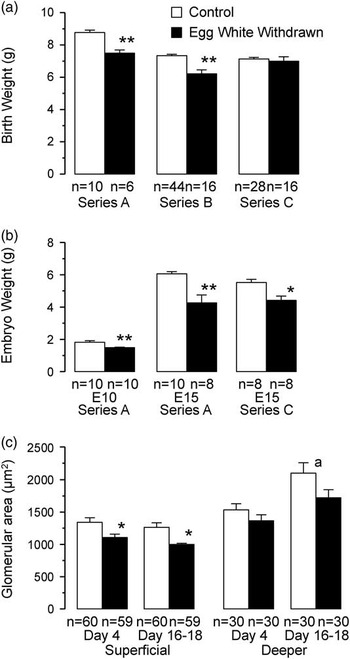
Undernutrition reduced birth (hatch) weight significantly in Series A (15.2%) and in Series B (15.4%) (Fig. 1a). In Series C, the birth weight of EwW quail showed a tendency to be lower than that of CTs, but the difference was not significant. This is presumably because the time lag between hatch and measurement of birth weight in series C was larger in EwW groups and thus some chicks start to grow, whereas all CT birds hatched nearly simultaneously. Embryonic weights on E15, however, were significantly lower in EwW quail in both Series A and C (Fig. 1b). In all series, the hatching rate of EwW groups was low (CT quail 75–80%, EwW quail 5–10%). Interestingly, approximately half of the eggs that did not hatch contained considerably grown embryos, suggesting that reduced nutrition failed to provide a sufficient energy source to proceed with maturation and hatching. The glomerular sizes (areas) of EwW quail were significantly lower on D4 and D16–D18 than those of CTs in the superficial region (Fig. 1c) (considered as more newly developed glomeruli), whereas no significant difference was noted in the deeper regions.
Effects of reduced nutrition on birth weight (a) and embryo weight (b) of control and egg white withdrawn quail from Series A, B and C. (c) Glomerular size/area during postnatal maturation from Series C. E10 and E15 indicate embryonic day 10 and 15, respectively. *P<0.05; **P<0.01 from control group; a P<0.01 from control postnatal day 4 age group by Tukey–Kramer method.

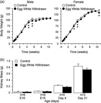
Body growth was similar in both groups except at a few time-points when EwW birds showed slightly lower body mass (Fig. 2a). We measured body mass in all series (showed similar results), and the growth curve of Series B (longest duration) is shown. The body mass of female quail is higher, partly due to holding eggs, than that of male birds. There was no significant difference in kidney weights between CT and EwW quail (Fig. 2b).
Body mass of control and egg white withdrawn (EwW) quail (Series B, males and females) as index of body growth (a) and kidney weight (b, Series A). There was no obvious difference in body growth curve between the control and EwW groups except for a few term points when EwW quail showed lower body mass. Numbers of birds used for measurement of body mass are the following. Control: male, n=4–15, female, n=19–28; EwW: male, n=4–11, female, n=3–7. The numbers vary because birds are used for examinations on a periodic schedule. *P<0.05; **P<0.01 from control group by Student’s t-test.

Number of glomeruli during development and histological and histochemical observations
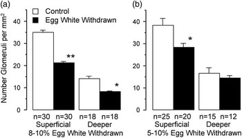
In both superficial (equivalent to cortex in mammalian kidney) and deeper regions (equivalent to juxtamedullary areas), the number of glomeruli was significantly lower in kidneys from young mature (3-week-old) EwW quail (Fig. 3a, Series A). For comparison, the reduction in the number of glomeruli in an earlier studyReference Nishimura, Yang and Lau 15 is shown in Fig. 3b (4.5 weeks). In contrast, the number of glomeruli measured in longitudinally cut sections was similar between CT and EwW kidneys during development (E10, E15 and D2).Reference Nishimura, Yang and Lau 15
The number of glomeruli in superficial (equivalent to cortical) and deeper (equivalent to juxtamedullary) regions of mature control and egg white withdrawn (EwW) quail kidneys counted in longitudinal histology sections. See Methods section for details of measurement. (a) The effect of 8–10% of EwW on the number of glomeruli (Series A). (b) For comparison, the result from previous studiesReference Nishimura, Yang and Lau 15 done by the same method is shown (4.5 weeks). *P<0.05; **P<0.01 from control group by Student’s t-test.

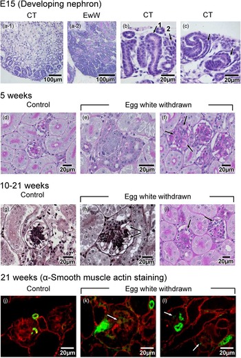
Histological views of kidneys were similar in all three series examined, and representative views are shown in Fig. 4. In condensate surface areas of E15 kidneys (nephrogenic zone) from intact (Fig. 4a-1) and EwW quail (Fig. 4a-2), ureteric bud-like structures (Fig. 4b), undifferentiated dark-stained mesenchymal cell mass (Fig. 4b), and S-shaped body stage glomeruli (Fig. 4c) were seen in both CT and EwW. More mature glomeruli with clear capillary loops were also seen in E15 and D4 kidneys, and the number further increased with age. In E10 and E15 kidneys, the condensate nephrogenic zone areas were irregular and larger in EwW kidneys than in CT ones (Fig. 4a-2) and mesonephros still remained in E15 and D4 EwW kidneys. Furthermore, the glomeruli were smaller in D4 EwW kidneys, particularly in the superficial area (less mature zone) (Fig. 1c).
Histological views of glomeruli from embryonic day 15 (PAS staining), 5-week-old (PAS staining) and 10- to 21-week-old (PAM staining) quail, and immunofluorescent view of α-smooth muscle (SM) actin in 21-week-old quail. (a) Condensate nephrogenic surface area from control (CT, a-1) and egg white withdrawn (EwW, a-2) containing undifferentiated mesenchymal cells. (b) Ureter bud (arrow-1) and mesenchymal mass (arrow-2); (c) S-shaped developing glomerulus; arrow indicates developing podocytes. (d) Mature glomeruli from control quail. (e) Possible glomerulus and tubules that immaturely stopped growing in egg white withdrawn (EwW) quail. (f) Expanded mesangial cells (deeper zone) from EwW quail with accumulation of lightly PAS-stained materials (arrows). (g) Glomerulus from control quail (21 weeks) with well-developed loop structure and Bowman’s capsule; PAM staining. (h) Enlarged glomerulus from EwW quail (21 weeks) with enlarged mesangium region; arrows indicate fused capillary loop space. (i) Glomeruli from EwW quail (10 weeks); PAS staining. Capillary loop structures are not clearly seen. Arrows indicate giant fused loops. (j) Immunoreactive α-SM actin staining of control glomeruli. Small arterioles show positive staining. (k and l) Positive α-SM actin staining in arterioles and glomerular mesangium (arrows) in EwW quail.

In 5-week-old CT kidneys, glomeruli showed distinct Bowman’s space and capillary loops and PAS-positive mesangium (Fig. 4d). In contrast, 5-week-old EwW kidneys revealed, in superficial regions, cell proliferation areas consisting of undifferentiated cell mass (Fig. 4e). Some of these masses appeared to contain incompletely differentiated glomeruli and renal tubules. Furthermore, after maturation, glomeruli in the deeper zone were larger in EwW kidneys (Fig. 4f), and mesangial cells were frequently expanded and contained substances only lightly stained with PAS (Fig. 4f, arrows). In 10- to 21-week-old or older EwW quail, glomeruli were often irregular in shape and larger than those of CTs in deeper areas (Fig. 5). The mesangial areas contained PAM-positive (also laminin-positive) substance and were larger in EwW kidneys (Fig. 4g and 4h). Fused giant loops were often seen (Fig. 4h and 4i, arrows). Possible adhesion between Bowman’s capsule and capillary loops was also seen (Fig. 4i).
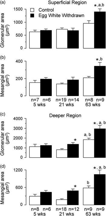
Glomerular area (size) and mesangial area of superficial (a, b) and deeper (c, d) regions from 5-, 21-, and 63-week-old quail kidneys [n=number of glomeruli from three birds per age per each control (CT) and egg white withdrawn (EwW) group] (Series B). Areas were measured using a Keyence fluorescent microscope and cell count program in longitudinally cut histology specimens (PAM staining) using well-shaped glomeruli with either vascular poles or urinary poles or both. In deeper regions, glomerular size and mesangial areas showed significant time- and treatment-dependent effects (ANOVA), whereas in superficial regions the difference between CT and EwW groups is clear only in 63-week-old quail kidneys. *P<0.05 from control group. Significantly higher (P<0.05) than 5 weeks (a) and 21 weeks (b) controls.

We examined α-SMA using an antibody that cross-reacts with avian α-SMA. SMA was clearly seen in the blood vessels from 21-week-old quail kidneys (Fig. 4j, 4k and 4l). In addition, the mesangium from EwW quail, but not from CT quail, showed immunoreactive SMA (Fig. 4k and 4l, arrows).
Size of glomeruli and mesangium
Glomerular size (area, µm2) and mesangium size (area, µm2) were measured in longitudinally cut and PAM-stained slices, using a Keyence HS All-in-One Fluorescence Microscope BZ-9000 (Dynamic Cell Count BZ-H1C) (Fig. 5). We selected well-shaped glomeruli showing vascular poles, urinary poles or both. In deeper regions of medullary cones, glomerular size and mesangial areas significantly increased with maturation/aging (5, 21, 63 weeks) (ANOVA, time effect). Also, glomerular size and mesangial areas were significantly larger in the EwW quail than in the CTs (ANOVA, treatment effect). In superficial areas, glomeruli also increased in size with aging, but the treatment effect was not as clear as in deeper regions. In the neonatal to early maturation period (D4 to D17–D18), in contrast, the glomerular size was smaller in EwW quail (Fig. 1c).
EM observations
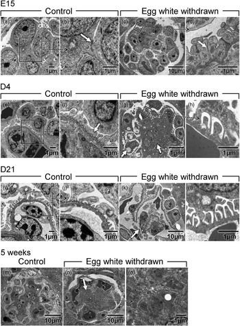
In the developing stage of CT glomeruli, the basal membrane was thin or irregular (Fig. 6a and 6b). Glomeruli from the EwW group also showed similar findings. Podocyte foot processes (FPs) often had irregular width (Fig. 6d–6h), and the cell junction complex was seen away from the basal membrane (Fig. 6a–6d) (arrows). In glomeruli that had distinct capillary loops, podocytes with well-developed FPs and slit diaphragms (Fig. 6e, 6f and 6j) and mesangium in the center area were seen (Fig. 6g and 6m). The regularity of the FPs increased with maturation in both CT and EwW kidneys. In EwW kidneys, vacuoles in the mesangium and podocytes (Fig. 6g), thin tall podocyte FPs (Fig. 6h and 6l), and basal membranes with irregular width (Fig. 6h) were seen. In EwW quail glomeruli, capillary loops were sometimes disrupted (Fig. 6k) or merged and showed ballooning. Mesangial areas were enlarged and contained cellular constituents with apparently complex interdigitation between mesangial processes and matrix and endoplasmic reticulum (Fig. 6n and 6o).
Electronmicroscopic views of glomeruli from embryonic day 15, postnatal day 4 (D4), D21, and 5-week-old quail from control and egg white withdrawn (EwW) groups. (b) is enlarged framed area from (a). Arrow in (b) shows cell junction apart from basement membrane. (d) is enlarged from (c) (framed) and shows well-developed foot processes (FPs) of podocytes with attachment to adjacent FP (arrow). Similarly, (f) and (h) are expanded areas from (e) and (g), respectively. (f) shows irregularly sized and fused FPs (arrow). In (g), numerous vacuoles are seen in the mesangial area (arrow) and some in podocytes. (i) and (j) [Enlarged from (i)] show well-developed FPs. In (k), arrow shows disrupted capillary loop and thin FPs in (l). (n) and (o) (expanded) indicate enlarged mesangium in EwW quail as compared to that of controls (m). For detailed observations, see Results section.

Junction protein markers
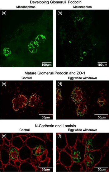
Immunoreactive podocin during kidney development (E10), double-staining of immunoreactive N-cadherin and laminin, and podocin and ZO-1 of young adult quail are shown in Fig. 7. Glomeruli of mesonephric kidneys were large in CT (Fig. 7a) and EwW kidneys (not shown), and podocin staining was seen along the capillary loops (Fig. 7a). In metanephric kidneys from E15 quail, podocin was expressed in glomeruli of the S-shaped body stage from both CT (Fig. 7b) and EwW groups. In mature glomeruli double-stained with podocin and ZO-1, immunoreactive podocin signals were seen as a continuous spot along capillary loops beneath the glomerular capsule outside the ZO-1 staining that is presumably basement membrane. There were no apparent differences in location and intensity of podocin staining between CT and EwW kidneys (Fig. 7c and 7d). In both groups, N-cadherin was strongly expressed in the superficial mesenchymal cell area (nephrogenic zone) and in the podocytes of S-shaped glomeruli and early capillary stage of metanephric glomeruli. N-cadherin staining was restricted to the capillary loop (podocytes) membrane immediately outside the basement membrane stained by laminin (Fig. 7e and 7f). There was no obvious difference between CT and EwW birds in terms of distribution and staining of N-cadherin. Clear laminin staining was seen in the mesangial area.
Immunofluorescent views of junction proteins. Podocin staining (FITC green) of large mesonephric (a) and developing metanephric (b) glomeruli from embryonic day 15 kidney from control (CT) group. (c) (CT) and (d) [egg white withdrawn (EwW)] are podocin (FITC green) and ZO-1 (TRITC red) double-staining of mature glomeruli from young adult quail. (e) (CT) and (f) (EwW) show N-cadherin (FITC) and laminin (TRITC) double-staining examined by confocal microscope (A1Rsi; Nikon; Tokyo, Japan). See Methods section for antibodies and staining conditions. For detailed observation, see Results section.

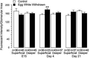
The fluorescent intensity of podocin signals determined by using a Keyence fluorescent microscope and cell count program is summarized in Fig. 8. There were no significant differences in intensity between CT and EwW groups in the superficial and deeper zones in three age groups (E15, D4 and D21) except in the D4 superficial area.
Fluorescent intensity of podocin signals determined by Keyence fluorescent microscope and cell count program in a total of 30–45 glomeruli from three birds from each control and egg white withdrawn group at embryonic day 15, postnatal day 4 (D4), and D21. Intensity was normalized by glomerular area in each glomerulus. **P<0.01 from control group by Student’s t-test.

Apoptosis
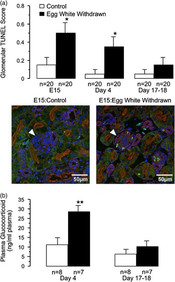
Apoptotic cells identified by TUNEL analysis are shown in Fig. 9a (upper panel). In all, 20 glomeruli were randomly selected from each CT and EwW group at three stages (E15, D4, D17–D18). The number of TUNEL signals (DNA fragment) per glomerulus was significantly higher in EwW than in CT groups on E15 (Fig. 9a, upper and lower panels) and D4 (Fig. 9a). The apoptosis level became lower with maturation in both groups, and no difference was noted between CT and EwW groups on Day 17–18.
(a) Glomeruli with positive terminal deoxynucleotidyl transferase-mediated dUTP-biotin nick-end labeling (TUNEL) marker that indicates possible apoptotic changes. In total, 20 glomeruli were randomly selected from control (CT) and egg white withdrawn (EwW) quail kidneys in three age groups each. Positive TUNEL signals (DNA fragments) in each glomerulus were counted, and the mean number of positive stainings per glomerulus is shown as the glomerular TUNEL score. Examples of TUNEL signals from embryonic day 15 CT and EwW kidneys are shown. Arrow head: glomerulus; bright green: TUNEL staining; red: wheat germ agglutinin lectin staining; blue: 4',6-diamidino-2-phenylindole dihydrochloride hydrate staining for nuclei. (b) Plasma glucocorticoid (corticosterone) determined by radioimmunoassay in developing quail at days 4 and 17–18. The minimum detection limit of the assay, defined as the apparent concentration at 95% B/B0 in the standard curve, was 3.0 ng/ml. No sample showed levels of corticosterone exceeding the maximum detection limit. *P<0.05; **P<0.01 from control group, Tukey–Kramer method.

Plasma corticosterone, glucose, insulin, creatinine, osmolarity and electrolytes
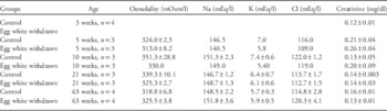
Plasma levels of corticosterone (major glucocorticoid in birds) determined by radioimmunoassay are shown in Fig. 9b. Data from Series A, B and C were combined. On D4, the glucocorticoid level was significantly higher in EwW quail. The level became lower on D17–D18, and no difference was noted between groups on D17–D18. Plasma levels of glucose (mg/dl) were high, but no significant differences were noted between CT and EwW groups on D4 (CT: 299.6±12.9, n=7; EwW: 269.3±20.3, n=7), on D17–D18 (CT: 331.6.±9.1, n=10; EwW: 370.9±8.6, n=8), or at 63 weeks (CT: 319.8±8.2, n=4; EwW: 337.8±14.4, n=4) of age. Plasma levels of immunoreactive insulin (μIU/ml) on D4 (CT: 1.96±0.30, n=5; EwW: 2.80±0.79, n=3) were higher than those on D17–D18 (CT: 1.19±0.19, n=10, P<0.01; EwW: 0.92±0.09, n=7, P<0.05), but no significant difference was noted between CT and EwW groups. Likewise, no differences in levels of plasma electrolytes, osmolality or creatinine were seen between CT and EwW groups at 3, 5, 10, 21 and 63 weeks of age (Table 1).
Plasma osmolality, electrolytes and creatinine in control and egg white withdrawn

Values are means±s.e. Those without s.e. indicate mean of n=2.
Discussion
Kidney development
The current study agrees with the general view of the avian kidney developmental stages of pro-, meso- and metanephric kidneys.Reference Davey and Tickle 26 We also noted the developmental process of avian glomeruli, including a ureteric buds stage, S-shaped body stage and capillary loop stage.Reference Davey and Tickle 26 The lower embryonic weights, smaller glomeruli and longer retention of mesonephric kidney seen in developing EwW groups indicate that EwW birds developed more slowly than CT birds. We reported previouslyReference Nishimura, Yang and Lau 15 that nephrogenesis continues after birth in the superficial condensate area consisting of mesenchymal cells; this nephrogenic zone was reduced in time-dependent fashion for ~3 weeks in CT quail. Also, at E10, embryos from the EwW group had wider nephrogenic zones (20.4±1.3% of medullary cone) than the CT group did (10.1± 2.4%, P<0.01)Reference Nishimura, Yang and Lau 15 (also, Fig. 4a-1 and 4a-2 in the present study); this difference was gone after hatch (D2), suggesting that in spite of delayed development, nephrogenesis was active in EwW birds. As we reported previously, surface glomeruli are smaller; and more mature, larger glomeruli are seen toward the deeper regions.Reference Nishimura, Yang and Lau 15
Effects of reduced nutrition during development and possible evidence of programming
Studies by Barker et al.Reference Barker, Eriksson, Forsen and Osmond 27 , Reference Barker, Winter, Osmond, Margetts and Simmonds 28 and numerous other epidemiological studies have indicated a strong inverse relationship between birth weight and the risk of coronary heart disease, hypertension, type 2 diabetes and other diseases in adulthood.Reference Fowden, Giussani and Forhead 1 – Reference Luyckx and Brenner 3 It is postulated that insufficient nutrient availability during gestation may lead to developmental adaptations and that a number of organ structures and functions undergo programming during embryonic and fetal life via hormonal influences.Reference Lau and Rogers 29 – Reference Taylor and Poston 30 These adaptive measures have short‐term benefits to the embryo and fetus, but subsequent catch‐up growth may create metabolic conflicts that predispose the adult to increased risks of adult disease susceptibility, including obesity.Reference Lau and Rogers 29 – Reference Taylor and Poston 30 Also, catch-up growth immediately after early malnutrition may program postprandial hyperleptinemia.Reference Coupe’, Grit, Darmaun and Parnet 31 A kidney with a reduced number of nephrons would have less renal reserve to adapt to dietary excesses or to compensate for renal injury.Reference Luyckx and Brenner 3 Impairment of nephrogenesis, renal sodium transport, the renin–angiotensin system, glucocorticoid levels (excessive increase) and the sympathetic nervous system appear to play a critical role in this fetal programming of adult diseases.Reference Alexander, Dasinger and Intapad 32 , Reference Baum 4 – Reference Moritz, Dodic and Wintour 6
We used a bird model because embryonic growth during incubation for hatching occurs with a predetermined nutrition source devoid of direct influence from maternal nutrition or circulation. In rodents and other mammals,Reference Fowden, Giussani and Forhead 1 , Reference Wu, Bazer, Cudd, Meininger and Spencer 33 reduced fetal nutrition is induced by decreasing the mother’s nutrition. The fetus is accordingly exposed to the metabolic and hormonal changes of the mother in addition to the effect of low nutrition. Also in birds, environmental factors such as temperature and humidity are well controlled during incubation. Furthermore, the present study indicates that so-called ‘fetal programming’ is not limited to mammalian species, but may be a phylogenetically important biological process.
The current study suggests that reduced nutrition during incubation causes delayed maturation and structural alteration of the quail kidney that are likely to program nephron disorders such as nephrosclerosis. The evidence includes (1) embryos were smaller and showed delayed embryonic growth and a lower hatching rate. The possible delay in kidney development agrees with the inhibition of ureteric bud branching in rats whose mothers had restricted nutrition.Reference Awazu and Hida 34 (2) The size of glomeruli in developing kidneys in the superficial area of EwW quail was smaller than that in CT kidneys. Birth (hatch) weight was smaller in most birds, but body growth soon caught up, as seen in the ‘catch-up growth’ of mammals.Reference Lau and Rogers 29 – Reference Coupe’, Grit, Darmaun and Parnet 31 , Reference Ibáñez, Suárez and Lopez-Bermejo 35 (3) The number of glomeruli was lower in the mature EwW group, particularly in the superficial region, agreeing with our previous resultsReference Nishimura, Yang and Lau 15 and with studies in mammals.Reference Baum 4 , Reference Manning and Vehaskari 5 In contrast, the number of glomeruli was similar in the two groups during development,Reference Nishimura, Yang and Lau 15 but the nephrogenic zone area was larger in low-nutrition quail. (4) The number of TUNEL signals in glomeruli was higher in EwW than in CT groups during the perinatal period, suggesting that more apoptosis takes place in nutrition-reduced kidneys. Therefore, although nephrogenesis actively occurs, apoptosis in glomeruli may exceed development/maturation in EwW quail, resulting in the reduced number of glomeruli after maturation by programming. (5) At 5 weeks of age (young adult), but not earlier, the mesangial cells of some EwW glomeruli frequently contain lightly PAS-positive edematous substance; and the capillary loops merge, forming giant loops partly similar to those seen in human nephrosclerosis.Reference Ikezumi, Suzuki and Karasawa 11 Furthermore, undifferentiated cell masses are seen in 5-week-old EwW quail kidney (Fig. 4e); we hypothesize that the biological clock for terminating nephrogenesis remains the same in growth-retarded kidneys as in CT kidneys. Immature glomeruli may lead to fibrotic degeneration in later life. The glomeruli of EwW quail became significantly larger with aging, suggesting hypertrophy compensating for the reduced number of glomeruli. This agrees with mammalian studies showing that overloading of the remaining nephrons causes compensatory enlargement and hyperfiltration.Reference Luyckx and Brenner 3
Role of mesangium in reduced nutrition-induced renal injury
In the nutrition-reduced group, we noted changes in the mesangium, such as vacuoles, edematous appearance, positive α-SMA, and proliferation/enlargement. It has been shown that glomerular cell death and accumulation of extracellular matrix correlate with the progress of nephrosclerosis and deterioration of renal function.Reference Sugiyama, Kashihara, Makino, Yamasaki and Ota 22 Extracellular matrix supports mesangial cell function and survival, and alteration in its constituents increases the susceptibility of mesangial cells to micro-environmental changes that lead to mesangial cell apoptosis and glomerular scarring. Also, the glomerular basement membrane and contractile mesangial cells establish a biomechanical unit that develops wall tension in the glomerular capillaries.Reference Sakai and Kriz 36 , Reference Sakai, Lemley and Hackenthal 37 It is thus possible that incomplete mesangial-basement membrane structure due to insufficient development in EwW quail may lead to the failure of the biomechanical unit, particularly in young birds, when blood pressure and thus glomerular hydrostatic pressure rapidly increase.Reference Nishimura, Xi and Zhang 38 It has been reported that mesangial flow (interstitial fluid in glomerulus) plays a role in the maintenance of mesangial structure and that function and flow disruption induced by occlusion of renal lymphatic and/or venous flow lead to mesangial and glomerular injuryReference Sugizawa 39 and aging.Reference Romano, Ferder and Inserra 40
In human nephrosclerosis, detachment of podocytes from the glomerular basement membrane seems to be the major mechanism of podocyte loss.Reference Kriz and Lemley 41 In the present study, histological or EM examination of EwW quail kidneys showed no clear morphological changes in podocytes, whereas ballooning or deformation of capillary loops was seen in EwW kidneys. Podocin and N-cadherin signals were expressed in mesonephros and metanephros, but no clear difference was noted in fluorescent visualization of signals between CT and EwW quail. Fluorescent intensity of podocin showed no difference between CT and experimental groups or among age groups, suggesting that cell junction proteins are unlikely to be impaired in the quail model of renal injury due to reduced nutrition. Immunoreactive podocin in quail kidneys was also confirmed using rabbit anti-podocin antibody (kindly provided by Dr. H. Tsukaguchi; Kansai Medical University; Osaka, Japan) raised against a synthetic peptide comprising the 17 carboxy-terminal amino acids of human podocin.Reference Yaoita, Nishimura and Nameta 20 Expression and distribution were similar to those in the present study. Genomic analysis revealed that the developing chick genome lacks nephrin and nephroproteins, whereas the structure of the slit diaphragms of podocyte FPs is maintained.Reference Miner 42
In the present study, plasma levels of creatinine (measured as an index for glomerular filtration rate), electrolytes and osmolality are similar between CT and EwW quail, suggesting that renal function is not impaired at this stage in spite of morphological injury of glomerular mesangium.
Mechanism of fetal programming
Two primary processes that drive programming are structural alteration and epigenetic changes in gene expression. Structural alteration includes low numbers of nephrons due to retarded development or to increased apoptosis, low numbers of cardiomyocytes and low deposition of elastin, leading to weak vascular walls.Reference Alexander, Dasinger and Intapad 32 , Reference Thornburg, O’Tierney and Louey 43 Intrauterine environmental changes (some may be permanent) trigger epigenetic changesReference Ingelfinger and Schnaper 44 that lead to modification of gene expression rather than changes in the DNA sequence or genetic code. DNA methylation can affect genome stability, viability, expression and imprinting. Several factors that trigger such changes include impaired maternal–fetal nutrition (deficit or excess), hypoxia, exposure to excess glucocorticoid and impaired placental function, such as reduced blood flow and low nitric oxide level.Reference Baum 4 , Reference Alexander, Dasinger and Intapad 32 , Reference Wu, Bazer, Cudd, Meininger and Spencer 33 , Reference Thornburg, O’Tierney and Louey 43 Such epigenetic modifications remain to be studied in the quail model.
We noted that neonatal EwW quail plasma corticosterone (major adrenocorticoid in birds that has both gluco- and mineralocorticoid actions) levels are also higher. It is possible that the impaired nutrition and low-energy source may be compensated by increased adrenocorticoid hormones. In humans, plasma cortisol levels were found to be higher; and the levels increased more in response to adrenocorticotropic hormone stimulation in the LBW group compared with the normal birth weight group, suggesting that prenatal programming of the hypothalamic–pituitary–adrenal (HPA) axis may be a fundamental mechanism underlying the development of metabolic syndrome in populations with LBW.Reference Luyckx and Brenner 3 , Reference Moisiadis and Matthews 45 , Reference Moisiadis and Matthews 46 Elevation of adrenal steroid hormones may also be a fetal response to stressful conditions such as insufficient nutrition or hypoxia. Prenatal stress and maternal exposure to glucocorticoid may lead to permanent modification of the HPA axis, possibly due to an impaired negative feedback mechanism,Reference Kapoor, Dunn, Kostaki, Andrews and Matthews 47 possibly via effects on the epigenome.Reference Moisiadis and Matthews 45 , Reference Moisiadis and Matthews 46 Prenatal exposure to increased glucocorticoid affects development of the fetal brain, neurotransmitter system and behavior.Reference Weinstock 48
Plasma levels of glucose in birds are in general higher than in other vertebrates of similar body size.Reference Braun and Sweazea 49 In the present study, we also noted that plasma glucose levels are over 300 mg/dl in both CT and EwW quail, whereas immunoreactive insulin levels are low in both the neonatal and maturing periods. We anticipated that plasma glucose levels would be higher in nutrition-reduced quail for energy supplementation, as shown in mammals.Reference Fowden, Giussani and Forhead 1 , Reference Portha, Chavey and Movassat 8 No differences in plasma glucose or insulin levels were seen, however, between the CT and EwW groups. It has been reportedReference Braun and Sweazea 49 that plasma insulin levels in birds are about one-tenth of those in rats, supporting the observation of lower numbers of β-cells in the avian pancreas; the reported levels agree with our current observation.
Recent studies show that an increased incidence of non-diabetic obesity in children is often associated with glomerulopathy, including glomerular hyperfiltration and renal injury, such as glomerulomegaly, mesangial expansion and nephrosclerosis.Reference Srivastava 50 It is not clear, however, whether these findings or the increase in FSGS is linked to the increased incidence of LBW or prematurely born babies. Niigata University Pediatric Department’s Nephrology Group, with which the authors collaborate, reportedReference Ikezumi, Suzuki and Karasawa 11 that of 16 children diagnosed with secondary FSGS by renal biopsy, 37.5% had LBW; LBW may be deemed a risk factor for FSGS and also a means of its early discovery. The results from the present animal experiments support these clinical observations.
Summary and perspectives
Major findings in Japanese quail include (1) undernutrition delayed embryonic growth and reduced the birth weight; (2) young adults exposed to reduced nutrition during development had a lower number of glomeruli due in part to increased apoptosis; and (3) glomeruli, specifically mesangium from undernutrition quail, were expanded with accumulation of laminin-positive substance, whereas podocyte junction proteins remained positive. This suggests that undernutrition may initially trigger the structural changes in the glomerular mesangium, possibly leading to nephrosclerosis. It remains to be determined whether hyperfiltration and increased glomerular pressure due to progressively rising blood pressure during maturationReference Nishimura, Xi and Zhang 38 cause mesangial injury and subsequent inflammatory responses in incompletely developed glomeruli or in those that have developmental defects such as lack of normal mesangial constituent substances. Particular attention may be needed to examine injury at the mesangial anglesReference Sakai, Lemley and Hackenthal 37 , Reference Kriz, Schiller, Kaissling and Taugner 51 of the glomerular capillary loop where the capillary wall directly faces the mesangium without a glomerular basement membrane and podocyte FPs. Our attempt to measure glomerular filtration rate by inserting a fine catheter chronically in aortae and veins of quail to see whether a low number of glomeruli affects renal functionReference Bailey and Nishimura 52 was halted due to considerable distress of the birds such as weight loss, limping and restricted movement. In a previous study,Reference Nishimura, Yang and Lau 15 we reported that EwW quail lost significantly more body mass by water restriction, suggesting impaired water homeostasis.
Although the current study does not show a difference in blood glucose and insulin levels between CT and EwW quail, further time-course study of catch-up growthReference Taylor and Poston 30 , Reference Ibáñez, Suárez and Lopez-Bermejo 35 is necessary. Indeed, preterm birth is a global problem that needs greater attention from policymakers, researchers, health care providers, the media, donor organizations and other stakeholders.
Acknowledgments
The authors thank Michiko Igashima and Harumi Kogami for excellent technical assistance in histology, Dr. Toshio Shimada for help in measurement of plasma corticosterone. Preliminary studies were presented at the Council for High Blood Pressure Research, Orlando, FL, 2011. The authors are entirely responsible for the scientific content of the manuscript.
Financial Support
This work was supported by the Ministry of Education, Culture, Sports, Science and Technology, Japan (H.N., Grant-in-Aid for Scientific Research No. 21390307).
Conflicts of Interest
None.
Ethical Standards
The authors assert that all procedures contributing to this work comply with the ethical standard of the relevant national guides of the care and use of laboratory animals (Japanese quail, Cotrunix Japanica). Animal protocols were reviewed and approved by the Institutional Animal Care and Use Committee of the Niigata University Graduate School of Medical and Dental Sciences, Niigata, Japan and University of Tennessee Health Science Center.