Introduction
Alzheimer's dementia (AD) is a slowly progressing disorder that passes through a prodromal stage when symptoms are mild or barely detectable (Jagust, Reference Jagust2008). This period has received several descriptions and definitions (e.g. Kral, Reference Kral1962; Hughes et al., Reference Hughes, Berg, Danziger, Coben and Martin1982; Blackford and La Rue, Reference Blackford and La Rue1989; Levy, Reference Levy1994; Ebly et al., Reference Ebly, Hogan and Parhad1995) which were primarily created to describe and to detect the prodromal stage of AD and did not focus on the pre-stages of other kinds of dementia.
In 1999, Petersen and colleagues proposed diagnostic criteria to describe subjects who were neither cognitively normal nor had dementia but who had mild cognitive impairment (MCI) characterized by memory deficits (later, amnestic MCI single; Petersen et al., Reference Petersen, Smith, Waring, Ivnik, Tangalos and Kokmen1999). Subsequently, these MCI criteria have been revised several times in order to identify specific characteristics of MCI subtypes that are at highest risk for progressing to AD (Palmer, Reference Palmer2010). Additionally, there were attempts to assign clinical heterogeneous MCI subtypes preceding different types of dementia (Petersen, Reference Petersen2001).
The most recent and most detailed clinical classification of MCI was given in 2004 (Petersen, Reference Petersen2004). Four MCI subtypes were defined: amnestic MCI (aMCI) single, aMCI multiple, non-amnestic MCI (naMCI) single, and naMCI multiple. These four MCI subtypes were assigned potential prodromes to several neurodegenerative disorders (such as AD, frontotemporal dementia, or dementia with Lewy bodies (DLB)) or to vascular dementia (VaD): aMCI is presumed to have a high likelihood of progressing to AD and the non-amnestic subtypes are assumed to have a higher likelihood of progressing to a non-AD dementia (Petersen et al., Reference Petersen2001; Petersen, Reference Petersen2004; Busse et al., Reference Busse, Hensel, Gühne, Angermeyer and Riedel-Heller2006).
An unambiguous assignment between clinical subtypes of MCI and subsequent kinds of dementia would be of great importance in clinical research and practice helping with early recognition of AD or non-AD dementias, e.g. VaD. Corresponding advice could be given and treatment could be started at an early stage.
To our knowledge, only two population-based studies have focused on this topic (Busse et al., Reference Busse, Hensel, Gühne, Angermeyer and Riedel-Heller2006; Manly et al., Reference Manly, Tang, Schupf, Stern, Vonsattel and Mayeux2008). Both studies included all four MCI subtypes according to Petersen for the analysis. Manly et al. reported AD as the only measured outcome. Thus we know the relative risk of different MCI subtypes concerning incident AD but we get no information concerning the risk of progression to other types of dementia. MCI with impairment of memory and at least one other cognitive domain was associated with greatest risk of progression to AD. Busse et al. analyzed the progression of the MCI subtypes to AD, VaD, and to other dementias. The authors showed that AD was the most common type of dementia at follow-up in all four subtypes of MCI with one exception (naMCI multiple). Therefore, an increased risk of a particular type of dementia could only partially be found. However, Yaffe et al. (Reference Yaffe, Petersen, Lindquist, Kramer and Miller2006) addressed this question in patients attending state-supported memory disorder clinics and found that subtypes of MCI have a major influence on subsequent types of dementia diagnosis. These inconsistent results induced us to investigate the prognostic accuracy of two clinical subtypes of MCI – aMCI and naMCI – on the development of AD, VaD, and mixed dementia. Analyses were done in a sample of subjects without dementia, aged 75 years at baseline, who were participants in a population-based cohort study (VITA study) with an observation period of 90 months.
Methods
Vienna Transdanube Aging (VITA) study
This study is based on data collected during the VITA study, which is a population-based cohort study of 75-year-old inhabitants of two districts of Vienna (Austria). The baseline investigation took place between May 2000 and November 2002. Three follow-up investigations were conducted 30 months after baseline (first follow-up), 60 months after baseline (second follow-up), and 90 months after baseline (third follow-up). Since age is the best predictor of cognitive decline in the elderly, the VITA age stratification design attempts to minimize its variation of age (SD: 0.2 years; mean: 75.8 years at baseline). For further details on methods and recruitment strategies, see Fischer et al. (Reference Fischer2002). The VITA study was approved by the local ethics committee and all participants gave written informed consent.
Sample
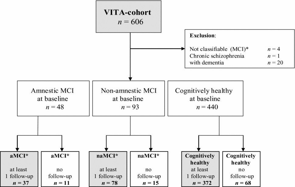
The VITA study involved 606 individuals born between May 1925 and June 1926. For this study, 25 subjects were excluded at baseline for different reasons: 20 subjects were excluded because of dementia (n = 20), one because of chronic schizophrenia (n = 1), and four subjects could not be classified concerning MCI because of missing psychometric test-performance (n = 4). Of the remaining 581 subjects, 94 subjects had no follow-up after baseline (aMCI: n = 11; naMCI: n = 15; cognitively healthy: n = 68).
Thus the final baseline sample of the current study comprised 37 subjects with aMCI and 78 subjects with naMCI, and 372 subjects who were cognitively healthy at baseline. Those 487 subjects had at least one follow-up investigation.
Mild cognitive impairment and cognitive health
Following the MCI criteria of Petersen (Reference Petersen2004), cognitive impairment was defined by 1.5 SD below the age-appropriate mean, derived from our total VITA cohort.
Each participant underwent neuropsychological testing with the Consortium to Establish a Registry for Alzheimer's Disease (CERAD; Morris et al., Reference Morris1989; standardized German translation: Berres et al., Reference Berres, Monsch, Bernasconi, Thalmann and Stähelin2000), and a test of divided attention – the Trail Making Test B (Reitan, Reference Reitan1956). The CERAD involved five cognitive tests: word list recall (verbal memory), figure recall (non-verbal memory), animal naming (verbal fluency), Boston naming test (naming), and figure copy (constructional praxis). Subjects were defined as cognitively healthy when all six psychometric tests scored within the 1.5 SD range. aMCI cases showed impairment in one or two memory tests exclusively, or showed impairment in one or two memory tests and additionally in one or more non-memory tests. naMCI cases showed impairment in one or more of the four non-memory tests, but both memory tests have to score within the 1.5 SD range.
The diagnosis of MCI was given to subjects without dementia with preserved activities of daily living (Lawton and Brody, Reference Lawton and Brody1969). We disregarded memory complaints as a MCI criterion, because its inclusion reduces the number of MCI diagnosed and underestimates the frequency of incident AD (Busse et al., Reference Busse, Hensel, Gühne, Angermeyer and Riedel-Heller2006).
Consensus diagnosis of dementia until the third follow-up
Diagnosis of dementia until the third follow-up was determined by applying the NINCDS-ADRDA criteria (for AD; McKhann et al., Reference McKhann, Drachman, Folstein, Katzmann, Price and Stadlan1984), the NINDS-AIREN criteria (for VaD; Román, Reference Román1993), and the Newcastle criteria (for DLB; McKeith et al., Reference McKeith1996). A panel of experts had to reach a consensus based on longitudinal information from psychometric results in combination with information from the Clinical Dementia Rating (Hughes et al., Reference Hughes, Berg, Danziger, Coben and Martin1982), instrumental activities of daily living (Lawton and Brody, Reference Lawton and Brody1969), and the Mini-Mental State Examination (MMSE; CERAD). Additional information was obtained from cerebral MRI (at baseline and at every follow-up); from FDG positron emission tomography available in 48 cases; from the diagnosis of depression (DSM-IV criteria; Saß et al., Reference Saß, Wittchen and Zaudig1996); and, for all cases, from relevant blood parameters (e.g. blood count, electrolytes including calcium, vitamin B12, and folic acid, and thyroid parameters including TSH). Information given by relatives was available for 197 subjects.
Statistics
Group comparisons were done by one-way ANOVA (age, MMSE), Kruskal Wallis test (education), t-test (age, MMSE), Mann-Whitney U-test (education), or the χ2 test (sex). All p-values less than 0.05 were considered to indicate statistical significance.
Sensitivity, specificity, positive predictive value (PPV), negative predictive value (NPV), and the corresponding 95% confidence intervals were specified as diagnostic values.
Analyses were performed using IBM SPSS Statistics 19.
Results
Prevalence of MCI subtypes and cognitively healthy participants at baseline
All 487 subjects had at least one follow-up investigation. Of those, 37 (7.4%) were diagnosed as aMCI and 78 (16.0%) as naMCI, and 372 (76.6%) were cognitively healthy at baseline (see Figure 1). There was no significant difference between aMCI, naMCI, and cognitively healthy participants regarding the proportion of subjects dropping out after baseline (χ 2 test: p = 0.411, χ 2 = 1.777, df = 2).
Study population at baseline.
*Modified MCI subtypes: exclusion of the criterion “subjective memory complaints.”

Sample characteristics
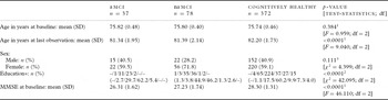
Table 1 summarizes the characteristics of the sample. There was no statistically significant difference in age and sex between subjects with aMCI and naMCI, and cognitively healthy subjects.
Characteristics of subjects at baseline

1 ANOVA.
2 Kruskal Wallis test.
3 χ2-test.
*Education: 1 = no school; 2 = elementary school; 3 = secondary modern school; 4 = professional school; 5 = secondary school; 6 = graduation diploma; 7 = university.
MMSE: Mini-Mental State Examination; aMCI: n = 1 missing value.
The three groups differed significantly regarding age at last observation (p < 0.0001), MMSE at baseline (p < 0.0001), and education (p < 0.0001; see Table 1 for test statistics).
Age at last observation
In terms of age, aMCI and naMCI did not differ significantly (t-test: p = 0.912, t = –0.111, df = 113) whereas both aMCI versus cognitively healthy (t-test: p = 0.005, t = –2.838, df = 407) and naMCI versus cognitively healthy (t-test: p = 0.002, t = –3.129, df = 99.042) were significantly younger. Subjects with aMCI were the youngest at their last observation. However, 37.8% (n = 14) subjects with aMCI, 46.2% (n = 36) with naMCI, and 70.2% (n = 261) cognitively healthy subjects had their last observation 90 months after baseline (third follow-up).
MMSE at baseline
Each group differed significantly from each other (t-tests: aMCI vs. naMCI: p = 0.007, t = –2.773, df = 72.704; aMCI vs. cognitively healthy: p < 0.0001, t = 8.536, df = 406; naMCI vs. cognitively healthy: p < 0.0001, t = 5.161, df = 96.281). Subjects with aMCI had the lowest MMSE score.
Education
aMCI and naMCI did not differ significantly (U-test: p = 0.084, U = 1185.0) whereas aMCI versus cognitively healthy (U-test: p = 0.005, U = 5189.5) and naMCI versus cognitively healthy (U-test: p < 0.0001, U = 8798.0) showed significantly less education.
Outcome: Alzheimer's dementia, vascular dementia, and mixed dementia
In the present study, the progression to any kind of dementia within 7.5 years was: 73.0% of aMCI, 41.0% of naMCI, and 17.5% of cognitively healthy.
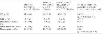
Table 2 summarizes the incidence rates of different kinds of dementias in relation to the MCI groups and to those who were cognitively healthy at baseline. The percentage of incident AD was significantly higher in aMCI compared to naMCI. aMCI and naMCI did not differ concerning incident VaD, mixed dementia, and other dementias.
Outcome of subjects within 7.5 years

*Fishers exact test.
Detailed information for the diagnosis group “other dementias” is not shown in Table 2: three individuals converted to pure DLB (one of each baseline group), 13 individuals converted to DLB in combination with AD (n = 3 of each MCI subtype, and n = 6 of cognitively healthy), and finally one of the naMCI group and one of the cognitively healthy group converted to a combination of AD, VaD, and DLB. None of the participants was diagnosed with frontotemporal dementia.
Amnestic MCI versus non-amnestic MCI
Alzheimer's dementia:
The percentage of incident AD in subjects with aMCI (46.0%) at baseline was twice as high as in subjects with naMCI (23.1%; χ 2 test: p = 0.013, see Table 2 for test statistics).
Vascular dementia:
The percentage of incident VaD in subjects with aMCI (5.4%) at baseline was more than twice as high as in naMCI (2.5%), though not showing statistical significance (Fisher's exact test: p = 0.593).
Mixed dementia:
The proportion of incident mixed dementia in the MCI groups at baseline was nearly identical. Overall, 10.8% of aMCI and 9.0% of naMCI developed a mixed dementia after baseline (Fisher's exact test: p = 0.744).
MCI versus cognitively healthy
Not necessarily as a primary goal but to complement the data, a comparison with cognitively healthy persons was performed, too. When subjects with aMCI were compared with subjects who were cognitively healthy, the proportion of all kinds of incident dementia were found to be significantly higher in the aMCI group (p-values not shown). This also applies to naMCI in comparison to cognitively healthy with the exception of incident VaD. The percentage of VaD in naMCI is higher but not statistically significant. Not developing dementia within 7.5 years was also significantly different in all three groups (see Table 2).
Diagnostic values of MCI subtypes
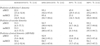
Sensitivity and specificity of aMCI and naMCI for the diagnosis of AD, VaD, or mixed dementia within 7.5 years after baseline are presented in Table 3.
Diagnostic values of aMCI and naMCI to predict the onset of AD, VD, and mixed dementia

Sensitivity rates were quite low for both MCI subtypes concerning all kinds of dementias (range 17.4%–33.3%). The subtype aMCI showed highest sensitivity concerning the prediction of AD (22.1%) and VaD (33.3%) whereas naMCI showed highest sensitivity concerning the prediction of VaD (33.3%) and mixed dementia (30.4%).
The subtype aMCI had high specificity for developing all three kinds of dementia: AD (95.1%), VaD (92.7%), and mixed dementia (92.9%). Specificity rates of naMCI were lower for all three types of dementia (range 84.2%–85.4%).
For both MCI subtypes the PPV was low (range 2.6%–45.9%). However, the NPV was higher (range 85.6%–99.1%).
Discussion
We investigated the prognostic accuracy of two clinical subtypes of MCI (aMCI and naMCI) in a population-based sample of subjects without dementia aged 75 years at baseline. The observation period was 7.5 years. In the present study, the proportion of incident AD in subjects with aMCI was significantly higher than in subjects with naMCI. The proportion of incident VaD and mixed dementia did not differ significantly concerning the MCI subtypes. Both MCI subtypes had a low PPV and high NPV for AD, VaD, and mixed dementia.
Comparing our results with other studies has several limitations: in each study MCI subtypes are defined differently, observation periods are of different duration, and sensitivity or specificity for MCI subtypes are rarely given. AD was the outcome most frequently used whereas the conversion to other kinds of dementia was not always specified. Therefore, the relevant design parameters are described in detail for each study.
The study by Manly et al. (Reference Manly, Tang, Schupf, Stern, Vonsattel and Mayeux2008) examined a community sample concerning the incidence rate of AD in MCI subjects with and without memory impairment (including subjective memory complaints) over a period of six years, which is comparable to our own. Overall, 20.44% of aMCI and 23.6% of naMCI were subsequently diagnosed with AD. Our results showed a double conversion rate of aMCI to AD and a nearly identical conversion rate of naMCI. Manly et al. presented a relative risk for developing AD between 3.2 and 4.3 for aMCI (single and multiple) and between 0.7 and 1.9 for naMCI (single and multiple).
Lopez et al. (Reference Lopez2007), after a mean follow-up period of only 4.3 years, found a conversion rate of aMCI to AD of 48.1%, which is comparable to our results. The conversion rate of MCI multiple cognitive domain type to AD was 41.3% and higher than that in our study. Both subtypes were independent of subjective memory complaints. Unfortunately, the authors did not state whether the MCI multiple cognitive domain type included memory impairment or not.
Rountree et al. (Reference Rountree, Waring, Chan, Lupo, Darby and Doody2007) evaluated the conversion of MCI subtypes to dementia in a sample of patients attending a memory disorder center over four years. They showed a conversion rate to AD that was comparable, even though a little higher, to our study. This was probably caused by recruiting more severely affected MCI patients in a health center for memory disorders than in a population-based cohort study. The authors differentiated aMCI (defined by Petersen), amnestic-subthreshold MCI (a test of delayed recall resulted in 1.5 SD under the mean), naMCI (single or multiple), and subjective memory loss without formal cognitive deficits. Combining aMCI and amnestic-subthreshold MCI to one single group, the conversion rate to AD was 53%; from naMCI, 32.2% of the subjects converted to AD.
Busse et al. (Reference Busse, Hensel, Gühne, Angermeyer and Riedel-Heller2006) compared the rate of progression across four MCI subtypes (aMCI single, aMCI multiple, naMCI single, naMCI multiple) within a follow-up period averaging 4.3 years. They described the outcome concerning the original MCI criteria (Petersen) and modified criteria (irrespective of memory complaints). Referring to the modified criteria as used in our study and combining aMCI single and multiple versus naMCI single and multiple, the conversion rates were in part different to our results (aMCI: to AD 33.9%, to VaD 1.7%, and to other dementias 11.9%; naMCI: to AD 22.4%, to VaD 7.1%, and to other dementias 3.5%). Sensitivity and specificity of MCI were given cumulatively for all kinds of dementia only. AD was the most common type of dementia in the follow-up of all subtypes of MCI with the exception of naMCI multiple. The assumption that each subtype is associated with an increased risk for a particular type of dementia was only partially shown by the authors.
Rasquin et al. (Reference Rasquin, Lodder, Visser, Lousberg and Verhey2005) investigated the prognostic accuracy of four different subtypes of MCI. Subjects without dementia from a memory clinic and a stroke cohort were included. The follow-up period was only 24 months. The results refer to the progression of the memory clinic sample to AD and of the stroke cohort sample to VaD separately. Probably because of the short observation period, the conversion rate of aMCI to AD was only 30.4% and that of naMCI was 10.2%. Within the stroke cohort sample, the conversion rate of aMCI to VaD was 15.4% and that of naMCI was 4.5%. To our knowledge, this study was the only one providing information concerning diagnostic values for all MCI subtypes separately: MCI multiple (all kinds) had the highest sensitivity for both AD (80.8%) and VaD (100%), which seems to be due to patient selection. aMCI single had the highest specificity (85.9% for AD and 100% for VaD). Comparable to our study, PPV was low for all subtypes and NPV was high. The authors noted that because of the low PPV other markers for the pre-dementia stage are needed. However, Yaffe et al. (Reference Yaffe, Petersen, Lindquist, Kramer and Miller2006) stated that subtypes of MCI have a major influence on subsequent types of dementia in participants enrolled by self-referral or by referral from family or physicians. They distinguished between aMCI single, naMCI single, and MCI multiple (amnestic and non-amnestic), and had a mean follow-up period of 3.1 years. Among the patients who progressed to AD, 76% had prior aMCI, 11% had prior naMCI single, and 13% had MCI multiple. Of those who progressed to VaD, 50% had prior aMCI, 8% had prior a naMCI single, and 42% were previously diagnosed as MCI multiple.
Summing up all findings, the conversion rate from each MCI subtype to different kinds of dementias varied. Different age of participants and different observation periods could confound this fact, because increasing age is accompanied by increasing incidence of dementia (Jorm and Jolley, Reference Jorm and Jolley1998). In addition, recruitment design (e.g. population-based cohort versus patients of a memory clinic or stroke unit) can be a major confounder.
It has to be considered that none of the studies presented above accounted for mixed dementia (AD/VaD) in their findings. It is not clear whether this kind of diagnosis was included in the AD or the VaD group or whether it has been excluded from analysis at all.
With regard to the prognostic accuracy of aMCI and naMCI, we did the analysis for AD, VaD, and mixed dementia (AD/VaD). Additionally, the group “other dementias” was established for the remaining subjects with other kinds of dementia (pure DLB, DLB+AD, DLB+AD+VaD). Diagnostic values were calculated for this group to avoid overlooking prognostic information of both MCI subtypes concerning dementias with DLB diagnosis: both MCI subtypes had a low sensitivity and PPV, and resulted in a high specificity and NPV (data not shown). This strengthens our main finding that only aMCI showed an increased risk for one particular type of dementia, namely AD.
Additionally, the enormous variation of prevalence rates of MCI subtypes at baseline in such studies has to be mentioned (Busse et al., Reference Busse, Hensel, Gühne, Angermeyer and Riedel-Heller2006; Manly et al., Reference Manly, Tang, Schupf, Stern, Vonsattel and Mayeux2008; Dlugaj et al., Reference Dlugaj2010; Petersen et al., Reference Petersen2010). The following specific reasons could explain those heterogeneous prevalence rates for MCI subtypes: first, different prevalence of MCI depends on the number of psychometric tests used to operationalize MCI criteria. Second, different age of participants, different definition of MCI (e.g. including subjective memory complaints or not) and MCI subtypes, and different criteria for defining cognitive impairment within MCI (1.0 SD or 1.5 SD under the age-appropriate mean) influence the prevalence rates in the literature.
There are limitations to our study that may have affected the results. One leading limitation of population-based studies concerns sample selection. The participation rate in the VITA study was only 40% of the complete age cohort (Fischer et al., Reference Fischer2007). However, medical information of non-participants was gathered at baseline. There was no indication that depression or cognitive problems were more frequent in non-participants. As expected, differences in the frequency of survival between participants and non-participants were identified (Fischer et al., Reference Fischer2008). Furthermore, although a same-age cohort, is of advantage in tackling the question investigated in this study, it reduces the generalizability of results across age cohorts. It must also be mentioned that subjects who were lost between the first and the third follow-up and who were cognitively healthy at that time may possibly skew the results.
Conclusion
In conclusion, an accurate attribution between clinical MCI subtypes and subsequent kinds of dementia could not be found. However, we agree with statements in the literature that the aMCI subtype showed an increased risk for AD.
Conflict of interest
None.
Description of authors’ roles
S. Jungwirth made a substantial contribution to the analysis and interpretation of data, wrote the paper, and gave final approval of the version to be published. S. Zehetmayer was responsible for the statistical design of the study and for carrying out the statistical analysis. She revised the paper critically for important intellectual content, and gave final approval of the version to be published. M. Hinterberger made a substantial contribution to conception and design, revised the paper critically for important intellectual content, and gave final approval of the version to be published. K. H. Tragl made a substantial contribution to conception and design, revised the paper critically for important intellectual content, and gave final approval of the version to be published. P. Fischer made a substantial contribution to conception and design, analysis and interpretation of data, and preparation and writing of the paper, along with revising the paper critically for important intellectual content and giving final approval of the version to be published.
Acknowledgment
The Vienna Transdanube Aging (VITA) study is supported by the Ludwig Boltzmann Society and is carried out at the Ludwig Boltzmann Institute of Aging Research.