Introduction
Along with cerebral malaria and metabolic acidosis, malarial anaemia is one of the major pathologies associated with malarial infections and estimated to be responsible for up to one-third of malaria deaths (Nweneka et al. Reference Nweneka2010). Severe malarial anaemia, defined as a haemoglobin concentration below 5 g dL−1 or a haematocrit below 0.15, most severely affects young children and pregnant women and is an important contributor to childhood mortality in malaria-endemic regions (Casals-Pascual et al. Reference Casals-Pascual2006; Ghosh and Ghosh, Reference Ghosh and Ghosh2007). The causes of malarial anaemia are multi-factorial, resulting from the loss of erythrocytes at the end of each intraerythrocytic developmental cycle, but also from immune-mediated destruction of infected and non-infected erythrocytes and an impaired erythropoietic response (Casals-Pascual et al. Reference Casals-Pascual2006; Ghosh and Ghosh, Reference Ghosh and Ghosh2007; Pradhan, Reference Pradhan2009; Pathak and Ghosh, Reference Pathak and Ghosh2016). The occurrence of severe malaria in cases of low parasitaemias or in ‘benign’ Plasmodium vivax infections and its persistence for weeks after parasite clearance from the system suggest that ineffective erythropoiesis may play a larger role than previously anticipated (Ekvall, Reference Ekvall2003; Helleberg et al. Reference Helleberg2005; Douglas et al. Reference Douglas2012; Panichakul et al. Reference Panichakul2012). The upregulation of a number of pro- and anti-inflammatory cytokines, growth factors and effector molecules by the host immune system in response to the parasite infection has been suggested to impair the production of new erythrocytes (Clark et al. Reference Clark2006; Haldar and Mohandas, Reference Haldar and Mohandas2009; Perkins et al. Reference Perkins2011). Tumour necrosis factor-α (TNF-α), which can induce cleavage of the main erythroid transcription factor GATA-1, interferon-γ and macrophage migration inhibitory factor (MIF) (Miller et al. Reference Miller1989; McDevitt et al. Reference McDevitt2006) in particular have been found to be associated with inefficient erythropoiesis. Furthermore, inflammation leads to an upregulation of the liver hormone hepcidin, which regulates iron homeostasis, resulting in a reduction of the amount of bio-available iron in circulation that can cause anaemia of chronic disease (Ghosh and Ghosh, Reference Ghosh and Ghosh2007; de Mast et al. Reference de Mast2009; Casals-Pascual et al. Reference Casals-Pascual2012).
The haemoglobin degradation product haemozoin, which is produced by the parasite in order to detoxify haem, has been found to stimulate the secretion of erythropoiesis-inhibiting proinflammatory factors but also to have direct inhibitory effect on erythroid development, impairing erythroid proliferation or inducing apoptosis at physiologically relevant concentrations between 1 and 10 µg mL−1 (Keller et al. Reference Keller2004b; Lamikanra et al. Reference Lamikanra2009). It remains a matter of controversy whether the haemozoin crystal itself is immunologically active or these effects are dependent on adherent DNA, proteins or lipids (Parroche et al. Reference Parroche2007; Jaramillo et al. Reference Jaramillo2009). In addition, studies have shown haemozoin to catalyse the conversion of fatty acids to cytotoxic long-chain aldehydes such as 4-hydroxynonenal (4-HNE), which are also implicated in inhibiting erythropoiesis (Schwarzer et al. Reference Schwarzer2003; Giribaldi et al. Reference Giribaldi2004; Skorokhod et al. Reference Skorokhod2010). Other parasite-derived products such as the glycosylphosphatidylinositol anchor found on numerous parasite proteins have been suggested to be responsible for the immunological response and the major pathologies of malaria infections (Schofield, Reference Schofield2007). Direct parasite-derived erythropoiesis inhibitors have however not yet been identified. Here we report erythropoiesis-inhibiting activity in conditioned medium (CM) from P. falciparum cultures, confirm an effect of parasite-derived crude (but not purified) haemozoin on both the proliferation and differentiation of erythroid cells, and elucidate further the nature of the parasite factors that impair erythropoiesis. The findings are an important step towards the identification of parasitic factors involved directly in erythroid inhibition, which could be a target for medical interventions.
Materials and methods
All chemicals were obtained from Sigma-Aldrich (Arklow, Ireland) unless stated otherwise.
Erythroid cell culture
Haematopoietic stem/progenitor cells
Mononuclear cells were isolated from peripheral blood buffy coats obtained from the Irish Blood Transfusion Services (Dublin, Ireland) using density gradient centrifugation with Histopaque®−1077. CD34+ cells were isolated from mononuclear cells via immunomagnetic separation using anti-CD34 magnetic beads (Miltenyi Biotec, Bisley, UK).
Cultures were initiated from frozen or freshly isolated mononuclear cells or CD34+ cells. For depletion of leucocytes and removal of monocytes/macrophages through adhesion, mononuclear cells were pre-cultured for 2 days in IMDM (Iscove's modified Dulbecco's medium) with stabilized glutamine (Biochrom, Berlin, Germany) containing 5% (v/v) heparinized human plasma (Irish Blood Transfusion Services) and 3 U erythropoietin mL−1 (EPO) (Janssen-Cilag, Dublin, Ireland). Non-adherent cells were washed and cultured in erythroid medium consisting of IMDM supplemented with 5% (v/v) heparinized human plasma, 330 µg human holo-transferrin mL−1 and 10 µg recombinant human insulin mL−1 and containing 100 ng stem cell factor (SCF) mL−1 (Cambridge Biosciences, Cambridge, UK), 5 ng interleukin-3 (IL-3) mL−1 (R&D Systems, Minneapolis, MN, USA) and 3 U EPO mL−1 and 10−6 m hydrocortisone for 1–2 days in a fresh culture plate. CD34+ cells were plated directly into the appropriate conditions without pre-culture. As mononuclear cells isolated using density gradient centrifugation were found efficiently to generate erythroid cells without the need for further purification of haematopoietic stem/progenitor cells, these cells were subsequently used to initiate erythroid cultures grown in 96-well plates as described below.
Erythroid expansion in multi-stage culture for cell count-based growth assessment
Erythroid cultures were expanded in erythroid differentiation medium (EDM) as previously published (Giarratana et al. Reference Giarratana2011). Briefly EDM was composed of IMDM containing 1% of stabilized glutamine, and was supplemented with 330 µg mL−1 iron-saturated human transferrin, 10 µg mL−1 recombinant human insulin, 2 IU mL−1 heparin and 5% of human plasma. The medium was supplemented with 100 ng mL−1 SCF, 5 ng mL−1 IL-3 and 3 IU mL−1 EPO in the first 11 days of culture and solely with EPO thereafter. Cells were seeded at 1 × 10⁴ cells mL−1 on day 0, diluted 1 in 5 in fresh medium on day 4 and were reseeded in fresh medium at 5 × 10⁴ cells mL−1 on day 7 or 8, at 7 × 10⁵ cells mL−1 on day 11, at 4 × 10⁶ cells mL−1 on day 14 and 10 × 106 cells mL−1 on day 18. Manual cell counting was performed using the trypan blue exclusion method with trypan blue diluted 1/6 in phosphate-buffered saline (PBS) and added to cells at 1:1 or 1:4 ratios.
Erythroid expansion in batch culture for haemoglobin-based growth assessment
Mononuclear cells were seeded in erythroid medium containing 100 ng SCF mL−1, 5 ng IL-3 mL−1 and 3 U EPO mL−1 as standard conditions. These conditions served as a positive control for erythroid proliferation. As in vitro erythroid expansion shows significant batch-to-batch variation in general, experiments always contained an ‘internal’ positive control in which cells were expanded under normal growth conditions without any parasite-derived compounds. These controls served as a reference point, and comparability between experiments was therefore achieved through comparison of a culture's behaviour compared with its respective control. As a negative control, cells were seeded in pure IMDM medium lacking growth factors and plasma.
An erythroid proliferation assay based on the absorbance of haemoglobin at 405 nm (Boehm and Bell, Reference Boehm and Bell2014) was used to assess cell haemoglobinization as a measure of erythroid development. While haemoglobin absorbance is determined by both cell proliferation (in early stages of culture) and erythroid differentiation/degree of haemoglobinization (in late stages of culture), this method has been demonstrated as a quantitative measure of total erythroid output relative to a control culture. In brief, cultures were set up in 100 µL volumes per well, and for each condition, a well containing the appropriate medium blank without cells was included. Absorbance measurements of plates with lids were taken at 405 nm using a Synergy H1 plate reader (Biotek, Potton, UK) preheated to 37 °C and following 2 min of linear shaking at 567 cycles per min. Results were determined as the average absorbance of triplicate wells at 405 nm and their standard deviation unless stated otherwise. The averages were corrected by the medium blank and presented as percentage and standard deviation of the positive control value referring to cultures grown in erythroid medium supplemented with growth factors and plasma. Negative control of cells grown in medium lacking all growth factors and plasma served to confirm that positive controls had accumulated sufficient amounts of haemoglobin to obtain statistically meaningful results when comparing different culture conditions. Negative controls reached very low absorbance at 405 nm due to lack of cell growth and high cell death under these conditions but showed some variation due to limited residual growth and effects of the presence of contaminating erythrocytes in the mononuclear cell preparation.
Supplementation of cultures with parasitic factors
CM from P. falciparum cultures was added at different concentrations (% v/v) to erythroid medium containing supplements (transferrin, insulin, plasma and growth factors) at 2× or 5× concentrations to obtain standard (1×) concentrations after addition of the fraction of CM. Crude natural or synthetic haemozoin was resuspended in pure IMDM and added at various concentrations to erythroid medium containing all factors at 2× concentration. All of the tested factors, i.e. CM, β-haematin or haemozoin, were added from the start and for the duration of the culture.
Erythroid cultures were seeded in 48- or 96-well flat bottom plates and cultured in a humidified incubator at 37 °C and 5% CO2 for up to 21 days and all conditions were set up in triplicate.
Results are expressed as the average of triplicates and standard deviation unless stated otherwise. Statistical analysis was performed using Prism software (La Jolla, CA, USA). A two-tailed t-test was used for comparison between two sets of data, and multiple datasets were compared using one-way analysis of variance. Asterisks refer to statistical difference to positive control unless indicated otherwise: *P ⩽ 0.05, **P ⩽ 0.01, ***P ⩽ 0.001.
Flow cytometry
Cells were stained with anti-CD45-fluorescein isothiocyanate (FITC), anti-CD71–FITC or anti-glycophorin A (GpA)–phycoerythrin (PE) antibodies or appropriate IgG isotype controls for 30 min at room temperature, washed with PBS and analysed using a Coulter Epics XL (Beckman Coulter, Brea, CA, USA) flow cytometer. Compensation was performed using single-stained samples and isotype controls, i.e. cells were stained with an FITC-coupled antibody (anti-CD45 or anti-CD71) and a PE-isotype control or a PE-coupled antibody (anti-GpA) and an FITC-isotype control, respectively. Gating of viable cells was based on low forward scatter (FSC)/high side scatter (SSC).
Plasmodium falciparum-conditioned medium
Plasmodium falciparum-conditioned medium (Pf-CM) was obtained from blood-stage cultures of mycoplasma-free P. falciparum 3D7 cultures grown in RPMI medium supplemented with 5% (w/v) Albumax® in a candle jar according to published methods (Fennell et al. Reference Fennell, Al-shatr and Bell2008). CM was obtained from asynchronous cultures comprising a mixture of parasites in different stages of intraerythrocytic development at 10–15% parasitaemia after overnight culture. The medium was passed through a 0.2 µm filter to remove haemozoin and cellular debris and used at up to 20% (v/v) in erythroid medium. Plasmodium falciparum cultures were also used to condition protein-free IMDM in order to complement erythroid cultures with higher fractions of CM (up to 80%) without causing a dilution of the erythroid medium and to reduce the protein complexity caused by large amounts of Albumax®. To this end, mycoplasma-free [tested using LookOut Mycoplasma PCR detection kit (Sigma Aldrich)] asynchronous P. falciparum cultures were grown to high parasitaemia, washed two times with wash medium consisting of RPMI supplemented with 0.18 g sodium bicarbonate L−1 and one time with IMDM and then resuspended in IMDM. For higher protein content, cultures were concentrated 6–8-fold by resuspension in lower volumes of IMDM after washing. Cultures were cultured for 3–4 h to obtain CM without causing stress responses (i.e. no visible crisis forms were detected; parasitaemia and parasite stages were comparable to RPMI). CM was obtained by centrifuging the culture supernatant for 10 min at 2000 rpm (825 g) followed by filtration through a 0.2 µm filter and stored at 4 °C for short-term or −20 °C for long-term storage. Proteins were concentrated using an Amicon Ultra (Millipore, Billerica, MA, USA) centrifugal spin filter with a molecular mass cut-off of 30 kDa. CM was treated with proteinase K immobilized on Eupergit C beads, washed three times with PBS, at 1 U mL−1 at 37 °C overnight or for 24 or 36 h. Beads were removed by centrifugation at 250 g for 10 min and the medium was passed through a 0.2 µm filter.
Haemozoin
Synthetic haemozoin (InvivoGen, Toulouse, France) was resuspended in PBS at 5 mg mL−1, vortexed extensively and stored at 4 °C. The stock solution was diluted into pure IMDM to obtain working concentrations.
Haemozoin was isolated from mycoplasma-free P. falciparum cultures with parasitaemias between 10 and 15% that were predominantly in the trophozoite stage using saponin lysis (0.5 g L−1) and extensive washes based on methods published by Keller et al. (Keller et al. Reference Keller2004a, Reference Kellerb). The haemozoin pellet was frozen with protease inhibitors at −80 °C. Haemozoin pellets were extensively washed with PBS before and after any treatment. Haemozoin pellets were treated with Triton® X-100 at concentrations of 0.1, 0.5, 1 and 5% (v/v) in PBS for 30 min on ice with occasional vortexing. For proteinase K treatment, pellets were incubated with proteinase K at concentrations of 0.5, 0.1, 0.05, 0.01 or 0.005 mg mL−1 in PBS for 1.5 h or with 0.2 mg proteinase K mL−1 (Qiagen, Venlo, The Netherlands) in PBS overnight (14–18 h), or for 24 or 36 h at 37 °C in a shaking incubator at 120 rpm. All treatments were followed by extensive washes with PBS at 10 000 rpm for 10 min and resuspension in pure IMDM. Haemozoin quantification was performed by dissolving the crystals in 20 mm NaOH for 2 h and determining the absorbance at 405 nm compared with a haemin standard curve. Supernatants from detergent or enzyme treatments were assessed for protein content by sodium dodecyl sulphate–polyacrylamide gel electrophoresis (SDS–PAGE). SDS–PAGE was performed using a 10% (w/v) polyacrylamide separating gel with 10 µL samples, run at 150 V and stained with Coomassie Brilliant Blue R250 or silver according to published methods.
Results
Parasite-CM inhibits erythroid proliferation and differentiation in a concentration-dependent manner
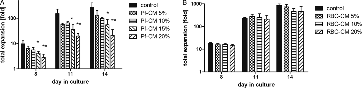
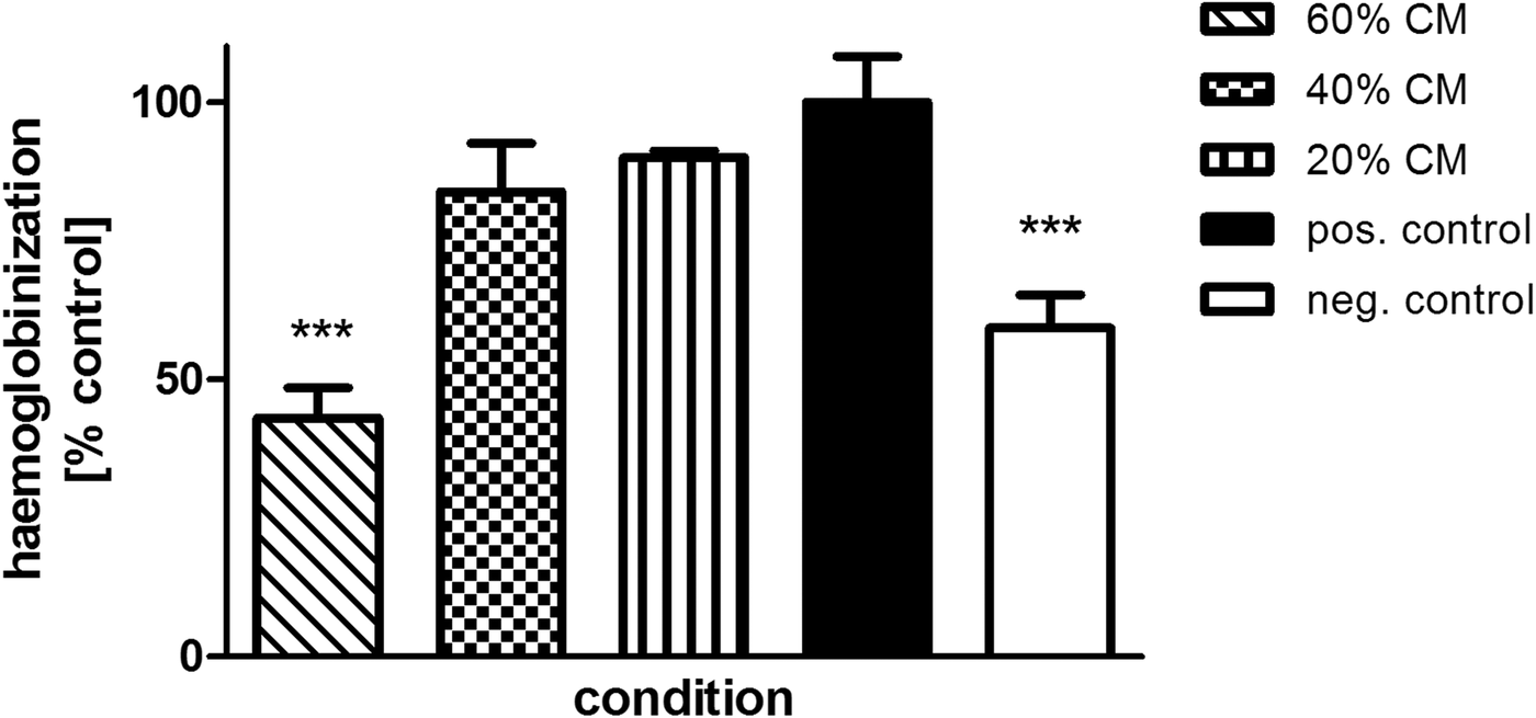
Cytokine imbalances resulting from the immune response to infections with Plasmodium have been proposed to be the principal problems underlying the development of malarial anaemia due to dysregulated erythropoiesis (Haldar and Mohandas, Reference Haldar and Mohandas2009). While many of the involved factors have been shown to affect the erythropoietic pathway, far less attention has been paid to a possible direct role of parasite-borne factors. Using an ex vivo erythropoiesis model which faithfully recapitulates the formation of erythrocytes from haematopoietic stem cells in culture (Giarratana et al. Reference Giarratana2005, Reference Giarratana2011; Boehm et al. Reference Boehm, Murphy and Al-Rubeai2009), the effect of CM from asynchronous P. falciparum blood-stage cultures on erythroid development was assessed. Parasite-CM (Pf-CM) was found to inhibit erythroid proliferation in a concentration-dependent manner, whereas medium conditioned with uninfected erythrocytes (RBC) did not elicit such a response (Fig. 1a and b). The effect of CM on erythroid output assessed using the multi-step expansion protocol and manual cell counting was confirmed using a spectrophotometric assay based on haemoglobin absorbance (Fig. 2). Haemoglobin absorbance and cell concentration have shown a linear correlation and similar amounts of sample-to-sample variation. Measurements in late stages of erythroid culture reflect not only the overall proliferation of erythroid cells but may also be the result of a delayed differentiation (with a delay in haemoglobinization) or indeed a blockage at a specific stage in the differentiation process, all of which can contribute to a reduced output of functional erythroid cells. As cells were grown in batch without medium replacement, overall expansion achieved with this method was lower and the degree of inhibition by parasite-CM therefore appears less severe than observed for the multi-step expansion protocol in Fig. 1.
(a) Inhibition of erythroid expansion by Plasmodium falciparum-conditioned medium (Pf-CM): erythroid expansion was reduced to a degree dependent on the proportion of Pf-CM that was used. (b) Conditioned medium from uninfected erythrocytes (RBC-CM) did not elicit this response. Cell expansion was assessed using manual cell counting with trypan blue and results are shown as averages of triplicates with vertical bars indicating standard deviations. Difference from positive control: *P ⩽ 0.05, **P ⩽ 0.01.

Inhibition of erythroid expansion by Plasmodium falciparum-conditioned IMDM. Proliferation/haemoglobinization of erythroid cells was inhibited in a concentration-dependent manner by conditioned medium (CM) from cultures of P. falciparum assessed on day 11 of culture using a spectrophotometric proliferation assay based on haemoglobin absorbance. Results are averages of triplicates presented as percentage of positive control and standard deviation (vertical bars). The negative control was a culture lacking growth factors and plasma. Difference from positive control: ***P ⩽ 0.001.

Conditioned IMDM (basal medium for erythroid expansion) elicited the same inhibitory effect as conditioned RPMI (with or without Albumax®) used for parasite culture and was subsequently used in order to be able to employ higher fractions of CM without diluting the erythroid medium and to avoid interference of proteins from the Albumax® supplementation (Figs 2 and 4). The degree of inhibition caused by CM varied between different batches of CM, most likely due to differences in parasitaemias or parasite stages, but showed the same concentration dependence. Cultures were grown asynchronously to obtain CM enriched in factors from all stages of parasite development. The inhibitory activity may be produced throughout intraerythrocytic development or at a specific stage; in the latter case, it is possible that concentrations in vivo (where parasites develop in more synchronous populations) might be even higher than those obtained in culture. While synchronized cultures would provide the advantage of being able to attribute potential inhibitory effects to certain parasite stages, results would depend strongly on achieving optimal sampling time points during these cultures.
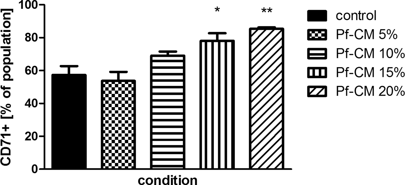
Cultures treated with Pf-CM showed not only reduced cell proliferation but also delayed differentiation suggesting an impairment of erythroid development in general. Cell samples taken at different time points in culture indicated that cells were significantly less mature as seen by the persistence of early markers of differentiation (e.g. CD45) and the presence of more CD71+ cells (Fig. 3). At the end of the culture, lower percentages of mature CD71-/GpA+ cells were found in cultures treated with CM than in controls.
Delayed differentiation in response to treatment with conditioned medium. Erythroid cells treated with Plasmodium falciparum-conditioned medium (Pf-CM) showed a delayed differentiation as seen by persistence of the erythroid marker CD71 (as measured by flow cytometry) in late stages of culture (day 18). Difference from positive control: *P ⩽ 0.05, **P ⩽ 0.01.

The erythropoiesis-inhibiting factor in CM appears to be proteinaceous
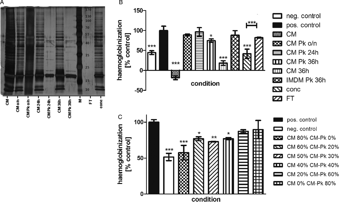
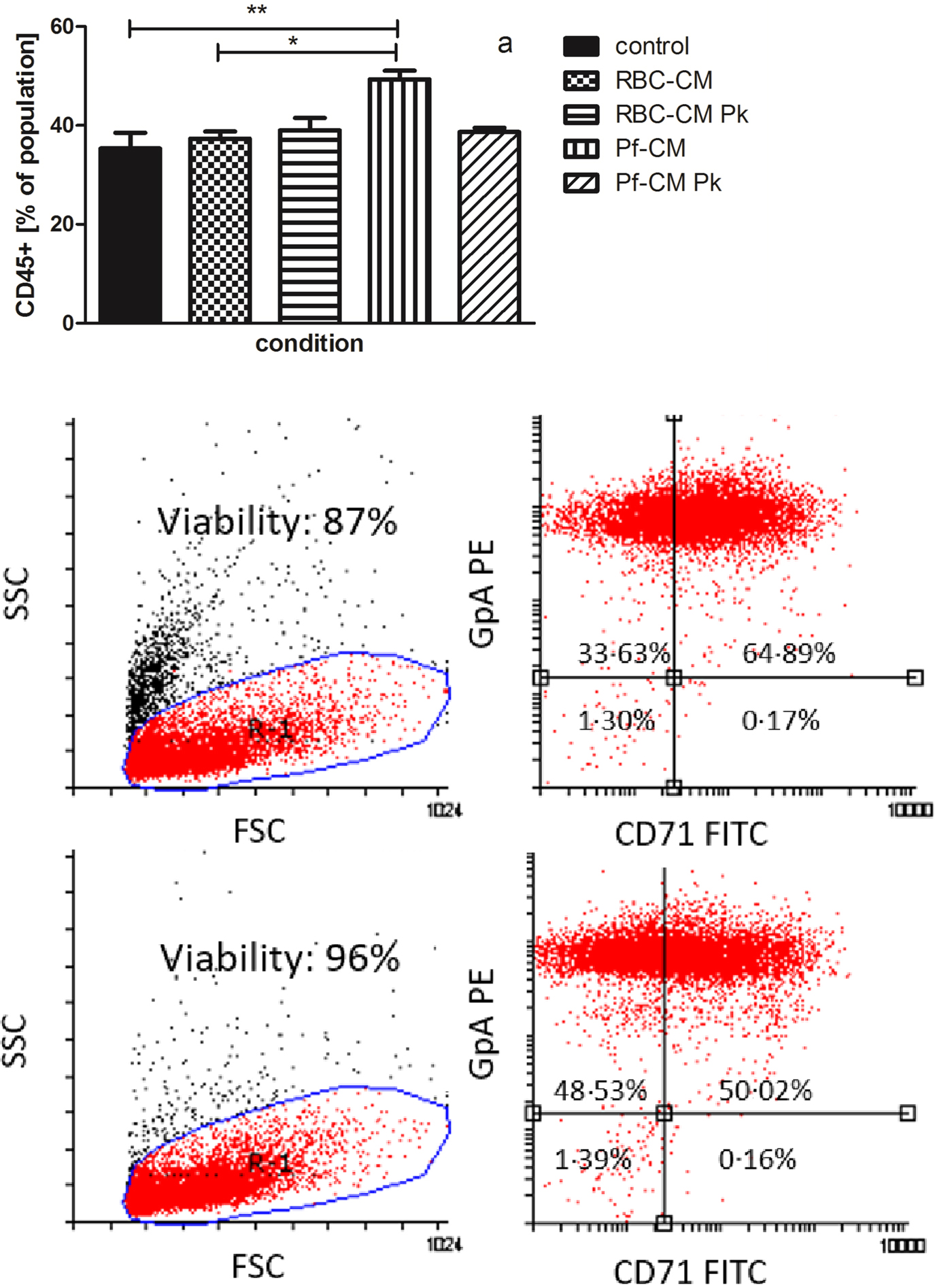
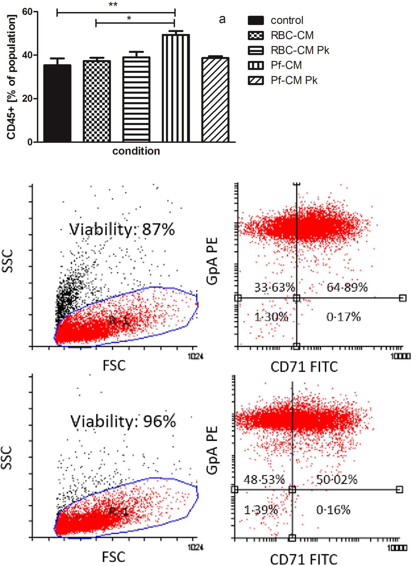
Treatment of CM with proteinase K immobilized on Eupergit® C beads resulted in an incomplete degradation of proteins after overnight incubation and degradation of a majority of the proteins after 24 or 36 h (Fig. 4a). The strong erythropoiesis-inhibiting activity observed for untreated CM was alleviated by all proteinase K treatments regardless of the incubation time, while CM incubated at 37 °C for 36 h without protease retained most of its activity (Fig. 4b). The effect of pure IMDM remained unchanged by proteinase K digestion and a concentration of proteins from CM using a centrifugal spin filter (30 kDa cut-off) and redilution to its original volume with pure IMDM showed the inhibitory activity to reside predominantly in the retentate rather than the flow through, providing support in terms of size and (adherent) nature for a proteinaceous factor. The fact that the rediluted concentrate displayed a weaker effect than the original CM, which completely inhibited erythroid growth, was mostly likely a result of some proteinaceous material remaining adherent to the filter, so that not the total amount of bioactive material could be recovered, an effect which might possibly be improved by using alternative means of protein concentration such as dialysis. Complementation of untreated CM with proteinase K-treated CM showed a concentration–response relationship to the fraction of the untreated CM and no inhibition by the proteinase K-treated CM (Fig. 4c). Protease digestion was not only able to revert the reduced proliferation of erythroid cells but also improved differentiation in early and late stages of erythroid culture. Higher percentages of immature CD45+ cells remained in Pf-CM-treated cultures than those treated with CM from uninfected erythrocytes or proteinase K-digested CM at day 4 in culture (Fig. 5a). As CD45 is present on haematopoietic stem/progenitor cells but lost during erythroid differentiation, it can be a valuable indicator of erythroid development even if it is not erythroid specific. Maturation in late stages of culture was also improved by proteinase K treatment of the CM, as found by an increase in mature CD71-/GpA+ cells over less mature CD71+/GpA+ cells (Fig. 5b). Flow cytometric analysis additionally indicated a larger population of dead cells/debris (low FSC and high SSC) in cultures with CM, suggesting decreased cell viability or higher cell fragility in late stages of culture.
Effect of proteinase K treatment of conditioned medium. (a) SDS-PAGE of Plasmodium falciparum-conditioned IMDM (CM) (batch a from Fig. 2) subjected to proteinase K (Pk) treatment overnight (o/n), for 24 or 36 h showing some protein degradation after overnight treatment (lane 3) and strong protein degradation after 24 h (lane 5) or 36 h (lane 7) treatment (left). M indicates molecular-weight standards. FT and conc indicate the flow through and retentate, respectively, of conditioned IMDM passed through a centrifugal filter unit with a molecular mass cut-off of 30 kDa. (b) Treatment with proteinase K overnight (CM Pk o/n), for 24 h (CM Pk 24 h) or 36 h (CM Pk 36 h) reverted the inhibitory action of conditioned medium from cultures of P. falciparum while incubation at 37 °C for 36 h (CM 36 h) without enzyme did not (top right). Treatment of pure IMDM with proteinase K (IMDM Pk) showed no effect. Proteins concentrated from conditioned IMDM with a centrifugal filter unit (conc) showed inhibitory effects while little inhibition was found with the flow through (FT) (P = 0.004). (c) Complementation of conditioned medium (80–0%) of P. falciparum cultures with proteinase K-treated conditioned medium showed a concentration–response relationship to the proportion of untreated conditioned medium and samples containing 80% proteinase K-treated conditioned medium showed no growth reduction on day 8 of erythroid culture compared with the untreated control (bottom right). Expansion was assessed using a haemoglobin-based absorbance assay and results are averages and standard deviation of triplicates presented as percentage of positive control grown without conditioned medium (vertical bars). In (b) and (c), the negative control was a culture lacking growth factors and plasma. Difference from positive control: *P ⩽ 0.05, **P ⩽ 0.01, ***P ⩽ 0.001.

Reversion of delayed differentiation by proteinase K treatment: (a) significantly higher percentages of immature CD45+ cells persisted in cultures treated with conditioned medium from Plasmodium falciparum cultures (Pf-CM) than in medium from non-parasitized erythrocytes (RBC-CM) (P = 0.006) and proteinase K digestion of the Pf-CM overnight reverted this fraction to control levels (top). *P < 0.05; **P < 0.01. (b) Delayed differentiation in cultures treated with Pf-CM (top) as seen in higher fractions of CD71+/GpA+ (right top quadrant) cells and lower numbers of CD71-/GpA+ (left top quadrant) cells was reverted by treatment of Pf-CM with proteinase K (bottom) (based on gate R1 = viable cell population). GpA PE, glycophorin A–phycoerythrin; CD71 FITC, CD71–fluorescein isothiocyanate. Proteinase K-treated Pf-CM also showed less dead cells and debris [low forward scatter (FSC), high side scatter (SSC)] in flow cytometric analysis. Experiments consisted of cells grown in three independent wells for each treatment and experiments were repeated twice; plots of representative cultures are shown here.
The effect of haemozoin on erythroid differentiation is due to adhering biomolecules
Haemozoin concentrations in the blood of malaria patients have been reported between 1 and 13 µg mL−1 (Keller et al. Reference Keller2004b). While this estimation is based on haemozoin concentrations found intracellularly within circulating erythrocytes, similar concentrations of free haemozoin have been measured in the plasma of malaria patients (Casals-Pascual et al. Reference Casals-Pascual2006). Haemozoin concentrations in the bone marrow could be significantly higher. Apart from the accumulation of malarial pigment in the macrophages of the bone marrow, parasitized RBC can become sequestered in marrow sinusoids (Wickramasinghe et al. Reference Wickramasinghe1987), leading to localized high concentrations of free haemozoin when the cells rupture. Haemozoin has been found to elicit strong immunological responses both in vivo and in vitro and directly to inhibit erythroid proliferation (Skorokhod et al. Reference Skorokhod2010) and cause apoptosis of erythroid cells (Lamikanra et al. Reference Lamikanra2009). It has been a matter of debate whether this activity is based on the haemozoin crystal itself (Jaramillo et al. Reference Jaramillo2009) or mediated by adhering biomolecules such as proteins (Shio et al. Reference Shio2010), lipids (Carney et al. Reference Carney2006) or DNA (Parroche et al. Reference Parroche2007).
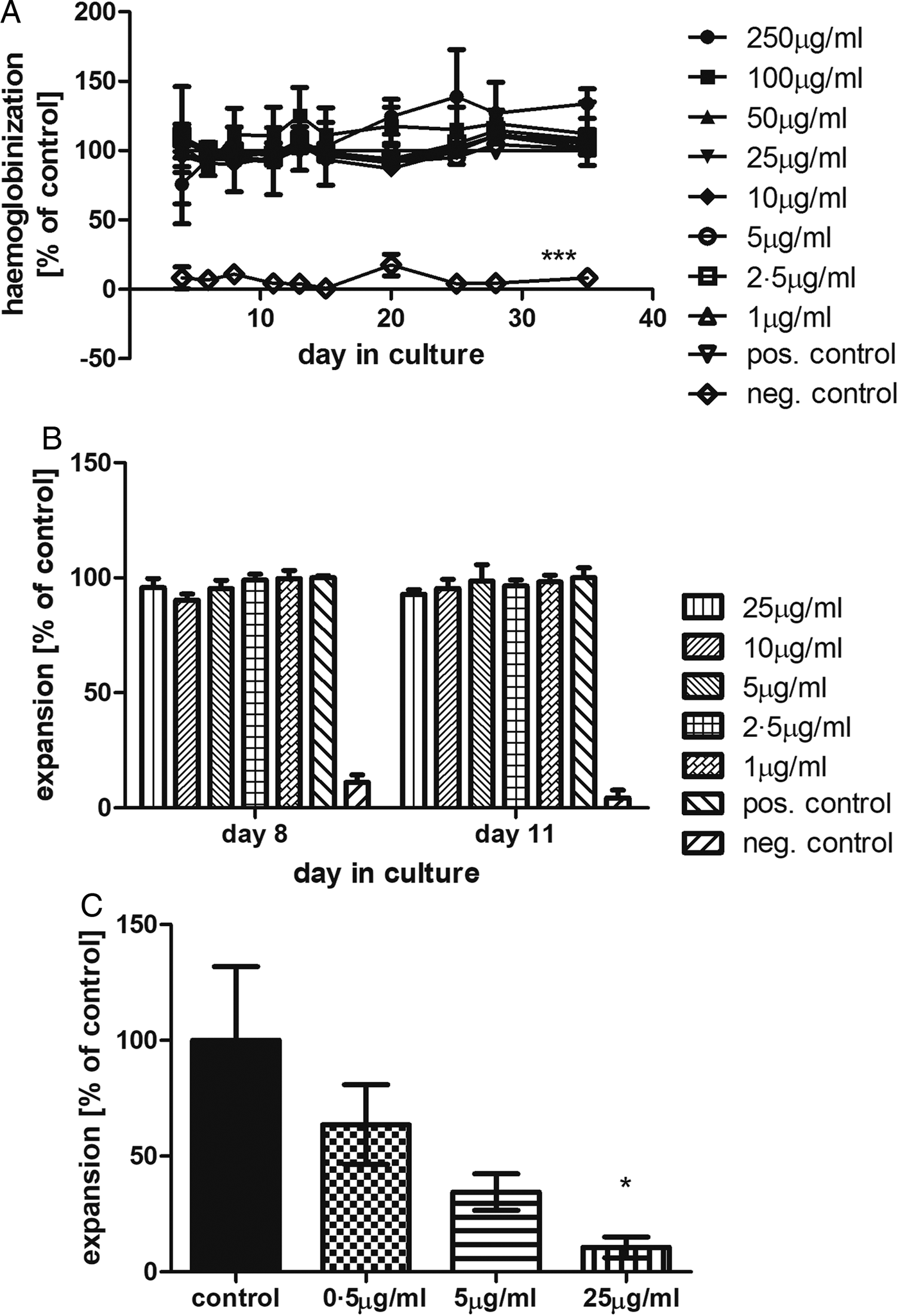
It has furthermore been suggested that the inhibitory effect of haemozoin on erythroid cells is a result of the non-enzymatic conversion of long chain fatty acids to toxic aldehydes such as 4HNE through catalytic action of haemozoin (Skorokhod et al. Reference Skorokhod2010). HNE has been found to form covalent, stable adducts with proteins via amino-, thiol- and histidyl-groups, and with DNA, which impact on cellular function, e.g. by delaying cell cycle progression (Poli et al. Reference Poli2008). In order to assess this hypothesis, we tested the effect of synthetic haemozoin (β-haematin) on ex vivo erythroid development. A number of different methods exist for the formation of β-haematin from haemin, but large differences have been found in the stability of the crystals obtained depending on the synthesis method. Synthetic haemozoin was therefore purchased from a commercial supplier to ensure product stability and avoid batch-to-batch variation. No inhibitory effect of synthetic haemozoin on erythroid growth could be detected in our system in the range of physiologically relevant concentrations (1–10 µg mL−1) (concentrations 1–25 µg mL−1 shown as excerpt in Fig. 6b) or even with concentrations up to 250 µg mL−1 (Fig. 6a). While the biological activity of haemozoin has been suggested to be influenced by particle size, control of crystal size is difficult due to the propensity to aggregate in aqueous media even after intense sonication (Coban et al. Reference Coban2010). Crude parasitic haemozoin on the other hand inhibited erythroid proliferation in a concentration-dependent manner with haemozoin at 25 µg mL−1 conferring a 90% reduction in cell growth (Fig. 6c).
The effect of haemozoin on erythroid proliferation: (a) synthetic haemozoin did not inhibit the proliferation of erythroid cells as determined using a haemoglobin-based absorbance assay. None of the tested haemozoin concentrations between 1 and 250 µg mL−1 showed a significant reduction of cell growth. (b) Excerpt of erythroid proliferation on days 8 and 11 for physiologically relevant concentrations between 1 and 25 µg mL−1. Results are averages of triplicates shown as percentage and standard deviation of positive control grown without synthetic haemozoin and the negative control culture lacked growth factors and plasma. To account for the absorbance of the synthetic haemozoin itself, each plate contained a blank for each β-haematin concentration in erythroid medium but without addition of cells. At each day of culture when absorbance was measured, the respective blank was subtracted. No significant difference was found between any of the different haemozoin concentrations and the positive control, while the negative control was significantly different from all tested conditions (P ⩽ 0.001). (c) Expansion of erythroid cells was inhibited by increasing concentrations of crude haemozoin isolated from Plasmodium falciparum cultures by saponin lysis. Expansion was determined by manual cell counting using trypan blue exclusion and results are averages of triplicates presented as percentage of untreated control and standard error of the mean (vertical bars). Difference from positive control: *P ⩽ 0.05, ***P ⩽ 0.001.

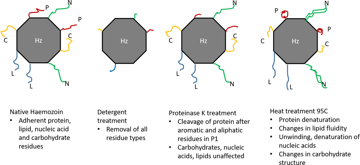
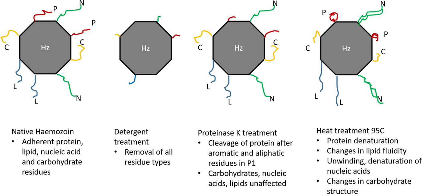
In order to address whether this direct erythropoiesis-inhibiting activity was mediated by biomolecules such as proteins, crude parasitic haemozoin was subjected to various treatments including detergent treatment with Triton® X-100, heat inactivation and proteolytic digestion (Fig. 7). Detergent treatment was aimed at complete removal of potential adhering biomolecules such as proteins, lipids, carbohydrates or nucleic acids. Heat inactivation on the other hand would not remove such biomaterial from the haemozoin crystal but denature protein structure, rendering any adhering proteins inactive. At temperatures of 95 °C, the structure and fluidity of most lipids would also be modified, nucleic acids would unwind and denature (melting of double strands) and most carbohydrates would undergo structural changes. The use of proteinase K for proteolytic digestion serves as an indicator of the adhering biomolecule being proteinaceous rather than a lipid or other moiety. However, as proteinase K preferentially cleaves at the peptide bond adjacent to the carboxyl group of aliphatic or aromatic amino acid residues with blocked α amino groups (Ebeling et al. Reference Ebeling1974), some peptide residues may remain on the haemozoin crystal even after the digestion.
Schematic presentation of treatments used to treat natural haemozoin: (a) natural haemozoin with potential adhering protein (red), lipid (blue), nucleic acid (green) or carbohydrate (yellow) residues; (b) Triton® X-100 treatment removed all residues types from the haemozoin crystal; (c) proteinase K treatment specifically cleaved peptides but left other residues unaffected; and (d) heat treatment resulted in conformational changes and/or inactivation of all residues.
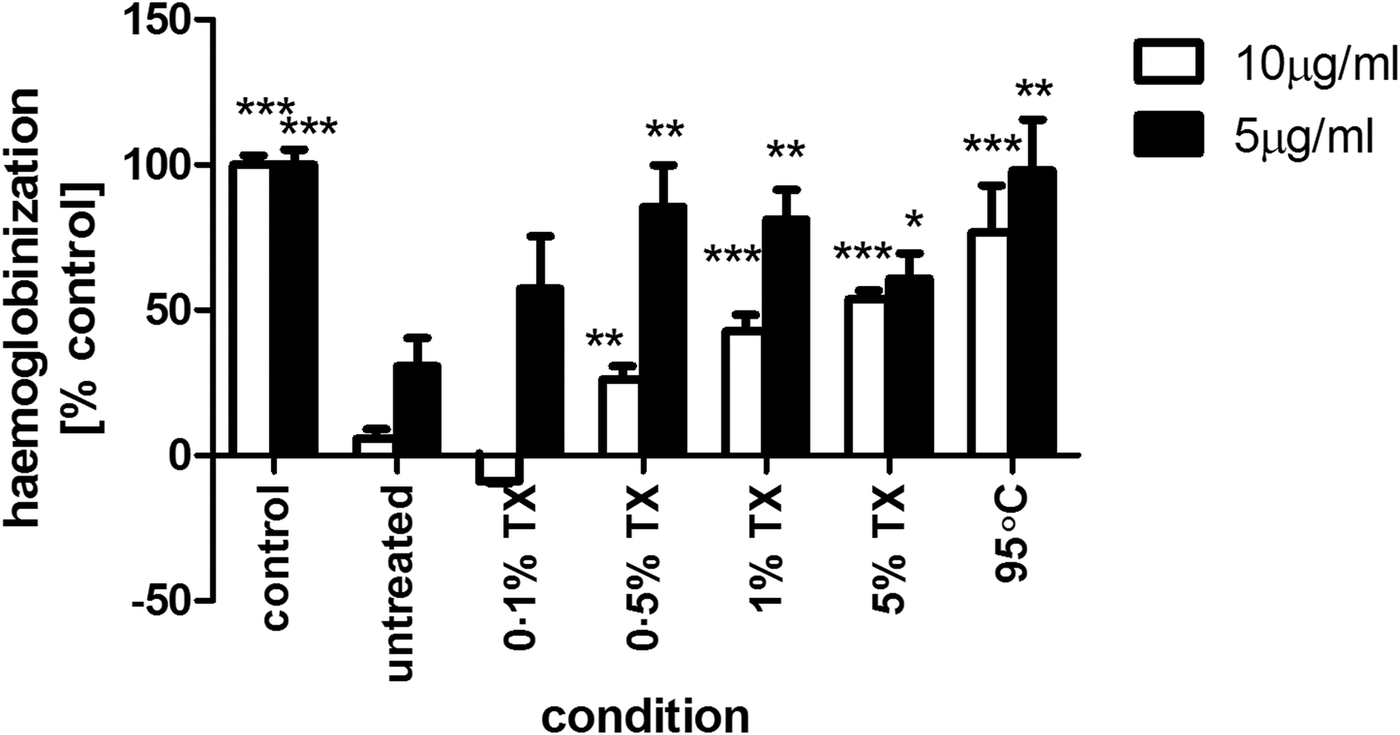
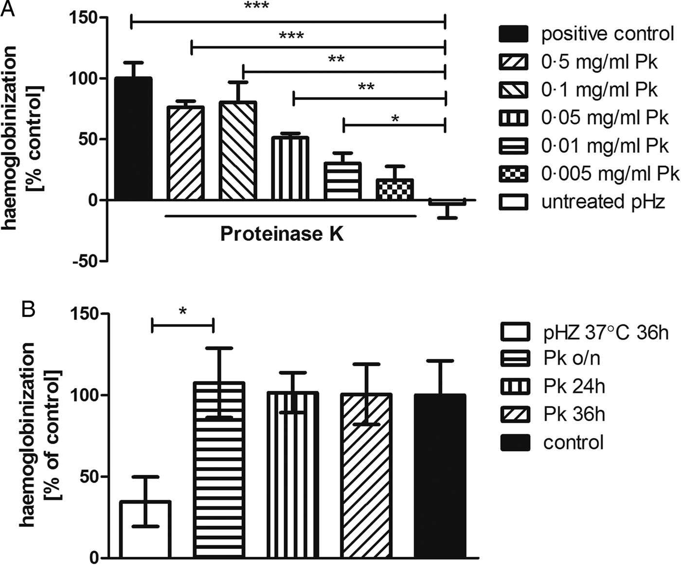
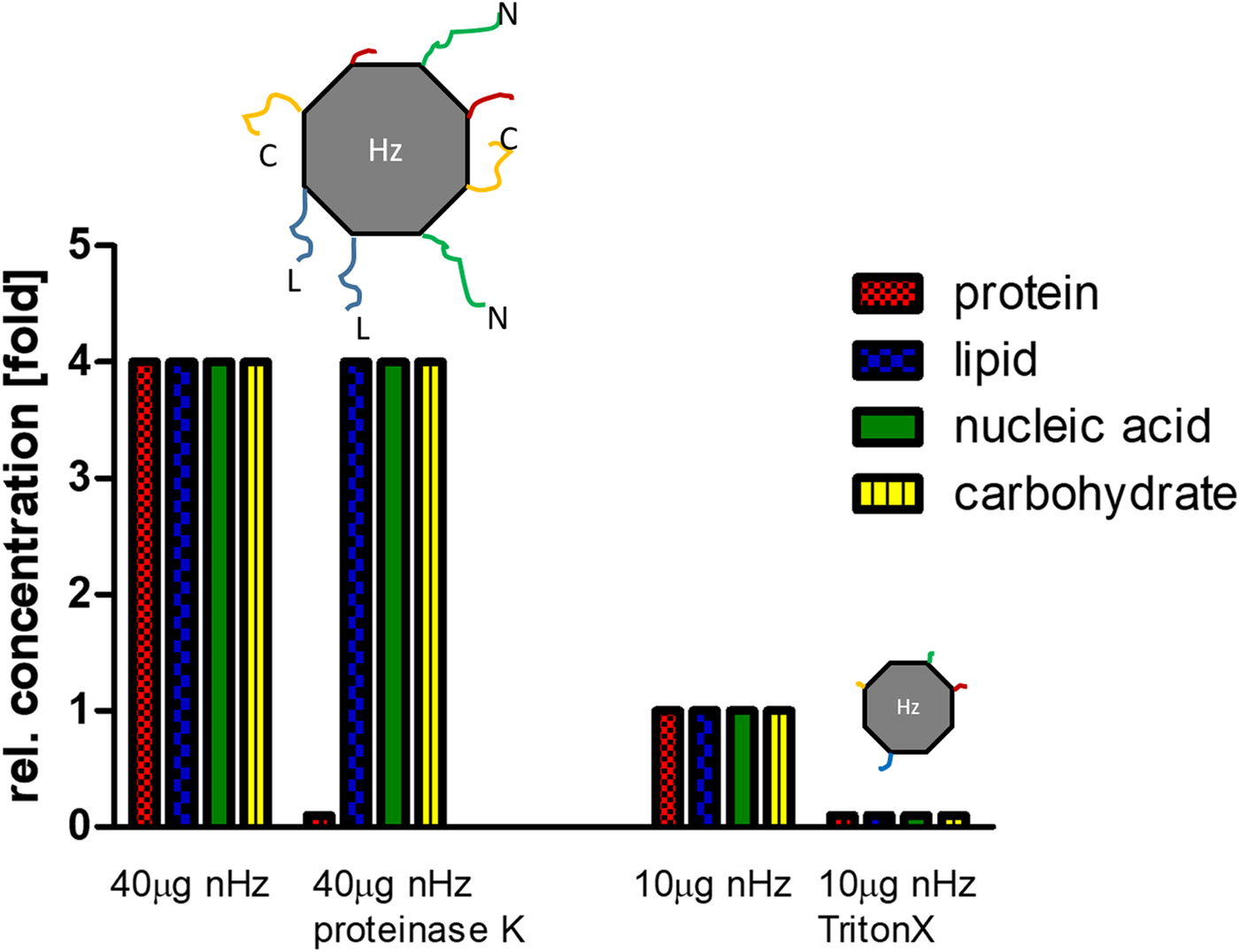
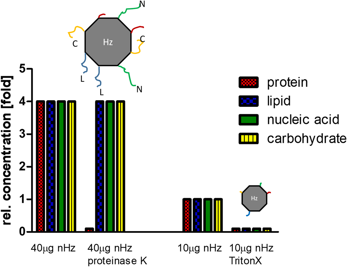
Crude parasite haemozoin showed strong inhibitory effects on erythroid development in physiologically relevant concentration ranges (5 and 10 µg mL−1) and treatment with increasing concentrations of Triton® X-100 was able to revert this inhibition in a concentration-dependent manner for detergent concentrations above 0.1% (v/v) (Fig. 8). Similar effects could be achieved by heating haemozoin to 95 °C for 30 min: heat-inactivated haemozoin showed no significant impact on cell haemoglobinization compared with control (Fig. 8). These observations suggest that the erythropoiesis-inhibiting activity found in haemozoin is mediated by one or more biomolecule(s), which is/are detergent-soluble and heat-labile. Crude haemozoin was therefore subsequently subjected to digestion by proteinase K (Fig. 9a). Supra-physiological concentrations (40 µg mL−1) of haemozoin were added to the cultures in order to establish dependencies on the proteinase K concentrations (and incubation time), which may not have been as apparent at lower pHz concentrations. Protease digestion (0.2 mg mL−1) was able to revert the inhibitory activity seen in crude haemozoin (Fig. 9b), confirming that this inhibition is at least partially mediated by protein(s)/peptide(s). Comparison of detergent and heat-treated haemozoin at 10 µg mL−1 in Fig. 8 and proteolytically digested haemozoin at 40 µg mL−1 in Fig. 9 shows that all of these treatments could revert the inhibitory activity observed for untreated haemozoin, where concentrations of 10 and 40 µg mL−1 completely inhibited erythroid development. The fact that proteinase K treatment completely alleviated the inhibitory activity caused by 4-fold higher concentrations of haemozoin supports that this inhibition may be protein mediated as non-proteinaceous material, removed by detergent treatment or inactivated by heat, would be unaffected by the protease and remain attached to the haemozoin crystals at 4-fold the concentration that is normally inhibitory (shown schematically in Fig. 10). Proteinaceous factors therefore appear to be more important in erythroid inhibition than other factors such as lipids, nucleic acids or carbohydrates as these would remain active after proteolytic digest.
Triton® X-100 treatment and heat inactivation of crude haemozoin resulted in a reduction of the growth inhibition caused by crude haemozoin. Expansion/haemoglobinization was assessed on day 10 of erythroid culture using a haemoglobin-based absorbance assay relative to control and crude haemozoin at concentrations of 5 or 10 µg mL−1 treated with the indicated concentrations (v/v) of Triton® X-100 (TX) in PBS for 30 min or heated to 95 °C for 30 min. Results are shown as percentage and standard deviation of control grown without haemozoin (vertical bars). Difference from untreated haemozoin: *P ⩽ 0.05, **P ⩽ 0.01, ***P ⩽ 0.001.

Proteinase K treatment reverted inhibitory activity of haemozoin: (a) Treatment of crude parasite haemozoin (pHz) (40 µg mL−1) with varying concentrations of proteinase K at 37 °C for 1.5 h showed reversion of inhibition in a concentration responsive manner. (b) Treating crude parasite haemozoin (pHz) with 0.2 mg mL−1 proteinase K (Pk) at 37 °C overnight (o/n), for 24 or 36 h resulted in complete reversion of the inhibition observed in cultures treated with crude haemozoin incubated for 36 h without enzyme. Cell expansion/haemoglobinization was assessed on day 8 of culture using haemoglobin-based absorbance measurement and results are averages of triplicates presented as percentages of the positive control and standard error of the mean (vertical bars). *P ⩽ 0.05, **P ⩽ 0.01, ***P ⩽ 0.001.

Schematic presentation illustrating the removal of protein residues from supra-physiological concentrations (40 µg mL−1) of natural haemozoin by proteinase K treatment leaving other residues adherent compared with the removal of protein, lipid, nucleic acid or carbohydrate residues from haemozoin at physiological concentrations (10 µg mL−1) by detergent treatment with Triton® X-100.
Discussion
A systematic analysis of data from 1980 to 2010 suggested that the mortality from malaria may have been grossly underestimated (Murray et al. Reference Murray2012). Malarial anaemia is likely to figure strongly amongst these numbers as it frequently fails to be diagnosed in resource-poor settings with limited access to healthcare, and due to confounding effects of other contributors to anaemia such as iron deficiency or helminthic infections (Ghosh and Ghosh, Reference Ghosh and Ghosh2007; Kinung'hi et al. Reference Kinung'hi2014). In children, mild anaemia can negatively impact on physical, mental and emotional development, while severe anaemia may be life-threatening. The causes of severe malarial anaemia are complex and multi-factorial (McDevitt et al. Reference McDevitt2006). Not only are erythrocytes lost due to parasite release and immune-mediated destruction of parasitized erythrocytes but at the same time removal of non-parasitized cells from circulation can exceed the destruction of parasitized RBC by up to 12-fold (Jakeman et al. Reference Jakeman1999; Pradhan, Reference Pradhan2009). A failure of the bone marrow sufficiently to replace the cells lost from circulation further contributes to the development and particularly the persistence of anaemia. The underlying mechanisms could involve both direct inhibition of erythropoiesis by parasite-derived factors released during erythrocyte invasion and lysis and indirect inhibition as a result of modulation of the host immune response. While impaired erythropoiesis is likely to result from a combination of inhibitory factors and mechanisms, an investigation of the effect of parasite-derived factors in an isolated manner could allow the elucidation of possible key contributors that may provide a more directly targeted approach to tackling this disease.
The effect of parasite-CM, as a source of factors that are secreted and released during P. falciparum blood stages, on the proliferation and differentiation of erythroid cells was assessed in an ex vivo erythropoiesis model based on peripheral blood haematopoietic stem cells (Giarratana et al. Reference Giarratana2005, Reference Giarratana2011; Boehm et al. Reference Boehm, Murphy and Al-Rubeai2009) as well as using a haemoglobin-based spectrophotometric assay for the quantification of erythroid development (Boehm and Bell, Reference Boehm and Bell2014). We identified direct erythropoiesis-inhibiting activity in the culture supernatant of P. falciparum that is of protein/peptide-containing nature and must either be a parasite product or a host factor modified by the parasitic infection process, as uninfected RBC did not induce a similar response. While the in vivo situation involves a myriad of other cells and factors and their interplay, the system used here is a simplified model that provides an indication of the role of inhibitory factors produced by P. falciparum and released from parasitized erythrocytes that could potentially be of relevance in vivo. A potential involvement of stress factors released by the parasites, particularly in the high haematocrit/high parasitaemia cultures used to generate concentrated CM needs to be considered. Further analysis of parasite morphology and physiology and the composition of the CM particularly with respect to possible quantitative and qualitative differences of secreted factors produced in CM from 15% haematocrit (6-fold concentrate) during 4 h and that from standard 2.5% haematocrit for 24 h would therefore have to be addressed in future work. Concentration of the factors from the obtained CM using spin filters posed some difficulties as not all of the bioactive material could be recovered most likely as a result of some proteinaceous material remaining adherent to the filter. Rediluted concentrate, which displayed a weaker effect than the original CM, thus differed in its composition from the non-concentrated CM. This effect might possibly be improved by using alternative means of protein concentration such as dialysis and also needs to be considered for future attempts at isolating the candidate proteinaceous factor(s). In a next step, plasma from malaria patients, and those suffering from malarial anaemia in particular, could be used in the ex vivo erythropoiesis model described here, to validate the presence of such erythropoiesis-inhibiting factors in the human circulation. Casals-Pascual et al. found an inhibition of erythroid expansion in vitro in response to intact and lysed parasitized erythrocytes but attributed this effect mainly to the corresponding amount of haemozoin (Casals-Pascual et al. Reference Casals-Pascual2006). Macrophage MIF produced by mononuclear cells has been identified as a factor which contributes to malarial anaemia by suppressing erythroid colony formation (McDevitt et al. Reference McDevitt2006). Interestingly, Augustjin et al. found a parasite homologue of this factor to be secreted into erythrocytes during blood-stage infection and released upon schizont rupture and infections with a PMIF knock-out strain of Plasmodium berghei resulted in increased reticulocyte production in rodent models (Augustijn et al. Reference Augustijn2007). A unique parasitic factor with erythropoiesis-inhibiting activity is unlikely to provide the sole explanation for this pathology as cytokine imbalances elicited by the immune response to the infection have already been shown to be key factors in inhibiting erythropoiesis. The finding of a direct parasite-borne erythropoiesis-inhibiting activity nonetheless offers a new direction to the elucidation of malarial anaemia and its treatment options as it may offer the possibility of raising neutralizing antibodies or small-molecule inhibitors as a therapeutic approach that might not interfere with the host immune system.
The deposition of parasitic haemozoin in the bone marrow has long been recognized as a feature of malarial infections but has only come into focus as a key influence on the host immune response in recent years. While haemozoin is rapidly ingested by neutrophils and monocytes/macrophages, free haemozoin has been measured in the plasma of malaria patients (1.8–10.9 µg mL−1) and found to be associated with lower haemoglobin concentrations and higher degrees of reticulocyte suppression (Casals-Pascual et al. Reference Casals-Pascual2006). Similar in vitro approaches employing erythropoiesis models have been used by other groups to investigate the effects of parasitic haemozoin on expansion, differentiation and apoptosis of erythroid cells using haemozoin concentrations in the range of 1–10 µg mL−1 believed to be physiologically relevant (Casals-Pascual et al. Reference Casals-Pascual2006; Lamikanra et al. Reference Lamikanra2009; Awandare et al. Reference Awandare2011) and in conjunction with inflammatory mediators and cytokines. Work by Lamikanra et al. showed that exposure to purified haemozoin induced gene expression changes in human erythroblasts, which were distinct to those caused by the pro-inflammatory cytokine TNF-α and are involved in the regulation of the integrated stress response and apoptosis (Lamikanra et al. Reference Lamikanra2015). It has also been suggested that the toxic effect of haemozoin on the erythroid system is not of biological nature but a purely chemical effect resulting from the peroxidation of fatty acids to toxic long-chain aldehydes via a non-enzymatic catalytic action of the haemozoin crystal (Schwarzer et al. Reference Schwarzer, Arese and Skorokhod2015). Measurements of aldehydes such as HNE confirmed this finding and erythroid cells showed susceptibility to HNE at concentrations of 7 µ m, which resulted in cell cycle inhibition via increased expression of p53 and p21 (Skorokhod et al. Reference Skorokhod2010). Ingestion of haemozoin by monocytes/macrophages and their activation by haemozoin-bound host fibrinogen (Barrera et al. Reference Barrera2011) is believed to modulate their production of cytokines and result in a transfer of peroxidation products to erythroid cells, effects which have been implicated in contributing to the inhibition of erythropoiesis within the erythroblastic islands (Giribaldi et al. Reference Giribaldi2004; Schwarzer et al. Reference Schwarzer2008; Perkins et al. Reference Perkins2011). In the absence of non-erythroid cells as mediators in the present study, the adhering factor itself must exert its activity directly on erythroid cells. While a role of fibrinogen in the erythropoietic pathway has not been documented, host fibrinogen associated with haemozoin has been shown to play an important role in the activation of cells such as monocytes through Toll-like receptor (TLR) 4 and integrin receptor signalling (Barrera et al. Reference Barrera2011), and experiments to elucidate a potential involvement of this factor could therefore provide a starting point towards identifying haemozoin-associated proteinaceous factors involved in inhibiting erythropoiesis.
In our study, we have not been able to confirm an erythropoiesis-inhibiting action of haemozoin in the absence of adhering biomaterial. Neither synthetic haemozoin nor naked, crude haemozoin showed the described effects. A similar discussion has been ongoing in regards to the more extensively studied immunomodulatory activity of haemozoin, where certain groups have found a clear effect of haemozoin obtained by chemical synthesis (Jaramillo et al. Reference Jaramillo2009), while others reported the need for DNA to be present on the crystal in order to activate TLR9 (Parroche et al. Reference Parroche2007). The effect of synthetic haemozoin crystals has been controversial and it has been argued that differences in crystal size and structure caused by varying synthesis techniques and the crystals’ propensity to aggregate are the cause for such discrepancy (Jaramillo et al. Reference Jaramillo2009; Coban et al. Reference Coban2010) or that a reduction of the inhibitory activity of haemozoin by various treatments is the result of modifications to the haemozoin crystal itself. Thawani et al. recently reported a suppression of EPO-induced proliferation of murine erythroid precursor cells by haemozoin from P. falciparum and Plasmodium yoelii both in vivo and in culture which exceeded inhibition caused by synthetic haemozoin (Thawani et al. Reference Thawani2014).
We cannot rule out that the protease-sensitive activity found in haemozoin is caused by certain adducts to the proteins such as residual haem, which had not yet been fully deposited into haemozoin or HNE generated by the haemozoin. However, if the HNE was derived from a non-enzymatic catalytic conversion of fatty acids and did not involve specific target proteins, we would expect to observe at least some erythropoiesis inhibition resulting from cultivation with synthetic and naked, crude haemozoin, particularly over extended periods of incubation time in protein- and lipid-containing media. While supra-physiological concentrations (40 µg mL−1) of haemozoin were used to establish the effects of proteinase K, the reversion of erythroid inhibition after protease treatment lends support to the theory that it is neither the pure haemozoin nor other adhering biomolecules that primarily mediated the inhibition as all of these would still have been present post-treatment in concentrations much in excess of those inhibiting erythropoiesis in other experiments (10 and 5 µg mL−1).
Despite global efforts and a UN Millennium Development Goal to halt and reverse the occurrence of malaria by 2015, malaria remains one of the most widespread human infectious diseases. As malaria therapy is increasingly faced with the emergence of resistant parasites, a novel approach lies in the targeting of pathogenic mechanisms, such as those causing malarial anaemia, rather than the malarial parasites themselves and might offer new treatment approaches that could be less prone to resistance than conventional antimalarial drugs (Bell and Boehm, Reference Bell and Boehm2013). With malarial anaemia accounting for an estimated one-third of malarial deaths and morbidity and mortality due to anaemia – including cases of what has been believed to be ‘benign’ vivax malaria – likely to be underestimated, a successful approach to the treatment of malarial anaemia would have immense implications. Autochthonous vivax malaria cases have occurred in Greece, Italy and France in recent years, and climate change is expected to cause malaria to return to other areas of Europe and previously malaria-free regions of the world in the future (Danis et al. Reference Danis2011). While anaemia is one of the main complications associated with P. vivax infections, the underlying causative mechanisms could differ between the species. Of note, P. vivax causes a 4-fold higher loss of uninfected erythrocytes than P. falciparum, and due to its strong preference for reticulocyte invasion, it greatly reduces the lifespan of RBC (Anstey et al. Reference Anstey2009; Castro-Gomes et al. Reference Castro-Gomes2014). Direct inhibition of erythroid growth and perturbation of cell division by P. vivax-infected RBC and their lysates was reported in in vitro cultures (Panichakul et al. Reference Panichakul2012), where parasite factors may inhibit erythroid cell growth through the downregulation of phosphorylation of the cytoskeletal protein ezrin (Panichakul et al. Reference Panichakul2015). It has furthermore been suggested to elicit a stronger inflammatory response than P. falciparum at comparable parasitaemia levels, all of which are factors that can influence the development and severity of anaemia in vivax malaria (Hemmer et al. Reference Hemmer2006; Gonçalves et al. Reference Gonçalves2012).
We demonstrate here that factors released by the parasite P. falciparum during blood-stage development in culture have a direct inhibitory effect on erythroid proliferation and differentiation ex vivo. A similar effect is caused by crude parasitic haemozoin. In both cases, the inhibitory factors are of protein/peptide-containing nature and it could be speculated that the same factor is found both in association with parasitic haemozoin and released into the culture supernatant upon host cell lysis or becomes detached from the haemozoin crystal. These findings are an important advance in determining the direct influence of parasite factors in erythroid development vs the role of the host immune response, both of which determine erythroid fate in vivo, and can form the basis for more targeted investigations. An identification of the actual effector protein(s)/peptide(s) may open up important new approaches to the treatment of malarial anaemia such as neutralizing antibodies or small molecules as a potentially ‘resistance-proof’ therapy in an era of increasing antimalarial drug resistance.
Financial support
DB is grateful for a postdoctoral fellowship from the Irish Research Council.
Conflicts of interest
None.
Ethical standards
Not applicable.