A variety of methods are available to assess global left ventricular function and volumes. Gated myocardial perfusion scintigraphy has become one of the most commonly used diagnostic modalities for the evaluation of cardiac diseases, providing values for left ventricular perfusion, asssessment of contraction, and left ventricular ejection fractions and volumes through R-wave gating.Reference Iskandrian, Hakki, Goel, Mundth, Kane-Marsch and Schenk1, Reference Fuster, Gersh, Giuliani, Tajik, Brandenburg and Frye2 Quantitative gated scintigraphy (Cedars-Sinai, LA, USA),Reference Germano, Nichols and Cullom3 and the Emory cardiac toolbox (Emory University, Atlanta, USA),Reference Faber, Akers, Peshock and Corbett4 are the two most widely used packages for the analysis of the parameters of myocardial perfusion and left ventricular function obtained with radionuclide cardiac imaging. Both methods have been validated extensively against other imaging modalities for calculation of ejection fractions and volumes, and have been compared simultaneously against the independent imaging modality of echocardiography.Reference Germano, Nichols and Cullom3–Reference Nichols, Lefkowitz and Faber5 The literature is rich on the use of gated myocardial perfusion scintigraphy in adults in clinical cardiology practice. Data is lacking, however, on its application in children, and its precision in quantifying left ventricular volumes and ejection fractions. Our present work, therefore, aims to assess the ability of gated myocardial perfusion, using quantitative gated scintigraphy and the Emory cardiac toolbox, to calculate ejection fraction and left ventricular volumes in children when compared to the well-known and validated technique of M-mode echocardiography tracings derived from cross sectional echocardiograms.
Materials and methods
Patients
We studied 64 children referred to our department of nuclear medicine for gated myocardial perfusion imaging for various reasons. All patients were evaluated in the department of paediatric cardiology. Laboratory and clinical data of the patients were obtained from their charts.
Protocol for gated myocardial perfusion scintigraphy
All patients underwent a 2 day protocol involving stress and rest with no medical intervention, except 5 patients who had studies performed only at rest. The data under conditions of stress was obtained at peak exercise using a modified Bruce treadmill protocol, giving a dose of 99 m-Technetium methoxyisobutyl-isonitrile adjusted for weight. If the stress study proved abnormal, the patients also underwent studies at rest. The scintigraphy was performed using a dual headed gamma camera (Siemens E-Cam, Erlangen Germany) equipped with low energy high resolution collimators. Detector heads were oriented at 90°, and 32 projections with 64 images at 30 seconds per projection were acquired over 180° on a matrix of 64 by 64. We used a zoom factor of 1.45, and gated the study at 8 frames per R-R interval. Gated images were reconstructed using a back projection algorithm and Butterworth filter, with a cut-off frequency of 0.45 cycles per pixel and order 5. The reconstructed slices were re-oriented along the left ventricular long axis.
Quantitative gated scintigraphy
We employed a widely used and fully automated method, which allows reselecting manual processing should the automatic mode fail to delineate the myocardial borders. The method works in three-dimensional space, and uses a set of data acquired in the cardiac short axis. The mid myocardial surface is determined by ellipsoidal fitting after initial estimation by a spherical sampling model. The method uses normal count profiles on the determined midmyocardial surface to estimate the epicardial endocardial offsets. This estimation is determined by fitting the count profiles with Gaussian curves, and calculating the corresponding standard deviations. For each profile, endocardial and epicardial surface points are defined by given percentages of the calculated standard deviations.Reference Germano, Kiat and Kavanagh6 Endocardial, epicardial, and valvar planes are calculated for each interval. The volumes of the left ventricular cavity are calculated for each gating interval by multiplying the individual pixel volume by the number of voxels contained in the three-dimensional space confined by the endocardial and valvar planes. The largest and the smallest volumes correspond to end-diastole and end-systole, and are used to calculate the ejection fraction.Reference Faber, Cooke and Folks7
Emory cardiac toolbox
This method works in three-dimensional space and again uses data acquired in the cardiac short axis. The software programme is fully automated, with the possibility to change the radius and centre of the short axis. The toolbox uses Fourier analysis for estimation of mural thickness, and detects a circumferential maximal count profile by applying an anatomically based model accounting for mural thickening to generate theoretical endocardial and epicardial surfaces.Reference Everaert, Bossuyt and Franken8 It uses two coordinate systems to sample the myocardium, a cylindrical coordinate for the mid and basal part of the myocardium, and a spherical coordinate for the apical portion. The program uses 8 frames per cardiac cycle.Reference Faber, Cooke and Folks7 We used the software obtained from the manufacturers of both systems to calculate end-diastolic and end-systolic volumes, and ejection fractions. In running both systems, left ventricular apical limits, valvar plane limits, and centres, epicardial radiuses, and computed endocardial and epicardial surfaces were all examined using available static and dynamic displays for quality control. Manual alterations in those parameters were used only if necessary, which was not more than one-twentieth of the studies, consistent with previous reports.Reference Faber, Cooke and Folks7, Reference Everaert, Bossuyt and Franken8
Echocardiography
The parametric information of left ventricular systolic and diastolic diameters and fractional shortening in parasternal long axis position was obtained from the echocardiographic reports, which were performed by experienced paediatric cardiologists using a Vingmend M-mode cross-sectional machine produced by General Electrics.
Statistical analysis
All statistical analyses were performed using the SPSS statistical program package (SPSS Inc., Chicago, USA). Cronbach’s alpha coefficient was used to assess the reliability of the three techniques. The data is presented as mean with standard deviations. When parametric test assumptions were satisfied, echocardiographic reports were compared to the date obtained with gated myocardial perfusion data using repeated measures analysis of variance. Pearson’s correlation coefficients were also calculated. The patients were subdivided according to their diagnosis, and data obtained from gated myocardial perfusion scintigraphy was compared with echocardiography using the Friedman test. A probability level of p less than 0.05 was considered to be significant.
Results
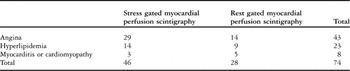
We studied 42 boys and 22 girls, aged from 1 to 18 years, with a mean of 12. years. The patients were grouped into those referred with angina, those referred with suspected hyperlipidemia, and those considered to have myocarditis or cardiomyopathy. We evaluated a total of 74 gated myocardial perfusion studies.
All patients referred with suspected angina had normal findings, while 2 patients with suspected hyperlipidemia, and all those with suspected myocarditis or cardiomyopathy showed either ischaemia or abnormalities of mural motion. The reliability between quantitative acintigraphy, the Emory toolbox and echocardiography for studies performed at rest to calculate end-diastolic and end-systolic volumes and ejection fractions proved to be 0.85, 0.86, and 0.97, respectively, when using Cronbach’s alpha coefficient. The same analysis in studies performed under conditions of stress produced values of 0.83, 0.86, and 0.90, respectively. In practical terms, values above 0.80 are taken to mean a high standard of agreement, and considered as being reproducible.Reference Brennan and Silman9 In Table 1, we show the distribution of studies according to the groups.
Distribution of the studies according to the groups of patients.

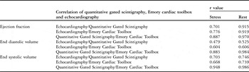
In Table 2, we show the relationship between the volumes measured with gated myocardial perfusion and echocardiography for studies performed both during stress and at rest for the overall population. There was a high correlation, with r values above 0.88, between quantitative gated scintigraphy and the Emory cardiac toolbox for ejection fraction, and ventricular volumes. The correlation between gated myocardial perfusion and echocardiography, in contrast, was weak, especially for ventricular volumes calculated under conditions of stress.
The relationship between the measured volumes with gated myocardial perfusion scintigraphy and echocardiography for studies both at stress and during rest in all patients.

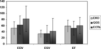
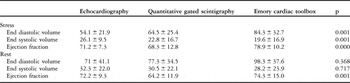
The differences among the means for end-diastolic volumes and ejection fractions during rest measured by these 3 methods (Table 3 and Fig. 1) were significant (p equals 0.002 and p equals 0.001 respectively).
The differences among the means for end-diastolic volumes and ejection fractions during rest measured by echocardiography, quantitative gated scintigraphy, and the Emory cardiac toolbox.

End diastolic-end systolic volumes and ejection fractions calculated by gated myocardial perfusion using the Emory cardiac toolbox and quantitative gated scintigraphy in studies performed at rest when compared to those measured by cross-sectional echocardiography.

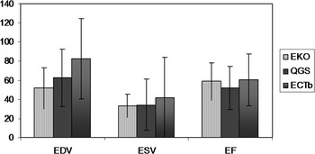
In the total group, the association between the volumes measured with gated myocardial perfusion and echocardiography under conditions of stress is shown in Figure 2, and summarized in Table 4. The differences among the means of volumes and ejection fractions during stress measured by these 3 methods were significant (p less than 0.001).
End diastolic-end systolic volumes and ejection fraction calculated by gated myocardial perfusion using the Emory cardiac toolbox and quantitative gated scintigraphy in studies performed under stress as compared to those measured by cross-sectional echocardiography.

Association between the measured volumes with stress gated myocardial perfusion scintigraphy and echocardiography.

Comparison of these 3 tests with the same parameters was also performed when the patients were divided according to their reasons for referral. In Table 5, we show mean values with standard deviations for end-diastolic and end-systolic volumes and ejection fractions during stress and rest for those referred because of suspected angina. Analysis of a total of 43 studies on the 29 patients in this group yielded significant differences among these 3 techniques with respect to determination of ventricular volumes and ejection fractions for studies carried out either at rest or under stress.
Mean values plus or minus standard deviations for end-diastolic and end-systolic volumes and ejection fractions during stress and rest for the group of patients referred with suspected angina.

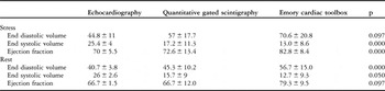
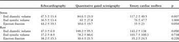
For those referred because of suspected hyperlipidemia, mean values for all three parameters during both stress and rest for all three techniques, and their comparisons with each other, are shown in Table 6. There were no significant differences among 3 tests with respect to end-diastolic volumes calculated during stress, and ejection fractions whilst at rest, whereas significant differences were found for the other parameters. When analysing those referred with suspected myocarditis or cardiomyopathy, there were no significant differences among the 3 modalities for all parameters, either performed under conditions of stress or at rest (Table 7).
Mean values for all three parameters during both stress and rest for all three techniques, and their comparisions with each other, in the patients referred with suspected hyperlipidemia.

Mean values plus or minus standard deviations for end-diastolic and end-systolic volumes and ejection fractions during stress and rest in the patients referred for investigation of myocarditis or cardiomyopathy.

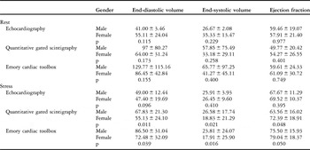
We show comparison of ventricular volumes and ejection fractions for all the patients according to gender in Table 8, which reveals significant differences between the genders for ventricular volumes and ejection fractions calculated during gated myocardial perfusion under conditions of stress.
Comparison of end-diastolic and end-systolic volumes and ejection fractions for all patients according to gender.

Discussion
Echocardiography is the most widely used method for measuring left ventricular volumes and functions, not only in adults but in children as well. The role of gated myocardial perfusion scintigraphy, as an alternative measure, has yet to be determined for children. Hence our purpose of assessing the technique as a tool for measuring these parameters using two well established commercial programmes, and comparing the findings to the values measured by cross-sectional echocardiography. The results generally reflected weak correlation and significant variation between the 3 methods when used in children. Unacceptable correlation was observed in studies performed under conditions of stress for quantitative gated scintigraphy and echocardiography for the measurements of all the parameters, with correlation coefficients no better than 0.48, 0.70, and 0.70 respectively. Likewise, the same measurements gave correlation coefficients of 0.60, 0.67 and 0.78 when the Emory cardiac toolbox was compared with echocardiography. The results were better for the studies performed at rest only for ejection fraction. The measured left ventricular volumes using the scintigraphic programmes were lower than values obtained using echocardiography (Table 2), while the calculations of ejection fractions were higher. These weak correlations can be attributed to the known scatter of photons from the myocardial walls of the left ventricular cavity, which usually leads to difference in estimation of ejection fraction by up to one-tenth in adults.Reference Germano and Berman10, Reference Iskandrian, Germano and VanDecker11 Differences in age and gender have been studied widely in adults, with gender being shown to have has a more important impact than age.Reference De Bondt, Van de Wiele, De Sutter, De Winter, De Backer and Dierckx12 This difference has been explained on the basis of smaller hearts, with a high rate of scatter, in female patients.Reference Bacher-Stier, Muller and Pachinger13 In our population, differences for gender were not observed in studies performed at rest. In those carried out under conditions of stress, however, the measured left ventriculer volumes were smaller, and the ejection fractions were higher, in girls, and the other way round in boys. This finding, although limited to studies performed under stress, may again be attributed to the fact that girls have smaller hearts than boys.
In children, the left ventricular cavity is much smaller than adults, which may result in worse resolution, thus creating difficulties for automatic programmes properly to define the endocardial surface. The extent of scatter may even lead to complete disappearence of the ventricular cavity in end-systole. In some cases, end-systolic volumes measured using the Emory toolbox were 16.6 plus or minus 19.6. This range starts from negative values, which raises doubt on the resultant measurements. Measuring end-systolic volumes by echocardiography provided higher values, with a highly significant difference between the techniques. These values were slightly better for the findings obtained using the automated programmes during studies performed at rest, but still the measurements were unacceptable. Others have reported that the calculated left ventricular volumes show more errors in the Cedars-Sinai programme than in the Emory programme when a patient had a small heart.Reference Toba, Kumita and Cho14
Comparison of the left ventricular volumes and ejection fractions obtained by gated myocardial perfusion scintigraphy using the two computer software programmes showed correlation coefficients of 0.89 to 0.99 for the left ventricular volumes and ejection fractions at rest and during stress. Similar observations were found by Nichols et al,Reference Nichols, Santana and Folks15 but they showed that, despite a strong correlation between the two methods of computing left ventricular functional values, statistical scatter was substantial and significant biases and trends were observed. They suggested that, when both software packages are used at the same time, it is important to take these differences into consideration, and to apply normal limits specific to each set of algorithms.Reference Nichols, Santana and Folks15 In our experience, we found strong correlations between the two methods of computing left ventricular functional values, but the agreement between was higher between the Emory toolbox and echocardiography for calculations of systolic volumes at rest. We performed the study using 8 frames per cardiac cycle, which made it possible to process the same gated data by both quantitative scintigraphy and the toolbox, as the commercial software programme accommodates only 8 frames per cycle of gated myocardial perfusion. Such comparison of gated data with echocardiography using 8 frames per cardiac cycle has previously been shown to be acceptable.Reference Germano and Berman10
When the patients were analyzed according to the reasons for their referral, measurement of left ventricular volumes at rest in those with suspected angina, end-systolic volumes and ejection fractions at rest in those suspected of hyperlipidemia, and all the other measurements except ejection fractions under stress for those referred for investigation of myocarditis or cardiomyopathy could be done by any of the 3 modalities, with similar values produced for each given parameter. The better findings for those with suspected myocarditis or cardiomyopathy could either be due to the relatively large size of the hearts secondary to their pathology, allowing better adjustments for size, or due to the relatively smaller size of the sample. We have not analyzed patients according to their findings observed on gated myocardial perfusion scintigraphy, as the majority of the studies were normal, which would make the statistical analysis of no value.
Our investigation has yielded negative results that demonstrate poor agreement between gated myocardial perfusion scintigraphy and echocardiography. This means that gated myocardial perfusion scintigraphy is less suitable for assessing left ventricular volumes, and is also less reliable in functional assessment of ejection fraction in children. Echocardiography, therefore, will still remain the most widely used and reliable technique in this part of the diagnostic work-up of children with suspected cardiac disease.
Conclusion
We have observed that calculations of ejection fractions and left ventricular volumes in both systole and in diastole when using echocardiography as opposed to quantitative gated myocardial perfusion methods are not well correlated. Given the high variance between the methods, it is possible that they may produce results that are clinically different from one another for the same data. This is important when ejection fractions are borderline. These methods investigated are not interchangeable, and should not be substituted arbitrarily for one another. Clinicians should not draw conclusions regarding improvement or deterioration of cardiac function when prior values for ejection fractions and left ventricular volumes were calculated using different methods.
Acknowledgement
We thank to Erdem Karabulut, PhD, for his help with the statistical analysis of our findings.