Introduction
Enzymes are the main class of nature’s catalysts. Within an organism, all chemical reactions proceed at rather constant temperatures, well below 50°C, and mostly at ambient pressure, therefore necessitating the need for catalysis. Although the structure and activity of enzymes have been optimized through evolution, some acceleration of enzymatic catalyzed reactions can still be achieved at elevated temperatures. This is a principle applicable to all chemical reactions, where increased thermal energy is favorable to overcome the activation energy of a reaction. However, heating of enzymes is limited by the temperature of unfolding of an enzyme, where denaturation (loss of activity) occurs.
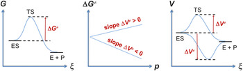
On the other hand, the activity of some enzymes can also be enhanced by elevated pressures. Reference Eisenmenger and Reyes-De-Corcuera1–Reference Luong, Erwin, Neumann, Schmidt, Loos, Schmidt, Fändrich and Winter5 In this case, the transition state, which is associated with a maximum of energy halfway between the reactants and the product, is favored under higher pressures because it has a lower volume than the reactants. The reaction path is then characterized by a negative activation volume, ΔV ≠ (≠ refers to the transition state of the chemical reaction), and the activation Gibbs energy, ΔG ≠, is lowered by dΔG ≠ = ΔV ≠ dp, where dp is the pressure increase ( Figure 1 ).
Simplified thermodynamic description of an enzymatic reaction, where the enzyme-substrate complex (ES) is turned over into the free enzyme (E) and product (P). Note: G, Gibbs energy; V, volume; p, pressure; ξ, reaction coordinate; TS, transition state; ≠, transition state of the chemical reaction.

On a molecular level, pressure effects include changes in the hydration and interaction of molecules. Hydrogen bonds are often strengthened, whereas ionic and hydrophobic interactions can be broken in aqueous solution under high pressures. Reference Luong, Kapoor and Winter6–Reference Boonyaratanakornkit, Park and Clark8 In this way, reaction paths are selected by pressure, where the transition state or the products are characterized by smaller volumes. Reference Kapoor, Triola, Vetter, Erlkamp, Waldmann and Winter9–Reference Schuabb, Kumar, Pataraia, Marx and Winter11 Moreover, pressure can induce a higher conformational flexibility of the reactants, which is usually favorable for enhanced reaction rates. Thus, the application and the investigation of high pressure in biosciences and biotechnology is promising. Reference Akasaka and Matsuki12
α-chymotrypsin (α-CT) is a well-known enzyme used as a model enzyme in many studies. Reference Sun, Winter and Winter13–Reference Marini and Wunsch17 α-CT has a molar mass of 25,000 g mol–1 and a positive net charge in neutral solution. This charge facilitates adsorption of α-CT on negatively charged surfaces. α-CT adsorption has been observed on a wide variety of surfaces. Reference Derr, Dringen, Treccani, Hildebrand, Colombi Ciacchi and Rezwan18–Reference Celej, D’Andrea, Campana, Fidelio and Bianconi21 α-CT hydrolyzes peptide bonds, ideally those formed by aromatic amino acids (Trp, Tyr, Phe). The enzymatic activity of α-CT can be approximately described by the Michaelis–Menten mechanism, where the substrate molecule binds reversibly to the enzyme in a first step. Reference Blow14 The conversion of the substrate to the product is then achieved in the bound state in an irreversible second step. For α-CT, a third step of product release must be added:
 $$\eqalign{ & \alpha {\rm{ - CT}} + {\rm{S}}{{{k_1}} \over {{k_{ - 1}}}}\alpha {\rm{ - CT}} \bullet {\rm{S}} \cr & \buildrel {{k_2}} \over \longrightarrow {{\rm{P}}_{\rm{2}}} - {\rm{\alpha - CT}} + {{\rm{P}}_{\rm{1}}} \cr & \mathrel{\mathop{\kern0pt\longrightarrow}\limits_{ + {{\rm{H}}_{\rm{2}}}{\rm{O}}}^{{k_3}}} {\rm{\alpha - CT}} + {{\rm{P}}_{\rm{1}}} + {{\rm{P}}_{\rm{2}}} \cr}$$
where k 1, k –1, k 2, and k 3 are the rate constants of the three reaction steps. The substrate (S) is cleaved into two products, P1 and P2. k 2 is small and rate-limiting when peptides are cleaved, Reference Blow14 as presented in this article. In several studies, it has been observed that k 2 of α-CT increases with increasing pressure, indicating a negative volume change of activation, ΔV ≠. Reference Akasaka, Nagahata, Maeno and Sasaki2–Reference Taniguchi and Suzuki4,Reference Luong and Winter22 This conclusion can be derived from Eyring’s theory: Reference Mozhaev, Heremans, Frank, Masson and Balny23
 $${k_2} \propto \exp \left( { - \Delta {G^ \ne }/RT} \right)$$
,
 $${\rm{d}}\ln ({k_2}/{k_{{\rm{ref}}}})/{\rm{d}}p = - \Delta {V^ \ne }/RT$$
,
 $${\rm{d}}\ln ({k_2}/{k_{{\rm{ref}}}})/{\rm{d}}T = + {E_{\rm{a}}}/R{T^2}$$
,
where k ref is the rate constant measured at a reference pressure (e.g., 1 bar) or reference temperature. E a is the activation energy, R the gas constant, and T the temperature. It must be noted, however, that there are also enzymes that are inactivated by pressure due to a positive activation volume (Figure 1). Reference Suladze, Cinar, Sperlich and Winter24–Reference Schuabb and Czeslik26
Equation 1a implies that ΔV ≠ can be obtained from the rate of product formation, v 0, because k 2/k ref = v 0/v ref, if the rate under pressure, v 0, is measured under identical conditions as the rate at 1 bar, v ref, and the enzyme is saturated with the substrate. The rate is derived from the initial linear increase in the product concentration with time, v 0 = dc P1/dt (c P1 is the concentration, c, of P1). The activation volume is the difference in the volumes of the transition state and the enzyme-substrate complex, ΔV ≠ = V ≠ – V ES. If the transition state and the enzyme-substrate complex have different compressibilities, which would not be an anomalous behavior, then the activation volume changes with increasing pressure.
When the rate of product formation, v 0, is measured as a function of substrate concentration, c S, at constant temperature and pressure, the kinetic parameters, K M = (k –1 + k 2)/k 1 (Michaelis constant) and k cat = k 2 (turnover number), are obtained by linear regression analysis (modified Lineweaver–Burk plot):
 $${{{c_{{\rm{E,tot}}}}} \over {{v_0}}} = {1 \over {{k_{{\rm{cat}}}}}} + {{{K_{\rm{M}}}} \over {{k_{{\rm{cat}}}}}} \cdot {1 \over {{c_{\rm{S}}}}}$$
,
where c
E,tot is the total enzyme concentration. The ratio k
cat/K
M is denoted as enzymatic efficiency. For large substrate concentrations,
 ${v_0} \propto {k_{{\rm{cat}}}}$
.
Effects of pressure and temperature on enzyme activity in aqueous solutions
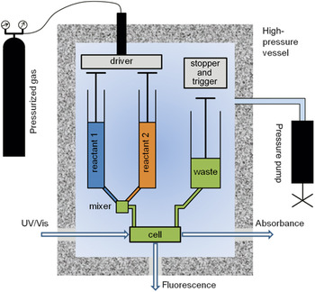
The enzymatic activity of α-CT in aqueous solution has been studied at high pressures and elevated temperatures. Reference Mozhaev, Lange, Kudryashova and Balny3,Reference Taniguchi and Suzuki4,Reference Luong, Kapoor and Winter6,Reference Luong and Winter22 When using N-succinyl-L-alanyl-L-alanyl-L-prolyl-L-phenylalanine-p-nitroanilide (SAAPPNA) as the substrate, the formation of the product, p-nitroaniline, can easily be recorded via measurement of optical absorbance at 400-nm wavelength. Reference Mozhaev, Lange, Kudryashova and Balny3,Reference Luong, Kapoor and Winter6 Here, we describe the use of the high-pressure stopped-flow technique ( Figure 2 ). Reference Luong, Kapoor and Winter6,Reference Luong and Winter22 In this experiment, the enzyme and the substrate solutions are mixed rapidly and are pushed through an optical observation cell into the collection syringe. When the filling of this syringe is stopped, the optical detection is started, and the formation of the product is recorded as light absorption or fluorescence intensity with a dead time of only a few milliseconds. The whole process occurs within a high-pressure vessel, meaning that the sample solution is held at constant pressure before, during, and after binding the substrate to the enzyme and in the course of the reaction.
Schematic drawing of the high-pressure stopped-flow instrument from TgK Scientific (Bradford-on-Avon, UK). Reactions under pressures up to 2000 bar can be followed by recording UV/Vis absorption or fluorescence intensity. The dynamic components include two reactant syringes and a collection syringe, which are mounted inside the high-pressure vessel that has pressure-stable sapphire windows for optical detection.

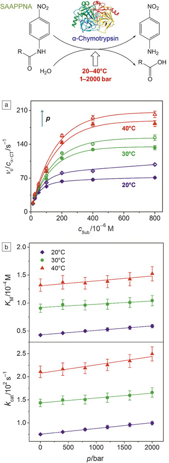
Figure 3 shows the Michaelis–Menten plots for the hydrolysis of SAAPPNA by α-CT. Reference Luong, Kapoor and Winter6 In the pressure and temperature range studied, the initial rate, v 0, increases with increasing substrate concentration and approaches the maximum value at 0.4 mM of SAAPPNA, where the enzyme is almost saturated with the substrate. Both high pressure and high temperature enhance the enzyme activity as shown by the higher reaction rates. Upon increasing the pressure from 1 to 2000 bar, v 0 increases by 40%, and upon increasing the temperature from 20 to 40°C, an almost threefold increase of v 0 is observed.
(a) Pressure- and temperature-dependent normalized rate, v 0/c α-CT, of N-succinyl-L-alanyl-L-alanyl-L-prolyl-L-phenylalanine-p-nitroanilide (SAAPPNA) hydrolysis (shown at the top) catalyzed by α-chymotrypsin (α-CT) (in TRIS buffer solution at pH = 8). (b) The Michaelis constant, K M, and the turnover number, k cat, have been determined by fitting Equation 2 to the data in (a). All data were obtained using the high-pressure stopped-flow method. Reference Luong, Kapoor and Winter6

The observed pressure and temperature effects can be related to the kinetic constants of the enzyme reaction. High pressure fosters both the turnover number, k cat, and the Michaelis constant, K M (Figure 3). As a result, the enzymatic efficiency, k cat/K M, remains almost constant, when the pressure rises from 1 to 2000 bar at 20°C. Reference Luong, Kapoor and Winter6 Elevated temperatures also enhance the turnover number, k cat, and the Michaelis constant, K M. However, the enzymatic efficiency, k cat/K M, still remains almost constant upon a temperature increase from 20 to 40°C at 1 bar. Reference Luong, Kapoor and Winter6
The deduced activation volumes, ∆V ≠, are all negative at the three temperatures studied, about –3.5 cm3 mol–1 at 20°C, –1.7 cm3 mol–1 at 30°C, and –2.1 cm3 mol–1 at 40°C. A negative activation volume can be explained by a smaller volume of the transition state relative to the enzyme-substrate complex. It is likely that the dense packing of hydrating water molecules around the emerging charges in the transition state is responsible for this volume change. Reference Low and Somero27 Apparently, this effect is most significant at 20°C, where ∆V ≠ has been found to be most negative. It is interesting to note that the pressure-induced enhancement of activity is associated with just a small reduction of the activation Gibbs energy, ΔG ≠. For example, at 20°C and 2000 bar, the activation Gibbs energy is reduced only by –3.5·cm3 mol–1 × 2000 bar = –0.7 kJ mol–1. The activation energy, E a, as deduced from the temperature dependence of k cat (Equation 1b, with k 2 = k cat), is almost unchanged in the pressure range covered and is found to be approximately 39 kJ mol–1. This value is typical for the hydrolytic activity of α-CT with low-molecular-weight substrates. Overall, the results show that both high pressures and high temperatures enhance the catalytic activity of the α-CT for the hydrolysis of SAAPPNA. Moreover, high pressure also induces stabilization of the enzyme against thermal inactivation. Pressure and temperature can thus be used simultaneously to drastically accelerate enzymatic reactions. Reference Luong, Erwin, Neumann, Schmidt, Loos, Schmidt, Fändrich and Winter5,Reference Luong, Kapoor and Winter6
Activation volumes of enzymes—Effects of adsorption and interfacial properties
In many biomedical and biotechnological applications, enzymes are immobilized on solid supports to be reused in many cycles. Reference Talbert and Goddard28 However, this immobilization of enzymes is often associated with lower degrees of stability and activity. Reference Mateo, Palomo, Fernandez-Lorente, Guisan and Fernandez-Lafuente29,Reference Rabe, Verdes and Seeger30 Here, we present the effect of pressure on enzymatic activity at aqueous–solid interfaces with different chemical modifications. Reference Schuabb, Cinar and Czeslik31–Reference Levin, Erlkamp, Steitz and Czeslik33 Using well-chosen model systems, these studies reveal how pressure could be helpful in biotechnological applications by favoring low-volume transition states.
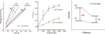
Silica particles have been used as a model adsorbent for proteins in several studies. Reference Czeslik and Winter34–Reference Cinar and Czeslik36 The pressure-dependent enzymatic activity of α-CT has also been studied, when this enzyme is adsorbed on silica particles, applying the high-pressure stopped-flow technique. Reference Schuabb, Winter and Czeslik32 The silica particles have a negative surface charge and bind to positively charged α-CT molecules in significant quantities. Moreover, no desorption of protein from the water–silica interface is expected under high pressures. Reference Koo, Erlkamp, Grobelny, Steitz and Czeslik37 The enzyme-particle solution and the substrate solution were filled into the two reactant syringes of the instrument (Figure 2). Figure 4 shows the results of the experiments. The increasing absorbance indicates an increasing concentration of product. The initial slope of each data trace is related to the rate of product formation, v 0. Rates are higher at higher pressures, and they also increase upon adsorption of α-CT on silica particles. Interestingly, in the adsorbed state, ln(v 0/v ref) does not change linearly with increasing pressure implying different compressibility of the enzyme-substrate complex (ES) and the transition state (TS). This finding is visualized in a volume diagram (Figure 4). At higher pressures, adsorbed α-CT has a less negative activation volume. It is plausible that the binding of the substrate to the adsorbed enzyme is sterically hindered, leaving some void volume in the ES complex with a higher compressibility.
High-pressure stopped-flow experiments to study the enzymatic activity of α-chymotrypsin (α-CT) to hydrolyze Ala-Ala-Phe-7-amido-4-methylcoumarin. The enzyme is adsorbed on silica particles or free in solution. Product formation has been detected by measuring the absorbance of light as a function of time. The slope of the logarithmic rate of product formation versus pressure is proportional to the negative activation volume (in mL mol–1), which is the difference in the volumes of the enzyme-substrate complex (ES) and the transition state (TS). Reference Schuabb, Winter and Czeslik32

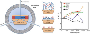
The disadvantages of using nanoparticles for studying immobilized enzymes include the need for full adsorption and the difficulty in modifying the surface of these particles. If there is only partial adsorption of the enzyme at an interface, an interface-sensitive technique has to be used, such as total internal reflection fluorescence (TIRF) spectroscopy, to probe only the adsorbed enzyme. Reference Axelrod, Burghardt and Thompson38,Reference Czeslik, Royer, Hazlett and Mantulin39 The excitation light is internally reflected in a quartz prism, which is in contact with the immobilized enzyme and the sample solution ( Figure 5 ). Only molecules that are in close proximity to the quartz prism surface are excited and fluoresce.
High-pressure total internal reflection fluorescence spectroscopy to study the enzymatic activity of α-chymotrypsin that is immobilized at chemically modified water–quartz interfaces (excitation at 380 nm, emission at 480 nm). The substrate was Ala-Ala-Phe-7-amido-4-methylcoumarin at pH = 7.8. Reference Schuabb, Cinar and Czeslik31 Note: PS, poly(styrene); PSS, poly(styrene sulfonate); PAH, poly(allylamine hydrochloride).

We have built a high-pressure cell for TIRF spectroscopy, where the fluorescence at the planar surface of a quartz prism is detected at pressures up to 2000 bar. Reference Koo and Czeslik40 The surface of the quartz prism can be modified easily, for example, by spin coating or layer-by-layer deposition of polyelectrolytes Reference Decher41 (Figure 5). Again, we used α-CT as the model enzyme, which was adsorbed on a bare hydrophilic quartz surface, a hydrophobic poly(styrene) film (PS), and polyelectrolyte multilayers terminated with the positively charged poly(allylamine hydrochloride) (PAH) and the negatively charged poly(styrene sulfonate) (PSS). Reference Schuabb, Cinar and Czeslik31 In addition, a so-called brush of poly(acrylic acid) (PAA) chains was prepared on the quartz prism surface (Figure 5). Reference Levin, Erlkamp, Steitz and Czeslik33 The main outcome of these studies is experimental proof that the pressure effect on adsorbed α-CT activity strongly depends on the physicochemical properties of the interface. Remarkably, α-CT activity can be enhanced by pressure (negative activation volume) when this protein is adsorbed at a negatively charged and hydrophilic interface, such as bare quartz, the PSS terminated multilayer, the PAA brush (Figure 5), and silica particles (Figure 4). In contrast, α-CT adsorbed on a hydrophobic PS film or a positively charged PAH-terminated multilayer is deactivated by pressure.
Owing to the complexity of the system, a molecular-level interpretation of these results is challenging. It is known that α-CT activity can be enhanced by a number of added co-solutes. Reference Kurinomaru, Tomita, Hagihara and Shiraki42–Reference Spreti, Mancini, Germani, Di Profio and Savelli45 In these cases, the altered environment of α-CT reduces the Gibbs energy of activation, ΔG ≠, which leads to larger rate constant, k 2 (Equation 1). It is suggested that a conformational selection caused by the added compound, which is the chemically modified interface here, constitutes the underlying mechanism. Free α-CT in bulk solution undergoes fluctuations between various conformational substates of the native state, whereas some fluctuations may be suppressed in the adsorbed state, and a certain substate will be preferred. If each substate is characterized by a different Gibbs energy of activation, different enzymatic rates will be observed at different interfaces. We envision that a molecular-level understanding of pressure effects on the active site of enzymes, as may, for example, be achieved by quantum mechanics/molecular mechanics simulations in concert with high-pressure nuclear magnetic resonance methodology, is within reach in the not too distant future.
Summary
The results obtained so far in the field of high-pressure enzymology provide two important insights. First, life under extreme conditions, such as high pressure in the deep sea, is possible, because nature has found strategies to stabilize proteins and other biomolecular systems, allowing them to operate even under harsh environmental conditions. Second, knowledge of these strategies is transferable to applications in biotechnology, biomedicine, and food processing, where enzymes are the central functional unit. By a combination of protein engineering, solution conditions (co-solutes, confinement, interfacial adsorption), and temperature and pressure, maximal robustness and performance of enzymes can be achieved. A molecular-level understanding of the interplay of all these parameters is still largely missing, however, but is on the agenda of several laboratories worldwide.

Claus Czeslik has been a professor of physical chemistry at TU Dortmund University, Germany, since 2011. He studied chemistry at Ruhr-Universität Bochum, Germany, and received his PhD degree in 1997 from TU Dortmund University. He completed postdoctoral research in physical chemistry at the University of Illinois at Urbana-Champaign, and at TU Dortmund University, where he received the Venia Legendi in 2006. His current research focuses on the interfacial structure and functionality of proteins. Czeslik can be reached by email at claus.czeslik@uni-dortmund.de.

Trung Quan Luong has been a postdoctoral fellow of the Cluster of Excellence RESOLV at TU Dortmund University, Germany, since 2015. He studied chemistry at Ruhr-Universität Bochum, Germany, where he received his PhD degree in 2012. He then worked for the Research Department Applied Competence Cluster Terahertz and joined Ruhr-Universität Bochum in 2014. His research interests include terahertz spectroscopy, confined water, protein dynamics, and enzyme activity under high-pressure and confinement conditions. Luong can be reached by email at trung.luong@rub.de.

Roland Winter has been chair of physical chemistry at TU Dortmund University, Germany, since 1993. He studied chemistry at the University of Karlsruhe, Germany. After earning his PhD degree, he joined the University of Marburg, Germany, as a postdoctoral fellow, and received his Habilitation degree in physical chemistry. After spending one year at the University of Illinois at Urbana-Champaign, he was appointed professor at the University of Bochum, Germany. His research interests focus on the study of the structure, dynamics, and functions of biomolecular systems, with a particular focus on high-pressure biophysics. Winter can be reached by phone at +49 231 755 3900 or by email at roland.winter@tu-dortmund.de.