Introduction
Insulin and insulin like growth factor-1 (IGF-1) are critical fetal growth hormones that have overlapping anabolic actions Reference Fowden1–Reference Lassarre, Hardouin and Daffos3 . Concentrations of these hormones are impacted by the health and nutritional status of the fetus, especially in the third trimester of gestation. Common pregnancy complications impacting nutrient transfer to the fetus and resulting in fetal overgrowth or growth restriction are characterized by high or low cord blood concentrations, respectively, of insulin and IGF-1 Reference Lassarre, Hardouin and Daffos3–Reference Ostlund, Bang, Hagenäs and Fried5 . IGF-1 is more strongly correlated with fetal weight than insulin Reference Christou, Connors and Ziotopoulou4,Reference Brown, Palmer and Teynor6,Reference Ong, Kratzsch and Kiess7 . Fittingly, IGF-1 has been studied in sheep as a fetal intervention for growth restriction Reference Jensen, Harding, Bauer and Gluckman8–Reference Eremia, de Boo, Bloomfield, Oliver and Harding10 . However, the impact of experimentally increasing IGF-1 on fetal insulin concentrations, glucose and amino acid metabolism, and β-cell development is incompletely understood.
IGF-1 is synthesized by the fetus and is present in most fetal tissues Reference Han, Lund, Lee and D’Ercole11,Reference Agrogiannis, Sifakis, Patsouris and Konstantinidou12 . In circulation, endogenous IGF-1 is largely bound to one of six major IGF binding proteins which regulate bioavailability. To isolate the effects of IGF-1 on target tissues without the influence of IGF binding proteins, recombinant long arginine 3 IGF-1 (IGF-1 LR3) is commonly used in animal models and has been shown to increase growth in fetal sheep Reference Lok, Owens, Mundy, Robinson and Owens13–Reference Stremming, Heard and White15 . Compared to endogenous human or sheep IGF-1, which contain 70 amino acids, IGF-1 LR3 is a larger protein, containing 83 amino acids, with the additional 13 amino acids on the N-terminus of the peptide. IGF-1 LR3 also substitutes the positively charged amino acid arginine as the third amino acid, replacing the negatively charged glutamic acid, found in human and sheep IGF-1 Reference Rotwein16,Reference Francis, Ross and Ballard17 . As a result, this IGF-1 analog has a high affinity for IGF-1 receptors but a low affinity for IGF binding proteins.
Several studies show that while IGF-1 preserves pancreatic β-cell mass and protects against β-cell damage Reference Mabley, Belin, John and Green18–Reference Robertson, Lu and De Jesus20 , IGF-1 exposure can also inhibit insulin secretion Reference Pørksen, Hussain and Bianda21–Reference White, Stremming and Boehmer23 . We previously infused IGF-1 LR3 into normally grown late gestation fetal sheep for one week and found that IGF-1 LR3 infusion resulted in lower fetal plasma insulin concentrations and attenuated fetal glucose-stimulated insulin secretion (GSIS), which persisted in isolated fetal pancreatic islets Reference White, Stremming and Boehmer23 . While fetal weight increased by ∼15%, demonstrating growth-promoting effects, plasma glucose and amino acid concentrations decreased in IGF-1 LR3 fetuses by the end of infusion Reference White, Louey and Chang14,Reference Stremming, Heard and White15,Reference White, Stremming and Boehmer23 . Notably, there were no changes in β-cell mass, but pancreatic insulin content was significantly higher after IGF-1 LR3 infusion suggesting that insulin was being produced but not secreted Reference White, Louey and Chang14 . This was possibly related to the concomitant low fetal plasma glucose concentrations as the same phenomena were observed with experimental fetal hypoglycemia Reference Rozance, Limesand and Hay24,Reference Rozance, Limesand, Zerbe and Hay25 . The divergence between groups in plasma glucose concentrations, however, did not occur until IGF-1 LR3 infusion day five Reference White, Stremming and Boehmer23 , suggesting that a shorter infusion duration may have different effects on insulin secretion. While this has not been studied in the fetal pancreas, insulin secretion from perfused adult rat pancreases decreased during an acute IGF-1 exposure but returned to normal once IGF-1 administration was stopped Reference Leahy and Vandekerkhove22 . This suggests that the persistent inhibition of insulin secretion by IGF-1 may depend on the dosing and kinetics of administration. Therefore, we investigated in the current study whether an acute IGF-1 LR3 infusion would also reduce circulating insulin concentrations and result in an intrinsic insulin secretion defect. We hypothesized that fetal glucose-stimulated insulin secretion would be attenuated during the 90-min IGF-1 LR3 infusion, but insulin secretion would return to normal in isolated fetal islets once the IGF-1 LR3 exposure was removed.
Materials and methods
Animal preparation
All experiments were conducted at the Perinatal Research Center, University of Colorado School of Medicine, with the approval of the Institutional Animal Care and Use Committee. This center is accredited by AAALAC International. Experimental details are reported in compliance with the ARRIVE 2.0 guidelines Reference Percie du Sert, Hurst and Ahluwalia26 .
Studies were conducted in pregnant late gestation Columbia-Rambouillet mixed-breed sheep (Nebeker Ranch, Lancaster, CA & University of Arizona Sheep Unit, Tucson, AZ) carrying a singleton fetus (n = 12; 7 males and 5 females). All ewes were derived from the same flock and were part of a synchronized breeding system. Ewes were acclimatized to the facility for 21 ± 1 days and then fasted for 24 hours and thirsted for 12 hours before surgery. Surgeries were performed at 122 ± 1 days gestational age (dGA; term = 147–152 dGA, average 149 dGA) to place indwelling polyvinyl catheters as previously described Reference Hay, Sparks, Quissell, Battaglia and Meschia27–Reference Boehmer, Brown, Wesolowski, Hay and Rozance31 . Briefly, pregnant sheep were given diazepam (0.2 mg·kg−1) and ketamine (20 mg·kg−1) through a superficial vein to induce anesthesia and then were maintained on isoflurane inhalation anesthesia (2–4%) for the duration of the surgical procedure. Depth of anesthesia was determined and maintained in response to maternal corneal reflex, toe pinch, assessment of jaw tone, continuous pulse oximetry, heart rate monitoring, and exhaled CO2 monitoring; anesthetic effect in the fetus was assessed by muscle tone. Sheep were given penicillin G procaine (600,000 units intramuscularly) prior to surgery. A midline incision was made along the linea alba and the uterine horn containing the fetus was exposed. A hysterotomy was performed and the fetal hindlimbs were exteriorized. Polyvinyl catheters were placed in the bilateral fetal pedal arteries with the tip positioned in the external iliac artery and bilateral saphenous veins with the tip positioned in the common femoral vein. Ampicillin (500 mg) was injected into the amniotic fluid prior to surgical closure of the abdomen. Catheters were then placed in the maternal femoral artery and vein. Maternal and fetal catheters were tunneled subcutaneously to the maternal flank. Before skin closure at maternal midline, Marcaine was applied for local analgesic (3 mL, 5 mg·ml−1 Marcaine, 0.5% bupivacaine hydrochloride). Sheep received flunixin meglumine on the day of surgery (2.2 mg·kg−1 divided twice per day intramuscularly) and for the two following days (2.2 mg·kg−1 per day intramuscularly). The sheep also received Probios (10 g by mouth twice per day) for 2 days post-operatively. Animals were allowed to recover for a minimum of five days following surgery prior to initiating experimental infusions.
Experimental design
Ten of the twelve animals underwent the following protocol, as the additional two animals were included for islet studies only. Fetal sheep received an intravenous infusion of either IGF-1 LR3 (IGF-1) (GroPep Bioreagents, Thebarton, ASTL) at 6.6 μg·kg−1·hr−1 based on an estimated fetal weight of 3.5 kg or a vehicle control (CON) infusion (saline with 0.5% bovine serum albumin, BSA) at 0.2 ml·hr-1 to match the delivery rate of IGF-1 LR3. IGF-1 LR3, an IGF-1 analog with very low affinity for IGF-binding proteins and high affinity for the IGF-1 receptor, was used to isolate the actions of IGF-1 on its target tissues Reference Sundgren, Giraud and Schultz32 . The dose was selected based on previous direct infusion studies into fetal sheep resulting in increased organ weights and lower insulin concentrations Reference Lok, Owens, Mundy, Robinson and Owens13–Reference Stremming, Heard and White15,Reference White, Stremming and Boehmer23,Reference Sundgren, Giraud and Schultz32 . Each animal received both infusions spaced 2–3 days apart. The initial fetal intravenous experimental infusion (either IGF-1 or CON) was alternated such that five animals received IGF-1 first, and five received CON first. Therefore, animals that received IGF-1 for the initial infusion received CON for the second infusion, while animals that received CON first received IGF-1 for the second infusion. This methodology was utilized to limit the potential effects of animal variability, increasing gestational age, and exposure priming. The first infusion was performed at 130 ± 1 dGA, and the second infusion was performed at 132 ± 1 dGA. Fetal blood was sampled for pre-infusion blood gas, hormone, and nutrient concentrations immediately before the experimental infusions began. To measure the response to the experimental infusion and to determine insulin and glucose concentrations prior to the hyperglycemic clamp, fetal blood was then sampled 70 and 80 min after the experimental infusion began, averaged, and noted as Post-75 min Reference Boehmer, Baker Ii, Brown, Wesolowski and Rozance33 . After 90 min of experimental infusion, fetal GSIS was measured using a primed, continuous, variable-rate hyperglycemic clamp Reference White, Stremming and Boehmer23,Reference Boehmer, Baker Ii, Brown, Wesolowski and Rozance33–Reference Gadhia, Maliszewski and O’Meara35 . The hyperglycemic clamp was initiated with a 33% dextrose (wt:vol in saline) bolus (825 mg glucose) into the fetus. This was followed by a constant infusion of 33% dextrose, titrated to keep the plasma glucose concentration near 2.5 mmol·L−1, which elicits 90% maximal insulin concentrations in vivo in this breed of fetal sheep Reference Green, Macko and Rozance36 . The dextrose infusion rate was held constant from minute 45 until the completion of the GSIS study. Fetal arterial samples were collected for measurement of glucose and insulin concentrations at 5, 10, 15, 20, 30, 45, 60, 75, 90, and 105 min. Experimental infusions (IGF-1 or CON) were continued throughout the GSIS studies. All infusions were stopped after minute 105.
Biochemical analysis
Biochemical analyses were performed as previously described Reference Culpepper, Wesolowski and Benjamin30,Reference Boehmer, Baker Ii, Brown, Wesolowski and Rozance33 . Blood was immediately analyzed for hematocrit, pH, PaCO2, PaO2, and O2 saturation using an ABL 825 blood gas analyzer (Radiometer, Copenhagen, Denmark). O2 content of the blood was calculated by the ABL 825 analyzer. Glucose and lactate were immediately measured from plasma samples (Yellow Springs Instrument 2900, Yellow Springs, OH, USA). Additional arterial plasma samples were frozen at −80 °C for measurement of hormone and amino acid concentrations. Insulin, endogenous IGF-1, and cortisol were measured by an enzyme-linked immunosorbent assay [ELISA; Insulin: ALPCO, Salem, NH, USA; intra- and inter-assay coefficients of variation (CVs) = 5.6% and 4.7%, respectively; sensitivity = 0.14 ng·ml-1; IGF-1: ALPCO; intra- and inter-assay CVs, 3.1% and 5.6%, respectively; sensitivity, 0.09 ng·ml-1; cortisol: ALPCO; intra- and inter-assay CVs = 4.6% and 5.8%, respectively; sensitivity = 1.0 ng·ml-1] and norepinephrine by high-performance liquid chromatography (HPLC; Model 2475, Waters, Milford, MA, USA; intra- and inter-assay CVs = 9.2% and 9.0%, respectively; sensitivity = 170 pg·ml-1). Amino acid concentrations were measured using a Dionex 300 model 4500 amino acid analyzer (Thermo Fisher Scientific, Waltham, MA, USA) after deproteinization with sulfosalicyclic acid. Any results below the lower limit of detection were as assumed to be half of the lower limit of detection value. We were unable to directly measure circulating IGF-1 LR3 concentrations.
Fetal pancreatic islet isolation
Prior to necropsy, the above ten fetuses received a third and final IGF-1 or CON infusion at 138 ± 1 dGA, which was the same infusate as received for the second infusion, but this time without the GSIS study. The goal of this study was to test the impact of in vivo IGF-1 LR3 infusion on isolated pancreatic islet insulin secretion. One additional animal was included to receive IGF-1, and another was included to receive CON infusion at time of necropsy to increase the power to detect differences in islet insulin secretion. Neither of these animals received the prior GSIS studies. Necropsies were performed at the 90-min mark of the final experimental infusion while the infusion was still running. Pancreases were left in situ for perfusion with a collagenase solution and islet isolation Reference White, Stremming and Boehmer23,Reference Limesand, Rozance, Zerbe, Hutton and Hay37 . Islet isolation and in vitro insulin secretion studies were performed as previously described (IGF-1, n = 6; CON, n = 6) Reference White, Stremming and Boehmer23,Reference Limesand, Rozance, Zerbe, Hutton and Hay37,Reference Benjamin, Culpepper and Brown38 . Islets were isolated from the fetal pancreas after collagenase perfusion and digestion and then purified. They were then cultured in Roswell Park Memorial Institute (RPMI) 1640 medium (Thermo Fisher Scientific, Waltham, MA) containing 2.8 mmol·L-1 glucose supplemented with 1% fetal bovine serum and 1X Penicillin-Streptomycin-Amphotericin B solution (Sigma-Aldrich, St. Louis, MO) to provide adequate nutrients and prevent antimicrobial growth. They were then incubated overnight at 37 °C in 95% O2 and 5% CO2 to allow recovery from the isolation process Reference White, Stremming and Boehmer23 .
Static islet incubation
Islet insulin secretion was assayed in static incubations of 10 islets Reference White, Stremming and Boehmer23,Reference Benjamin, Culpepper and Brown38 . Each in vitro incubation condition for each animal was tested in four replicates. After overnight incubation, the islets were washed twice in Krebs-Ringer buffer (KRB) with 0.5% BSA (wt:vol) media and once in KRB-BSA media supplemented with 10 μmol·L-1 forskolin (Sigma-Aldrich). Forskolin was added to the incubation media to activate adenylate cyclase and augment insulin release. Following this step, 10 islets were handpicked and placed into a 1.7 mL tube, and static incubations were performed for 30 min in KRB-BSA-forskolin media to allow for equilibration. An aliquot of this media was removed after 30 min to determine basal insulin secretion in the absence of glucose or potassium chloride (KCl) test media. Supplemental glucose or KCl was then added to each tube to reach final supplement concentrations of 1.1, 2.7, or 11 mmol·L−1 glucose or 30 mmol·L−1 KCl. Islets were incubated in these conditions at 37 °C for 1 hour and then placed on ice and pelleted at 2400 × g for 5 min at 4 °C. Medium was then aspirated and frozen for eventual determination of insulin concentration. The islet pellet was frozen at −80 °C until insulin was acid-ethanol extracted with 1 mol·L−1 HCl in 70% ethanol at −20 °C for 24 h. Cellular debris was removed by centrifugation (30 min at 15,000 × g) and the supernatant was saved for determination of insulin concentration. Insulin concentrations were measured by ELISA (ALPCO, Salem, NH) on dilutions of the basal media, test media, and acid-ethanol extract to determine insulin released (test media minus basal media) and total islet insulin content (test media plus islet extract). Insulin secretion was calculated as the fraction of the total islet insulin content released in response to glucose or KCl stimulation Reference White, Stremming and Boehmer23,Reference Rozance, Limesand and Hay24 . All KRB media used was equilibrated to 37 °C and 95% O2/5% CO2.
Statistical analysis
Statistical analysis was performed using GraphPad Prism 9 (San Diego, CA). Results were analyzed in a mixed models ANOVA with terms to account for repeated measures within the same fetus and based on infusion received. Time, experimental infusion (IGF-1 or CON), and their interaction were included as fixed terms. If interaction terms in the overall ANOVA were P<0.1, then protected Fisher’s least significant difference was used for individual means comparisons. For in vitro insulin secretion, the terms in the mixed models ANOVA were incubation condition (1.1, 2.7, or 11 mmol·L−1 glucose or 30 mmol·L−1 KCl), in vivo experimental infusion (IGF-1 or CON), and their interaction. Measurements made once to compare IGF-1 and CON infusions were analyzed by Student’s t-test. Results are expressed as means ± SEM. P values of ≤ 0.05 were accepted as significant. One KCl test media static islet incubation replicate out of four total from an animal that received IGF-1 infusion at time of necropsy was deemed an outlier based on a physiologically improbable value (80% secretion) and excluded from analysis; all other data points were included in the analyses.
Results
Fetal biochemistry, hormones, and metabolites
Fetal arterial biochemistry and hormones following vehicle control versus IGF-1 LR3 infusions are shown in Table 1. Fetal arterial blood gas values and lactate did not differ based on infusion. Plasma cortisol and norepinephrine concentrations were also similar.
Fetal arterial biochemistry and hormones

Values are means ± SEM. Measurements were made immediately before infusion start (Pre-infusion) and just prior to the fetal in vivo insulin secretion study (Post-75 min). CON, control infusion; IGF-1, IGF-1 LR3 infusion. Each animal (n = 10) received both infusions spaced 2–3 days apart. Statistical analysis was performed by mixed-model ANOVA. Significant P-values where P < 0.05 are bolded.
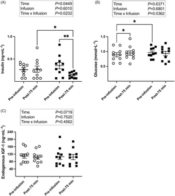
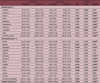
Fetal plasma insulin, glucose, and endogenous IGF-1 concentrations are displayed in Fig. 1. Plasma insulin concentrations decreased during IGF-1 infusion but not during the CON infusion (Time × Infusion P = 0.0232). At Post-75 min, just prior to the hyperglycemic clamp, insulin concentrations were 48% lower after IGF-1 infusion compared to CON infusion (individual means comparisons CON Post-75 min vs IGF-1 Post-75 min P = 0.0475 and IGF-1 Pre-infusion vs IGF-1 Post-75 min P = 0.0052). Fetal glucose concentrations also changed based on infusion (Time × Infusion P = 0.0362). However, this appears to be predominantly due to a slight increase in plasma glucose concentration with CON infusion (individual means comparisons P = 0.0468; CON Pre-infusion: 0.89 ± 0.07 mmol·L−1; CON Post-75 min: 0.95 ± 0.07 mmol·L−1). CON Pre-infusion glucose also started lower than IGF-1 Pre-infusion glucose (individual means comparisons P = 0.0187). IGF-1 Post-75 min glucose was not different than IGF-1 Pre-infusion glucose or CON Post-75 min glucose. Endogenous IGF-1 concentrations were not affected by infusion. Fetal amino acids are displayed in Table 2. Several amino acids—notably all three branched-chain amino acids—decreased with IGF-1 infusion more than with CON infusion (Time × Infusion P<0.05). Summed essential (Time × Infusion P = 0.0084), non-essential (Time × Infusion P = 0.0074), and total (Time × Infusion P = 0.0049) amino acids decreased with IGF-1 infusion but not with CON infusion.
Plasma insulin decreases with IGF-1 LR3 infusion. Fetal plasma insulin (A) decreases with IGF-1 but not with CON infusion. Glucose (B) started lower with CON infusion but was similar to IGF-1 by the end of infusion. Endogenous IGF-1 concentrations (C) did not change based on infusion. CON, control infusion (open circles, n = 10); IGF-1, IGF-1 LR3 infusion (closed squares, n = 10). Means ± SEM are shown. Statistical analysis was performed by mixed-model ANOVA; protected Fisher’s least significant difference test was performed for individual means comparisons if interaction P < 0.1. *Indicates P < 0.05 and **indicates P < 0.01 for individual means comparisons.

Fetal plasma amino acids

Values are means ± SEM. Measurements were made immediately before infusion start (Pre-infusion) and just prior to the fetal in vivo insulin secretion study (Post-75 min). CON, control infusion; IGF-1, IGF-1 LR3 infusion. Statistical analysis was performed by mixed-model ANOVA and protected Fisher’s least significant difference test for individual means comparisons if interaction P < 0.1. Significant P-values where P < 0.05 are bolded; interaction P < 0.1 are italicized. *Indicates P < 0.05 for individual means comparisons between CON Post-75 min and IGF-1 Post-75 min. #Indicates P < 0.05 for individual means comparisons between IGF-1 Pre-infusion and IGF-1 Post-75 min.
Fetal in vivo insulin secretion & fetal islet responsiveness in vitro
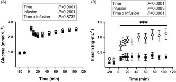
Fetal insulin secretion was measured in vivo with a primed, continuous, variable-rate hyperglycemic clamp. The dextrose infusion rate required to maintain the hyperglycemic clamp (minutes 60–105) was similar between groups (P = 0.1197; CON: 206.07 ± 13.12 μmol·min-1; IGF-1: 228.97 ± 15.09 μmol·min−1). During the GSIS study, glucose concentrations remained similar between IGF-1 and CON, but mean plasma insulin concentrations during the hyperglycemic clamp were 66% lower with IGF-1 infusion compared to CON infusion (P<0.0001; CON: 1.05 ± 0.16 ng·mL−1; IGF-1: 0.35 ± 0.08 ng·mL−1) demonstrating impaired in vivo GSIS (Fig. 2).
Glucose stimulated insulin secretion (GSIS) is attenuated with a 90-min IGF-1 LR3 infusion. Fetal plasma glucose (A) and insulin (B) during measurement of GSIS are plotted relative to the start of the hyperglycemic clamp at time 0. CON, control infusion (open circles, n = 10); IGF-1, IGF-1 LR3-infusion (closed squares, n = 10). Group means ± SEM are shown. Statistical analysis was performed by mixed-model ANOVA; protected Fisher’s least significant difference test was performed for individual means comparisons if interaction P<0.1. ***Indicates P<0.0001 for individual means comparisons.

Fetal islet insulin secretion was then measured in vitro. Total pancreatic islet insulin content was similar based on infusion (P = 0.8182; CON: 26.43 ± 6.70 ng·islet−1; IGF-1: 50.75 ± 26.04 ng·islet−1). Insulin secretion from isolated fetal islets, measured as basal insulin (Fig. 3) and fractional insulin release in response to glucose or KCl stimulation (Fig. 4), did not differ based on IGF-1 or CON infusion.
Basal insulin secretion is not different based on infusion at necropsy. Pancreatic islets isolated from control-infused (CON, open symbols, n = 6) and IGF-1 LR3-infused (IGF-1, closed symbols, n = 6) fetuses were evaluated for basal insulin secretion. One additional animal was included for islet studies to receive CON (open triangle), and another was included to receive IGF-1 LR3 infusion (closed triangle) at time of necropsy to increase the power to detect differences in islet insulin secretion. Means ± SEM are shown. Statistical analysis was performed by Student’s t-test.

Glucose stimulated insulin secretion is not different in isolated fetal islets exposed to IGF-1 in vivo at the time of necropsy. Pancreatic islets isolated from control-infused (CON, open symbols, n = 6) and IGF-1 LR3-infused (IGF-1, closed symbols, n = 6) fetuses at time of necropsy were tested for fractional insulin release in Krebs-Ringer bicarbonate buffer with 1.1, 2.7, or 11 mmol·L−1 glucose, or 30 mmol·L−1 KCl for 1 hour. One additional animal was included for islet studies to receive CON (open triangle), and another was included to receive IGF-1 LR3 infusion (closed triangle) at time of necropsy to increase the power to detect differences in islet insulin secretion. Means ± SEM are shown. Statistical analysis was performed by mixed-model ANOVA.

Discussion
In this study, we tested the impact of a 90-min IGF-1 LR3 infusion on in vivo fetal insulin secretion and in vitro insulin secretion from isolated fetal pancreatic islets. We confirmed the insulin- and amino acid-lowering effects of a direct fetal IGF-1 LR3 infusion and demonstrated that these changes occur within 75 min of infusion initiation. Importantly, we found that while fetal GSIS is attenuated during IGF-1 LR3 infusion, the attenuation does not persist in isolated fetal islets following the acute infusion. This in vitro finding is in contrast to a prior study in which we demonstrated that IGF-1 LR3 infusion for one week directly into fetal sheep circulation impairs GSIS in isolated fetal islets Reference White, Stremming and Boehmer23 . Thus, we have established not only that an acute IGF-1 LR3 infusion rapidly suppresses insulin secretion in vivo during exposure, but also that the fetal β-cell in vitro retains the ability to recover GSIS thereafter. Therefore, it is likely that the duration of IGF-1 LR3 exposure during critical developmental windows dictates the β-cell’s acute and long term ability to appropriately secrete insulin.
Several factors associated with the nutritional status and overall well-being of the fetus can influence the insulin/IGF-1 axis and fetal insulin secretion. Pregnancy complications that result in fetal overnutrition and overgrowth have elevated cord blood concentrations of insulin and IGF-1, while those resulting in poor fetal nutrient delivery and growth restriction have low concentrations of these critical fetal growth hormones Reference Ostlund, Bang, Hagenäs and Fried5,Reference Ong, Kratzsch and Kiess7 . While experimental insulin infusion has been shown to increase IGF-1 concentrations Reference Andrews, Brown and Thorn34,Reference Oliver, Harding, Breier and Gluckman39 , multiple studies in various animal models and insulin-secreting cell lines have demonstrated that IGF-1 exposure reduces circulating plasma insulin concentrations and GSIS, highlighting the complex interplay of these hormones Reference White, Louey and Chang14,Reference Leahy and Vandekerkhove22,Reference White, Stremming and Boehmer23,Reference Liechty, Boyle and Moorehead40 . Moreover, glucose and amino acids are key drivers of insulin secretion. In the current study, while glucose concentrations were similar at the end of control versus IGF-1 LR3 infusion, plasma amino acid concentrations decreased during IGF-1 LR3 infusion. Because acute and chronic amino acid infusions directly into fetal sheep circulation stimulate insulin secretion Reference Boehmer, Brown, Wesolowski, Hay and Rozance31,Reference Boehmer, Baker Ii, Brown, Wesolowski and Rozance33,Reference Gadhia, Maliszewski and O’Meara35,Reference Brown, Davis and Wai41 , it is possible that reduced circulating amino acid concentrations may reduce insulin secretion. While the effect of low amino acid concentrations on insulin secretion has not been directly tested in fetal sheep, a low protein diet during pregnancy in rats resulted in decreased insulin secretion from the progeny’s isolated fetal islets suggesting that normal plasma amino acid concentrations are crucial to establishing appropriate insulin secretion during development Reference Dahri, Snoeck, Reusens-Billen, Remacle and Hoet42 . We previously demonstrated that IGF-1 LR3 infusion for one week results in decreased umbilical amino acid uptake Reference Stremming, Heard and White15,Reference White, Stremming and Boehmer23 . Other studies acutely infusing recombinant human IGF-1 (rhIGF-1) directly into fetal sheep circulation have shown decreased fetal protein breakdown and thus decreased fetal amino acid bioavailability Reference Liechty, Boyle and Moorehead40,Reference Liechty, Boyle and Moorehead43 . In the current study, several amino acids decreased with the 90-min IGF-1 LR3 infusion, but it is unclear whether the decreases were due to decreased umbilical amino acid uptake, decreased protein catabolism, or increased amino acid utilization, all of which may vary with chronicity of IGF-1 LR3 exposure. This requires further investigation.
Other factors known to influence in vivo insulin secretion, such as oxygen content, lactate, cortisol, and norepinephrine, did not differ based on infusion making it unlikely that changes in aerobic metabolism, catecholamines, or glucocorticoid signaling played significant roles. This is important due to the fact that hypothalamic–pituitary–adrenal axis factors can fluctuate in the late gestation fetus. However, such changes are most notable in the final week leading up to delivery Reference Fowden, Li and Forhead44 , which would occur after the gestational age at which we completed our study. This further strengthens the argument for the contribution of concurrent decreased amino acid supply to reductions in fetal insulin secretion in response to an IGF-1 LR3 infusion.
Despite the attenuated in vivo fetal insulin secretion exhibited during IGF-1 LR3 infusion, fetal islets recovered insulin secretion to control values once the IGF-1 LR3 exposure was removed. While the effects of other endocrine hormones such as glucagon and somatostatin on islet insulin secretion were not tested, such effects are likely to be minimal as fetal sheep islets isolated using our protocol contain >90–95% β-cells Reference Rozance, Limesand, Zerbe and Hay25 . This recovery in insulin secretion is consistent with results from in vitro studies exposing the perfused adult rat pancreas to IGF-1 Reference Leahy and Vandekerkhove22,Reference Kawai, Suzuki and Takano45 . We previously demonstrated that islets isolated from fetal sheep exposed to a one-week IGF-1 LR3 infusion had similar insulin content but lower in vitro insulin secretion in response to glucose stimulation after an overnight incubation. In that study, islet incubation overnight in media containing rhIGF-1 10 nmol·L-1 did not have any effect on in vitro GSIS compared to overnight media without rhIGF-1 Reference White, Stremming and Boehmer23 . In the current study, islets exposed to IGF-1 LR3 for 90 min just prior to isolation display a similar response after overnight incubation to in vitro GSIS at all glucose concentrations tested as compared to CON. We speculate that a 90-min IGF-1 LR3 infusion is not long enough in duration to program the fetal islet to secrete less insulin.
Though we are unable to measure plasma IGF-1 LR3 concentrations, changes in fetal insulin secretion and amino acid concentrations suggest supraphysiologic total IGF-1. Our prior study demonstrated 43% lower endogenous IGF-1 concentrations following a one-week IGF-1 LR3 infusion compared to control infusion Reference Stremming, Heard and White15,Reference White, Stremming and Boehmer23 . This was presumably due to suppression of endogenous IGF-1 secretion in response to exogenous IGF-1 LR3 infusion. It is possible that an acute IGF-1 LR3 infusion may not provide the necessary time for endogenous IGF-1 suppression. Additional studies are needed to better understand the β-cell’s response to different IGF-1 dosing regimens and whether glucose or amino acid supplementation would improve fetal insulin secretion.
Our study was also not powered to detect sex differences. While prior fetal sheep studies have not detected sex differences in glucose-insulin responsiveness, β-cell mass, or plasma basal insulin or IGF-1 concentrations Reference Brown, Palmer and Teynor6,Reference Green, Macko and Rozance36 , this deserves further investigation. Additionally, studies are needed to evaluate the contribution of neurovascular networks and other in vivo factors that may influence fetal insulin secretion.
In conclusion, the present study demonstrates that an acute IGF-1 LR3 infusion during late gestation decreases circulating insulin and impairs fetal glucose-stimulated insulin secretion. These effects, however, resolve in vitro once the exogenous IGF-1 exposure is removed. This contrasts with our prior study that demonstrated an intrinsic islet defect after prolonged and continuous fetal exposure to IGF-1 LR3 Reference White, Stremming and Boehmer23 . Establishing and supporting an optimal insulin/IGF-1 axis during fetal development is critical to normal pancreatic development and long-term β-cell function. Based on the findings herein, we speculate that chronically, but not acutely, elevated IGF-1 during the late gestation fetal period in pregnancies complicated by gestational diabetes or maternal obesity may contribute to the risk of β-cell failure and diabetes later in life. Furthermore, our findings suggest that care must be taken to closely monitor plasma insulin, glucose, and amino acid concentrations when considering treatments for intrauterine growth restriction that may increase IGF-1-mediated signaling to promote growth. Therefore, this study significantly contributes to an important gap in our understanding about the implications of increased IGF-1 during pancreatic development.
Acknowledgements
The authors thank David Caprio, David Goldstrohm, Jenai Kailey, Dan LoTurco, Gates Roe, Larry Toft, and Karen Trembler for their technical support.
Author contributions
Conception or design of the work: AW, JS, LDB, PJR.
Acquisition of data: AW, JS, LDB, PJR.
Data analysis/interpretation of data: AW, JS, LDB, PJR.
Drafting and revising the work critically for important intellectual content: AW, JS, LDB, PJR.
All authors approved the final version of this manuscript and agree to be accountable for all aspects of the work.
Financial support
This work was supported by the National Institutes of Health (P.J.R, grant numbers R01DK088139, R01HD093701, T32HD007186 (J.S. and A.W. trainees)), (L.D.B., grant numbers R01HD079404, S10OD023553).
Conflicts of interest
None.
Ethical standards
The authors assert that all procedures contributing to this work comply with the ethical standards of the relevant national guides on the care and use of laboratory animals (sheep) and have been approved by the institutional committee (University of Colorado Institutional Animal Care and Use Committee). All experiments were conducted at the Perinatal Research Center, University of Colorado School of Medicine, with the approval of the Institutional Animal Care and Use Committee. This center is accredited by AAALAC International. Experimental details are reported in compliance with the ARRIVE 2.0 guidelines.