INTRODUCTION
Early instars of species with indirect development in many taxa are characterized by their own, species-specific features, and thus descriptions of their morphological features are among the primary tasks of taxonomy and systematics. Detailed larval descriptions are therefore available for numerous species in some taxa, e.g. decapod crustaceans (review in Ingle, Reference Ingle1992).
Contrary to this in Pycnogonida, comprising approximately 1350 species worldwide, knowledge of larvae is more limited comprising only approximately 70 species (Bain, Reference Bain2003a). A few species were studied in great detail, such as Pycnogonum litorale (Ström, 1762), a species that has become a kind of pycnogonid model species in many aspects (Behrens, Reference Behrens1984; Tomaschko et al., Reference Tomaschko, Wilhelm and Bückmann1997; Wilhelm et al., Reference Wilhelm, Bückmann and Tomaschko1997; Vilpoux & Waloszek, Reference Vilpoux and Waloszek2003). The only bigger, comprehensive study on the larvae is that of Bain (Reference Bain2003a), in which four postembryonic developmental lines are described and named: (1) typical Protonymphon; (2) atypical protonymphon; (3) encysted larva; and (4) attaching larva. Bogomolova & Malakhov (Reference Bogomolova and Malakhov2006) and Bogomolova (Reference Bogomolova2007) propose and describe a fifth postembryonic development type, the lecithotrophic protonymphon. A summary of earlier literature of larval development of different species is also given in Bain (Reference Bain2003a). In addition, the postembryonic development was studied, e.g. in Nymphon distensum Möbius, 1902 in Munilla (Reference Munilla1988); Phoxichilidium femoratum (Rathke, 1799) in Malakhov & Bogomolova (Reference Malakhov and Bogomolova2001); Austropallene cornigera (Möbius, 1902) in Bain (Reference Bain2003b); Tanystylum bealensis Gillespie & Bain, 2005 in Gillespie & Bain (Reference Gillespie and Bain2006); Phoxichilidium femoratum (Rathke, 1799) in Lovely (Reference Lovely2005); Nymphon grossipes Kroyer, 1845 in Bogomolova & Malakhov (Reference Bogomolova and Malakhov2006); Nymphon brevirostre Hodge, 1863, Nymphon micronyx Sars, 1888, and Nymphon grossipes Fabricius, 1780 in Bogomolova (Reference Bogomolova2007); and Ammothea glacialis (Hodgson, 1907) in Cano & Lopez-Gonzalez (Reference Cano and Lopez-Gonzalez2009). In the latter, description of the protonymphon is missing. Therefore, the present study is the first description of an ammotheid protonymphon using the resolution of the scanning electron microscope. Detailed descriptions of pycnogonid larvae may be helpful for a deeper understanding of their sets of characters and variability, and also to understand their phylogeny.
Studying the pycnogonid fauna of the Chilean fjords in cooperation with the Huinay Scientific Field Station, we found Achelia assimilis (Haswell, 1884) to be the most abundant fjord pycnogonid (Melzer et al., Reference Melzer, Schrödl, Häussermann, Försterra and Montoya Bravo2006). We caught male specimens carrying eggs, and a few carrying newly hatched protonymphon larvae used for the present study. As protonymphon larvae are very small, the scanning electron microscope was used in this connection as a powerful tool for studying their minute characteristics.
MATERIALS AND METHODS
The specimens were collected at the Huinay fjord from hydrozoans. Determination was made according to Hedgpeth (Reference Hedgpeth1961) and Gonzales & Edding (Reference Gonzales and Edding1990). Altogether four males with protonymphon larva were studied, two with the scanning electron microscope and two with the light microscope.
The light microscopic pictures were taken under an Olympus SZX stereo microscope with a Jenoptic Prog-Res C12 digital camera and a Leica DMRBE microscope with a Visitron Spot Insight colour digital camera. In addition several dozen larvae were processed for the scanning electron microscope using methods described in Meyer & Melzer (Reference Meyer and Melzer2004), Meyer et al. (Reference Meyer, Wehrtmann and Melzer2006) and Montoya Bravo et al. (Reference Montoya Bravo, Müller, Arango, Tigreros and Melzer2009). Protonymphon larvae were removed from the body and ovigera of adult males. Specimens were cleaned with 30% H2O2 in 70% ethanol (see Bolte, Reference Bolte1996 for details). Afterwards, specimens were dehydrated in a graded acetone series, critical-point-dried in a BAL-TEC CPD030, mounted on scanning electron microscope-stubs using self-adhesive carbon stickers, sputtered with gold in a Polaron ‘sputter coater’, and examined in a Leo 1430VP scanning electron microscope at 15 kV. Multiple protonymphon larvae of two different males were measured, using the measurement utility of the scanning electron microscope. Due to the drying process a shrinking of larvae occurred. Measurements showed that the body of the larvae shrunk about 15%. At the extremities and setae no significant shrinking was observed, due to their thicker cuticle. This means that proportions of these structures, e.g. of articles of appendages are the same in hydrated and dehydrated specimens.
The studied material is deposited at the Zoologische Staatssammlung München (ZSM) with the registration numbers ZSM A20100001, ZSM A20100002, ZSM A20100003, ZSM A20100126, ZSM A20100127, ZSM A20100128 and ZSM A20100129.
RESULTS
Description of the protonymphon
GENERAL SHAPE OF THE BODY
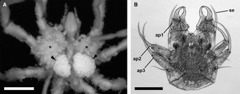
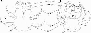
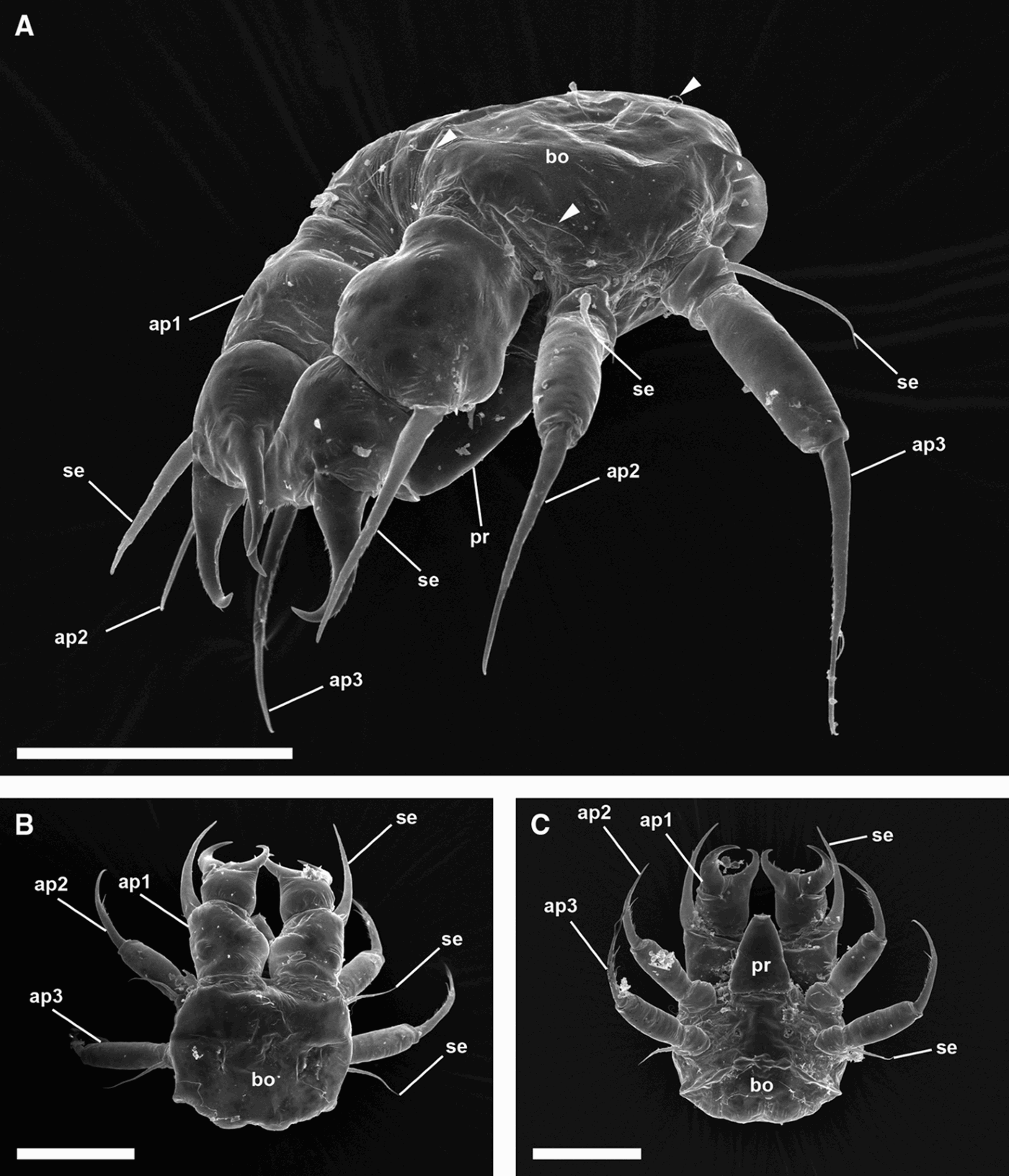
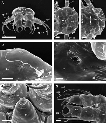
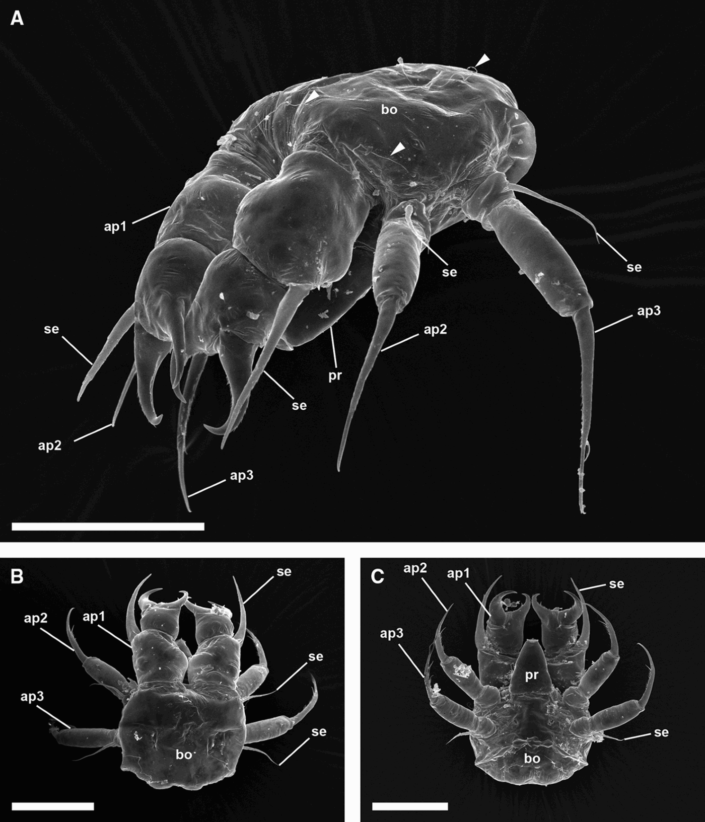
The body is almost circular (length: 143 µm, width: 148 µm; Figures 1B, 2A, B & 3A, C), slightly flattened dorsoventrally (height: 102 µm; Figure 3A). Deprived of segmentation lines, cuticle is smooth, with 3 pairs of appendages (anlagen of chelifores, pedipalps and ovigera), plus the proboscis. The eye turbercle is absent (Figures 1B, 2A, B, 3A, B & 4A, B). Ventral side has one pair of depressions located between the posteriormost appendages (Figure 4C, E). There are deepenings of the cuticle each containing a bifurcate setule laterally and medially above the chelifore bases (Figures 2A, B, 3A & 4B). There are similar setules in deepenings of the cuticle located posterior to the 3rd appendages of the body, at their ventral and dorsal sides, respectively (Figures 2A, B, 3A & 4B, C, D). The anus and yolk are absent (Figures 1B, 3B, C & 4B, C).
Light microscopy of Achelia assimilis. (A) Ventral view of adult male Achelia assimilis, carrying eggs (arrowheads) and newly hatched protonymphon larvae (asterisks); bar: 1 mm; (B) overview of protonymphon of Achelia assimilis; bar: 100 µm. ap, appendage; bo, body; pr, proboscis; se, seta.

External morphology of a protonymphon of Achelia assimilis (scheme from scanning electron microscopy data). (A) Dorsal view; (B) ventral view; ap, appendage; pr, proboscis; pt, spatulate protrusion; se, seta; st, setules.

Protonymphon of Achelia assimilis, overview. (A) Lateroventral view of the protonymphon; note setules on the body, medially above the bases of the 1st appendage, laterally above the 1st, and posterior to the 3rd appendage (arrowheads); bar 100 µm; (B) dorsal view of the protonymphon; bar 100 µm; (C) ventral view of the protonymphon; bar 100 µm. ap, appendage; bo, body; pr, proboscis; se, seta.
Protonymphon of Achelia assimilis, details. (A) Frontal view of the protonymphon; bar 100 µm; (B) dorsal view of the body; note setules on the body, medially above the bases of the 1st appendage, above each 1st appendage, and posterior to each 3rd appendage (arrowheads); bar 20 µm; (C) ventral view of the body; note setules on the body, posterior to each 3rd appendage (arrowheads); note pair of depressions between the 3rd appendages (arrows); bar 20 µm; (D) detail of a setule on the ventral side of the body; bar 10 µm; (E) detail of depression between the 3rd appendages on the ventral side of the body; bar 5 µm; (F) proboscis with the mouth opening; bar 20 µm; (G) dorsal view of the 1st appendages; note three protrusions on the 1st article (asterisks); bar 20 µm. ap, appendage; ar, article; bo, body; mo, mouth opening; pr, proboscis; se, seta.

PROBOSCIS
The proboscis is as long as the basal chelifore article (68 µm). It is of triangular shape, inserted between the bases of the 2nd appendages, ventral to the chelifores (Figure 3C). Diameter of insertion site: 58 µm. Surface smooth; mouth opening oval, located terminally on the proboscis, with edges bent outwards (Figure 4F).
LEG-DERIVED APPENDAGES OF THE BODY
The 1st appendage is already in the first instar of the typical shape of a pycnogonid chelifore. The 2nd (future pedipalps) and 3rd appendages (future ovigera) are very similar, of the shape of a short locomotion organ. All 3 appendages have 3 articles and a long seata on a protrusion at the basal article.
FIRST APPENDAGE (FUTURE CHELIFORES)
The 1st appendage is forward turned, as long as body (148 µm; Figures 4A, G & 5A–C). Contact areas with body are very wide, directly neighbouring each other (Figure 5A). The 2nd and 3rd articles form a fully developed chelifore (Figure 5B).
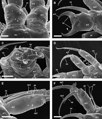
Protonymphon of Achelia assimilis, details. (A) 1st article of the 1st appendages; note three protrusions on the ventral side (asterisks); bar 20 µm; (B) 2nd article of the right 1st appendage; note single long robust spine (arrowhead); bar 20 µm; (C) close up of the chelifore with spines; note single long robust spine (arrowhead); bar 20 µm; (D) ventral view of 2nd and 3rd appendage; bar 20 µm; (E) 2nd article of 2nd and 3rd appendage; note spatulate protrusions; bar 20 µm; (F) 2nd and 3rd article of 2nd and 3rd appendage; note spatulate protrusion; bar 20 µm. ap, appendage; ar, article; bo, body; pr, proboscis; pt, spatulate protrusion; se, seta; sp, spines.

The basal article is as long as wide (length: 67 µm; width: 66 µm), with three basal roundish protrusions (Figures 4G & 5A). Of these, one is located mediobasally, and the two others further distally at the sides of the article. In addition, a long seta is inserted laterally (Figures 4G & 5B). With a length of 82 µm and a width of 15 µm this seta is almost as long as chelifore; distally with some small spines and a terminal pore, eventually a gland pore.
The 2nd article forms an immovable finger of the chelifore (Figure 4B). It is as long as basal article, but more slender (length: 80 µm; width: 48 µm). A movable finger (3rd article) is inserted at 2/3 of length of the immovable finger. The flattened upper and lower sides and also proximal section are deprived of spines. The outer edge has some spines arranged in 1–2 rows, the terminal spine is the biggest (Figure 5b, c). The inner edge is acute, with a row of 36–37 short spines (Figure 5B, C). Between spine No. 28 and No. 29, a single long robust spine is inserted, 4 times as long as short spine, terminally flattened, with apical pore, eventually a gland pore.
The 3rd article forms an outer, movable finger of chelifore (length: 49 µm; Figure 5B). Articulation is at about 2/3 of the length of the immovable finger (width: 24 µm). It is equipped with more spines than 2nd article: outer edge with numerous irregularly arranged spines and one pronounced terminal spine. The inner edge has at least two rows of up to 46 small spines (Figure 5B, C).
SECOND AND THIRD APPENDAGES (FUTURE PEDIPALPS AND OVIGERA)
The 2nd and 3rd appendages have cramping legs, inserted on not well pronounced lateral protrusions of trunk, turned sidewards, composed of 3 articles (Figure 5D–F).
The basal article is smooth, as long as wide (length: 27 µm; width: 29 µm; Figure 5D), ventral side longer than dorsal side, the articulation between 1st and 2nd articles is therefore in a peripheral position. Dorsally it has long, pointed seta (length: 54 µm; diameter at the base: 4 µm; Figure 5E, F) with numerous short secondary spines on the median section.
The second article is longer than wide (length: 71 µm, width: 26 µm; Figure 5E), more slender than basal article, surface smooth except for short spatulate protrusion, eventually representing a gland pore, inserted at 2/3 of the article's length.
The distal article is claw-like, contorted, terminating in a pointed tip (length: 90 µm, diameter at the base: 10 µm, Figure 5F). The outer side has some very small ribs, the inner side has a row of spines of various sizes: from proximal to distal, (1) some short, (2) 6–8 medium-length, (3) a large, blunt spine similar to that on the 2nd chelifore article (length: 9 µm), and (4) 2 apical spines are found.
DISCUSSION
The present paper on Achelia assimilis is the first detailed scanning electron microscopy study of a protonymphon larva of the family Ammotheidae Dohrn, 1881. The advantage of this technique is that, due to the high resolution power of the scanning electron microscope, even minute structures could be located allowing a very detailed description of the larval features. As larvae of only a few pycnogonid species were studied this way until present, our study is restricted to the features detailed in our description and comparison with the extant knowledge about protonymphons that is still at a somewhat general level. A true differential diagnosis is at the moment not possible, especially for our species that does not only represent the first detailed analysis of a representative of its genus, but even of a whole family of pycnogonids. The only other scanning electron microscopy analysis of ammotheid larvae is that by Cano & Lopez-Gonzalez (Reference Cano and Lopez-Gonzalez2009), where a description of the first larval instar is missing.
According to the classification suggested by Bain (Reference Bain2003a) the larvae of Achelia assimilis resembles a ‘typical protonymphon’ development: the first instar of Achelia assimilis is attached on the male's ovigers, it leaves the eggs as protonymphon, with a short, anterior proboscis, a pair of large chelifores as the first pair of appendages and two pairs of larval appendages used for locomotion, and later instars are free living and found on hydrozoans (see Melzer et al., Reference Melzer, Schrödl, Häussermann, Försterra and Montoya Bravo2006). In the subsequent larval instars the 1st and 2nd larval appendages develop directly to the chelifore and the palps, respectively. The 3rd larval appendages develop indirectly to the ovigera. They undergo a radical change being reduced to small unsegmented knobs and they subsequently become segmented ovigers (Bain, Reference Bain2003a; Vilpoux & Waloszek, Reference Vilpoux and Waloszek2003; Gillespie & Bain, Reference Gillespie and Bain2006). Further arguments for the classification as ‘typical protonymphon’ are the similar size, the similar structure and shape of the larval appendages and the similar arrangement of setules, which have been described in detail in studies of other ‘typical protonymphon’ larvae (e.g. Behrens Reference Behrens1984; Gillespie & Bain Reference Gillespie and Bain2006; Bogomolova, Reference Bogomolova2007). Finally the absence of yolk makes it reasonable to say that Achelia assimilis demonstrates the developmental pattern ‘typical protonymphon’ according to the classification of Bain (Reference Bain2003a).
On the other hand, Bamber (Reference Bamber2007) considered that pycnogonids have only one type of larva: the protonymphon. The eggs hatch as a protonymphon or as a postlarva (the larval stage having been passed in the egg). Bamber also distinguishes between basic protonymphon and variants, e.g. without chelifore spine or with lamellae on the larval legs. If this classification is applied, the larva of Achelia assimilis is a basic protonymphon.
In Pycnogonum litorale particular pores on the body are described (8 pairs of ‘Buckelporen’ by Behrens (Reference Behrens1984); 1 pair on the ventral side by Vilpoux & Waloszek (Reference Vilpoux and Waloszek2003)). We found similar structures in Achelia assimilis (Figure 4C, E). By comparison, Bogomolova (Reference Bogomolova2007) did not find such structure in her scanning electron microscopy study of three Nymphon species. However, in Pycnogonum litorale they are located on a protuberance and in Achelia assimilis they are found in a depression. Their function is unknown.
The setules in deepenings of the cuticle located laterally and medially above the chelifore bases and posterior to the 3rd appendages of the body, at their ventral and dorsal sides, respectively (Figures 2A, B, 3A & 4B, C, D) are also described by Behrens (Reference Behrens1984) in Pycnogonum litorale. Behrens found deepenings of the cuticle with bifurcate or trifurcate setules; in Achelia assimilis we found only deepenings with bifurcate setules. Bogomolova (Reference Bogomolova2007) observed the same number and spatial distribution of these setules on Nymphon brevirostre and Nymphon micronyx.
The pore on the seta on the basal article of the chelifores (Figures 4G & 5B) is also reported in different species (e.g. Meisenheimer, Reference Meisenheimer1902; Vilpoux & Waloszek, Reference Vilpoux and Waloszek2003; Bogomolova & Malakhov, Reference Bogomolova and Malakhov2006; Bogomolova, Reference Bogomolova2007). According to these authors the pore is the opening of a gland located in the ventral side of the basal article of the chelifore. It produces long clear strands to attach on the cocoon on the oviger of the male or on the hydrozoans (Dogiel, Reference Dogiel1911; Russell & Hedgpeth, Reference Russell and Hedgpeth1990; Bogomolova & Malakhov, Reference Bogomolova and Malakhov2006).
Vilpoux & Waloszek (Reference Vilpoux and Waloszek2003) and Bogomolova (Reference Bogomolova2007) found on the 3rd article of the chelifore, and on the 2nd and 3rd articles of the chelifore, respectively, several spines with a gland pore. Here, in Achelia assimilis we found only one spine with a pore on the 2nd article (=immovable finger; Figure 5B, C). Their function is unknown.
Hence, the protonymphon of Achelia assimilis can be clearly attributed to what is called a ‘typical protonymphon’ introduced by Bain (Reference Bain2003a). Until present, we have only cursory knowledge about the distribution of these types among the pycnogonid families (survey in Bain, Reference Bain2003a). More detailed scanning electron microscopy studies are necessary in order to determine sets of characters that may be characteristic for pycnogonid taxa on a higher level, such as families and genus, and also to find out if species-specific features exist that allow distinguishing between closely related species already in the early larvae as in larvae of other marine arthropods.
ACKNOWLEDGEMENTS
We thank Stefan Friedrich (Munich) for expert technical assistance. We also thank the team of the Huinay Scientific Field Station for their support with the diving basics. We gratefully acknowledge a grant given by the GeoBiocenterLMU (Munich). This paper is publication No. 47 of the Huinay Scientific Field Station.