Introduction
Epidemiological studies of wildlife infections are becoming more relevant due to the outbreaks of new haemosporidian infections which are frequently recorded in wild and domestic birds (Cardona et al., Reference Cardona, Ihejirika and McClellan2002; Valkiūnas, Reference Valkiūnas2005; Atkinson et al., Reference Atkinson, Thomas and Hunter2008; Ortiz-Catedral et al., Reference Ortiz-Catedral, Brunton, Stidworth, Elsheikha, Pennycott, Schulze, Braun, Wink, Gerlach, Pendl, Gruber, Ewen, Perez-Tris, Valkiūnas and Olias2019). These studies are useful for better understanding the causes of parasite spread and the emergence of new diseases. Avian haemosporidian parasites are widespread, and they cause severe disease or even mortality in non-adapted birds (Garnham, Reference Garnham1966; Donovan et al., Reference Donovan, Schrenzel, Tucker, Pessier and Stalis2008; Cannell et al., Reference Cannell, Krasnec, Campbell, Jones, Miller and Stephens2013; Tostes et al., Reference Tostes, Martinele, Vashist, Castañon, Pinto Pde, Daemon and D'Agosto2015; Ilgūnas et al., Reference Ilgūnas, Bukauskaitė, Palinauskas, Iezhova, Dinhopl, Nedorost, Weissenbacher-Lang, Weissenbock and Valkiūnas2016; Jia et al., Reference Jia, Valkiūnas, Yang, Zheng, Pu, Zhang, Dong, Suo and Zhang2018; Kelly et al., Reference Kelly, Baldwi, Frame, Childress and Wellehan2018; Groff et al., Reference Groff, Lorenz, Crespoc, Iezhova, Valkiūnas and Sehgal2019). In the past 30 years, the number of studies on the prevalence and diversity of haemosporidian parasites has increased, but the vectors transmitting these parasites have been insufficiently investigated (Valkiūnas, Reference Valkiūnas2005; Santiago-Alarcon et al., Reference Santiago-Alarcon, Palinauskas and Schaefer2012; Žiegytė et al., Reference Žiegytė, Markovets, Bernotienė, Mukhin, Iezhova, Valkiūnas and Vaidas Palinauskas2017; Bukauskaitė et al., Reference Bukauskaitė, Iezhova, Ilgūnas and Valkiūnas2019). The great majority of studies on avian haemoproteids address questions relating to molecular characterization, genetic diversity and the phylogenetic relationships of these parasites (Bensch et al., Reference Bensch, Hellgren and Perez-Tris2009, Reference Bensch, Canbäck, DeBarry, Johansson, Hellgren, Kissinger, Palinauskas, Videvall and Valkiūnas2016; Clark et al., Reference Clark, Clegg and Lima2014; Outlaw and Ricklefs, Reference Outlaw and Ricklefs2014). Determination of natural vectors is important for better understanding the transmission of invasive parasite species in the wild, as well as for predicting possibilities of disease spread due to climate change and for the protection of endangered species of birds. The prevalence of haemosporidian parasites in European birds is especially high after the birds have arrived from their wintering grounds (Valkiūnas, Reference Valkiūnas2005; Bensch et al., Reference Bensch, Jönsson and Copete2011). Seasonal bird migration is an important factor that increases haemosporidian parasite diversity in the Northern Holarctic (Ricklefs et al., Reference Ricklefs, Medeiros, Ellis, Svensson-Coelho, Blake, Loiselle, Soares, Fecchio, Outlaw, Marra, Latta, Valkiūnas, Hellgren and Bensch2017). Transmission of some pathogens occurs only in warm climate regions, including Southern Europe (Waldenström et al., Reference Waldenström, Bensh, Kibol, Hasselquist and Ottosson2002; Rojo et al., Reference Rojo, Hernandez, Campos, Santamaria, Dias and Casanueva2015; Valkiūnas et al., Reference Valkiūnas, Žiegytė, Palinauskas, Bernotienė, Bukauskaitė, Ilgūnas, Dimitrov and Iezhova2015), but there is often no information about the transmission of the same parasites in North Europe (Valkiūnas, Reference Valkiūnas1993).
Interestingly, the widespread and, for some birds, lethal malaria caused by Plasmodium relictum (genetic lineage pGRW4) is actively transmitted in warm climate countries, but not in temperate zones of Europe (Waldenström et al., Reference Waldenström, Bensh, Kibol, Hasselquist and Ottosson2002; Dimitrov et al., Reference Dimitrov, Zehtindjiev and Bensch2010; Marzal et al., Reference Marzal, Longoria, Callirgos and Sehgal2015). Recent experimental studies have shown that sporogony of this parasite can be completed in Culex pipiens mosquitoes at relatively low temperatures (Valkiūnas et al., Reference Valkiūnas, Žiegytė, Palinauskas, Bernotienė, Bukauskaitė, Ilgūnas, Dimitrov and Iezhova2015). Although this mosquito species is widespread in temperate climate zones (Earth System Research Laboratory, Colorado; website at http://www.esrl.noaa.gov/psd/), there are no data relating to the presence of P. relictum (pGRW4) in North European juvenile birds before their autumn migration (reports of this infection in juveniles would prove the presence of local transmission) or blood-sucking dipterans (natural vectors). Recent studies (Valkiūnas, Reference Valkiūnas1993; Waldenström et al., Reference Waldenström, Bensh, Kibol, Hasselquist and Ottosson2002; Ivanova et al., Reference Ivanova, Zehtindjiev, Mariaux and Georgiev2015; Palinauskas et al., Reference Palinauskas, Žiegytė, Ilgūnas, Iezhova, Bernotienė, Bolshakov and Valkiūnas2015; Rojo et al., Reference Rojo, Hernandez, Campos, Santamaria, Dias and Casanueva2015) have shown that European birds are infected with several species of malarial parasites: Plasmodium homocircumflexum (pCOLL4), P. delichoni (pCOLL6), P. ashfordi (pGRW2) and haemoproteids: Haemoproteus pallidus (hCOLL2), H. nucleocondensus (hGRW1) and H. payevskyi (hRW1), transmission of which takes place only in the tropics and/or southern regions, and these infections have never been detected in juvenile birds in North Europe. Factors preventing their transmission in the temperate zone of Europe remain unclear, but it is important to explore them, as they could play a crucial role in the emergence of new infections, especially in light of climate change in Europe.
The aim of this study was to define if the transmission of H. nucleocondensus (hGRW8) can be completed in Culicoides impunctatus, which are abundant in Northern Europe (Carpenter et al., Reference Carpenter, Groschup, Garros, Felippe-Bauer and Purse2013) and to discuss important factors for successful disease transmission in breeding areas. We conducted experimental infection of C. impunctatus biting midges, and followed resulting sporogony at temperatures close to natural. Culicoides impunctatus biting midges willingly take bird blood meals and thus, this species could be a natural vector for H. nucleocondensus.
Materials and methods
Study site, experimental birds and microscopic examination of blood
This study was carried out at the Biological Station ‘Rybachy’ of the Zoological Institute of the Russian Academy of Sciences on the Curonian Spit of the Baltic Sea (55°15′N, 20°86′E). Birds were captured by mist nets during fieldwork in May–August during 2015 and 2018. In all, 186 adult great reed warblers were caught and investigated for H. nucleocondensus infection after arrival from their African wintering grounds. Seventy-two juvenile great reed warblers were caught in July–August and investigated for the presence of this parasite before leaving for the wintering grounds. About 50 μL of bird blood was taken in heparinized microcapillaries by puncturing the brachial vein. Two blood slides were prepared from each bird. Residual blood was stored in SET-buffer for molecular analysis (Hellgren et al., Reference Hellgren, Waldenstrom and Bensch2004). Blood slides were air-dried, fixed in absolute methanol and stained with Giemsa stain solution, as described by Valkiūnas et al. (Reference Valkiūnas, Iezhova, Križanauskienė, Palinauskas and Bensch2008). Approximately 100–150 fields were examined at low magnification (400×), and then at least 100 fields were studied at high magnification (1000×). The intensity of parasitaemia was estimated as a percentage of the number of parasites per 1000 erythrocytes or per 10 000 erythrocytes if infections were light (<0.1%), as recommended by Godfrey et al. (Reference Godfrey, Fedynich and Pence1987). We used an Olympus BX51 light microscope (Olympus, Japan) with a digital camera and Olympus DP-SOFT software to analyse the blood slides. Haemoproteus nucleocondensus was identified according to Križanauskienė et al. (Reference Križanauskienė, Iezhova, Palinauskas, Chernetov and Valkiūnas2012). The morphology of the H. nucleocondensus (hGRW8) gametocytes was compared with hGRW1 lineage of the same species, the type material of great reed warbler A. arundinaceus (accession number 47792NS, Acrocephalus arundinaceus, collected by Križanauskienė) deposited in the Nature Research Centre, Vilnius, Lithuania. Voucher preparations of H. nucleocondensus gametocytes (hGRW8) from this study (accession nos. 49037–49040NS) were deposited in the Nature Research Centre, Vilnius, Lithuania.
One individual great reed warbler with a single H. nucleocondensus (hGRW8) infection was used as a donor to infect biting midges. The presence of a single infection was confirmed by the microscopic examination of blood slides and confirmed after sequencing parasite's DNA by visual inspection of double-base-calling on the electropherogram (Pèrez-Tris and Bensch, Reference Pèrez-Tris and Bensch2005). One great reed warbler uninfected with haemosporidian parasites (as confirmed by microscopy and PCR) was used as a donor to feed the group of control biting midges. Donor birds were kept indoors in a vector-free room under controlled conditions [20 ± 1°C, 50–60% relative humidity (RH), the natural light–dark (L/D) photoperiod], and were fed with a standard diet for insectivorous bird species. Both birds were released after the experiment.
Experimental infections of biting midges
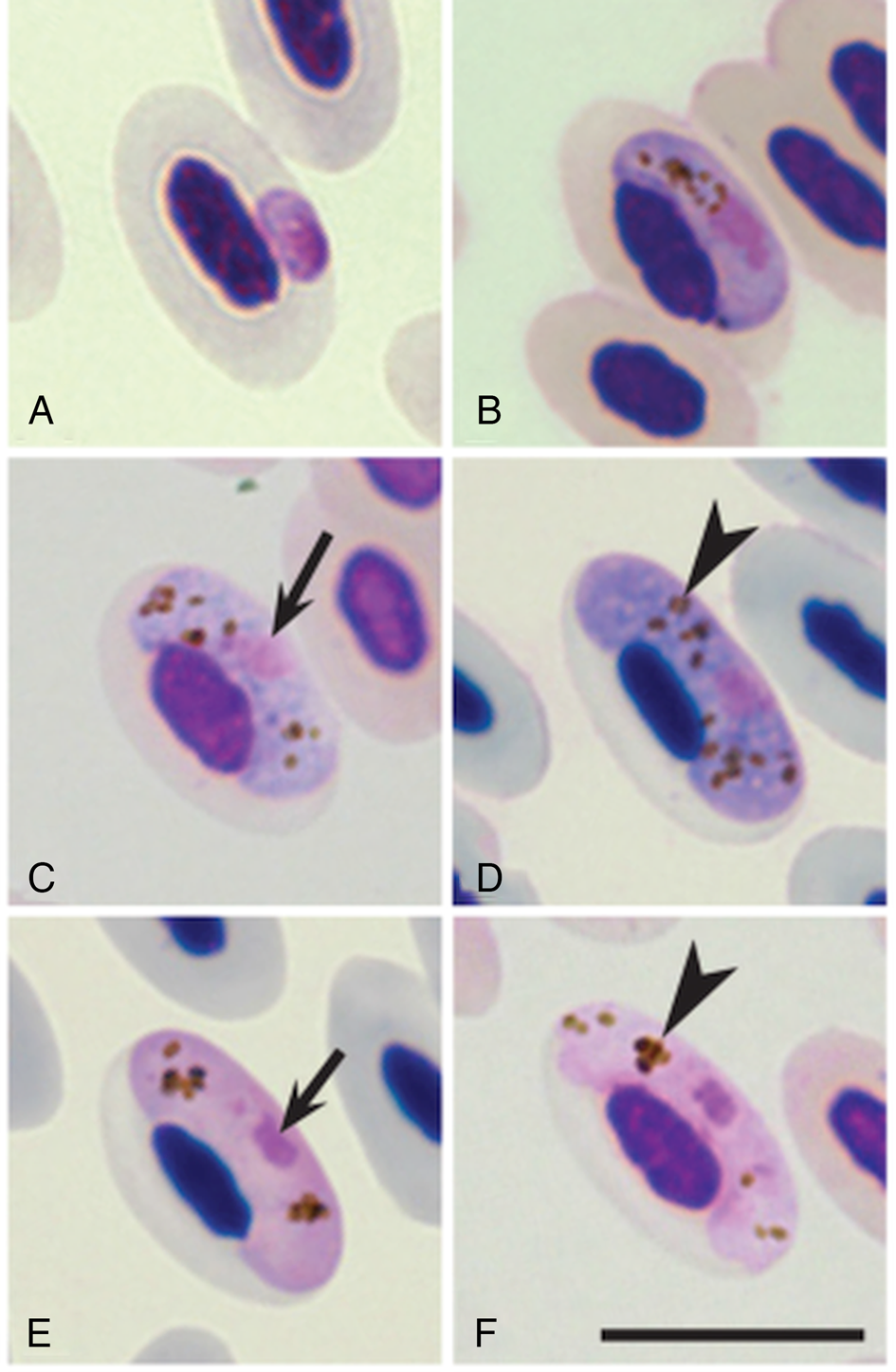
Experimental infection of wild biting midges was performed near Lake Chaika, located close to the village Rybachy (55°09′N, 20°49′E). The activity of C. impunctatus is high in this locality in June (Glukhova and Valkiūnas, Reference Glukhova and Valkiūnas1993). Experimental infections were performed between 10 and 25 June when the first generation of C. impunctatus predominates (Liutkevičius, Reference Liutkevičius2000; Žiegytė et al., Reference Žiegytė, Markovets, Bernotienė, Mukhin, Iezhova, Valkiūnas and Vaidas Palinauskas2017). Culicoides impunctatus females were infected by allowing them to take blood meals on one great reed warbler naturally infected with a single H. nucleocondensus infection. The parasitaemia of the mature gametocytes (Fig. 1) was 0.06% in the donor bird during the infection of biting midges. Twenty biting midges were fed on a non-infected great reed warbler and were used as negative controls. A detailed workflow of feeding and maintaining of the infected and control biting midges was described by Valkiūnas (Reference Valkiūnas2005) and Žiegytė et al. (Reference Žiegytė, Palinauskas, Bernotienė, Iezhova and Valkiūnas2014, Reference Žiegytė, Markovets, Bernotienė, Mukhin, Iezhova, Valkiūnas and Vaidas Palinauskas2017). The donor bird with H. nucleocondensus was used five times for infecting the biting midges. The cages with engorged flies were transported to the laboratory within 1 h of infection. The cages with insects were held at temperatures ranging from 14°C (at night) to 24°C (during the day), 60 ± 2% RH and photoperiod of 17:7 h L/D.
The stages of gametocytes of Haemoproteus nucleocondensus (genetic lineage hGRW8) in great reed warbler Acrocephalus arundinaceus (A–F). Young gametocytes (A, B), macrogametocytes (C, D), microgametocytes (E, F). All images are from the methanol-fixed and Giemsa-stained thin films. Short simple arrows – nuclei of parasites; arrowheads – pigment granules. Scale-bar: 10 μm

Additionally, 100 unfed Culicoides females were collected using an entomological net and were then fixed in 96% ethanol at the study site, for testing possible natural infections with Haemoproteus parasites using PCR-based methods (see description below). All insects were identified using morphological characters (Gutsevich, Reference Gutsevich1973), and the identification was supported by PCR-based methods in the laboratory (see description below).
Dissection of biting midges and preparation of ookinetes, oocysts and sporozoite specimens
The methodology of dissection, preparation of permanent preparations of ookinetes, oocysts and sporozoites, as well as their staining methods, were as described by Valkiūnas (Reference Valkiūnas2005) and Žiegytė et al. (Reference Žiegytė, Palinauskas, Bernotienė, Iezhova and Valkiūnas2014, Reference Žiegytė, Markovets, Bernotienė, Mukhin, Iezhova, Valkiūnas and Vaidas Palinauskas2017). We examined midgut contents for ookinetes in the period between 12 h post-infection (hpi) and 3 days post-infection (dpi), as well as the midgut wall for oocysts 2–5 dpi, and the salivary glands for sporozoites 4–10 dpi. The control group was examined for oocysts 2–5 dpi, and for sporozoites 4–10 dpi. After dissection, all residual parts of the biting midges were placed in 96% ethanol for PCR-based confirmation of the parasite lineage and insect species (see description below).
Representative preparations of zygotes (49041NS), ookinetes (49042NS), oocysts (49043NS) and sporozoites (49044NS) were deposited in the Nature Research Centre, Vilnius, Lithuania.
DNA extraction, PCR, sequencing and phylogenetic analysis
Total DNA was extracted from samples (bird blood stored in the SET buffer and biting midges placed in absolute ethanol) using an ammonium acetate extraction method (Richardson et al., Reference Richardson, Jury, Blaakmeer, Komdeur and Burke2001). For the identification of parasites, we used a nested PCR protocol (Bensch et al., Reference Bensch, Stjenman, Hasselquist, Ostman, Hansson, Westerdahl and Pinheiro2000; Hellgren et al., Reference Hellgren, Waldenstrom and Bensch2004). We amplified a segment of mitochondrial cytochrome b (cytb) gene using (1) initial primers HaemNFI and HaemNR3, which amplify DNA fragments of haemosporidians belonging to Haemoproteus, Plasmodium and Leucocytozoon and (2) HaemF and HaemR2 primers, which are specific to Haemoproteus and Plasmodium spp. The amplification was evaluated by running 3 μL of the final PCR product on a 1.5% agarose gel. One negative control (nuclease-free water) and one positive control (Haemoproteus sp. microscopy positive blood) were used per every 14 samples to control for false amplifications. Fragments of DNA from the positive samples were sequenced at the Nature Research Centre. The ‘Basic Local Alignment Search Tool’ (National Centre for Biotechnology Information website: http://www.ncbi.nlm.nih.gov/ BLAST) was used to determine genetic lineages of detected DNA sequences. Sequences were deposited in the GenBank (KY913917, KY913918).
DNA extracted from individual biting midges was used to support the morphological identification of C. impunctatus. We applied the insect-specific primers LCO149 and HCO2198 to amplify a fragment of mitochondrial cytochrome oxidase c subunit 1 (Folmer et al., Reference Folmer, Black, Hoeh, Lutz and Vrijenhoek1994). All obtained sequences corresponded to the C. impunctatus DNA sequences available in the GenBank (KJ627800).
For phylogenetic analysis, we downloaded 36 haemosporidian parasite cyt b sequences from the MalAvi database (http://mbio-serv2.mbioekol.lu.se/Malavi/; Bensch et al., Reference Bensch, Hellgren and Perez-Tris2009), which were previously linked to the morphological species. The GenBank accession numbers of sequences and codes of the parasite lineages are given in Fig. 3. The phylogenetic tree was constructed using MrBayes version 3.2.6 (Ronquist et al., Reference Ronquist, Teslenko, Van Der Mark, Ayres, Darling, Hohna and Huelsenbeck2012). The appropriate model of sequence evolution GTR + G was determined using the Akaike information criterion implemented in jModelTest 2.1.2 (Guindon and Gascuel, Reference Guindon and Gascuel2003; Darriba et al., Reference Darriba, Taboada, Doallo and Posada2012). Phylogenetic analysis was performed using MrBayes on CIPRES Science Gateway v. 3.3 (Miller et al., Reference Miller, Pfeiffer and Schwartz2010). Visualization of the phylogenetic tree was conducted using FigTree v 1.4.4 (software available at https://github.com/rambaut/figtree/releases).
Results
Microscopic and molecular examination of the parasite in the vertebrate host
Microscopic examination and PCR-based testing showed that (1) 29 of 186 investigated adult great reed warblers were infected with H. nucleocondensus; (2) none of the 72 juvenile great reed warblers were infected with H. nucleocondensus in their nesting areas before leaving for their wintering grounds.
Detailed analysis of H. nucleocondensus (hGRW8) revealed that the main diagnostic morphological features of gametocytes of this parasite lineage (Fig. 1) are the same as for the lineage hGRW1, which was used as the type lineage for the original description of H. nucleocondensus (Križanauskienė et al., Reference Križanauskienė, Iezhova, Palinauskas, Chernetov and Valkiūnas2012). Briefly, the earliest gametocytes are often seen in a lateral or sublateral position to the nucleus of the infected cell, and are usually roundish or oval (Fig. 1A). Young gametocytes (Fig. 1A and B) develop in mature erythrocytes and closely associate with the nuclei of infected erythrocytes. Macrogametocytes (Fig. 1C and D) grow alongside the nuclei of infected cells and are frequently closely associated with both the nucleus and the cell membrane of erythrocytes. Fully-grown macrogametocytes fill nearly all the space at the poles of erythrocytes (Fig. 1D). The nucleus of the parasite is variable in shape and compact, usually central or sub-central in position (Fig. 1C and D). Microgametocytes (Fig. 1E and F) have a rare character for bird haemosporidian parasites – their nuclei are markedly condensed and small, a distinctive morphological character of this species. The area of microgametocyte nuclei (Fig. 1E and F) is significantly less than that of macrogametocytes (Fig. 1D). The morphological characters of blood stages confirmed that the new lineage hGRW8 belongs to H. nucleocondensus.
Microscopic and molecular examination of the parasite in biting midges
None of the wild-caught biting midges was naturally infected with Haemoproteus parasites at the study site, indicating that our experimental insects were parasite free. PCR-based methods confirmed the morphological identification of investigated biting midges – all experimental midges belonged to C. impunctatus.
Thirty-six biting midges were experimentally infected with the H. nucleocondensus parasite. Twenty-two surviving biting midges were dissected for the detection of ookinetes (six midges), oocysts (four midges) and sporozoites (12 midges). Parasites were not detected in the midguts or salivary glands of biting midges fed on the non-infected great reed warbler used as the negative control.
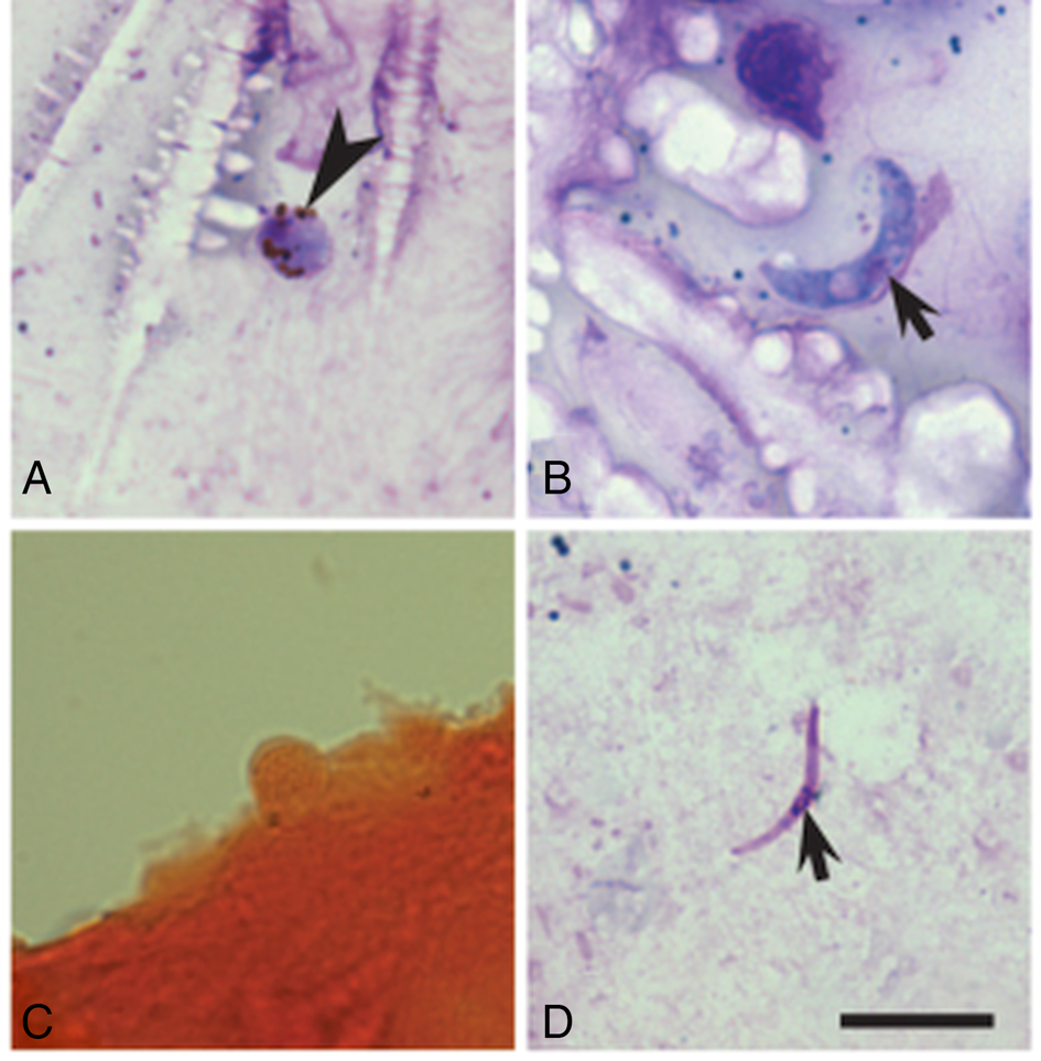
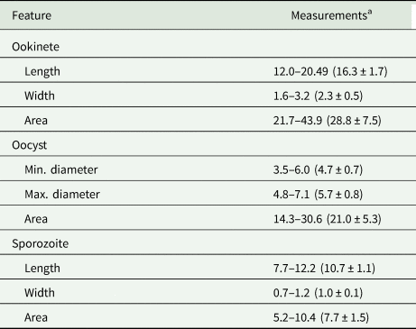
Numerous zygotes were seen in the midgut contents of the exposed midges 24 hpi (Fig. 2A). Mature ookinetes were seen in the midgut contents of experimentally infected midges between 24 and 36 hpi, but they were not seen 48 hpi. The ookinetes were elongated worm-like bodies with prominent, slightly off-centre located nuclei and visible vacuoles (Fig. 2B). Oocysts were first seen in the midgut wall 2.5 dpi and they were reported in the midgut preparations until 3 dpi. In formalin-fixed preparations, oocysts appeared as small roundish bodies (Fig. 2C). Pigment granules were visible in some oocysts. Sporozoites were seen in the salivary glands from 7 to 9 dpi (Fig. 2D). They look like fusiform cells with slightly off-centre nuclei and approximately equally pointed ends. The morphometric characters of the ookinetes, oocysts and sporozoites are given in Table 1. In accordance with microscopy data, PCR-based analysis confirmed the presence of the lineage hGRW8 in the experimentally infected biting midges.
The stages of sporogony of Haemoproteus nucleocondensus (hGRW8) in biting midge Culicoides impunctatus midgut (A, B, C) and salivary glands (D). Zygote with pigment granules 24 h post infection (hpi) (A), mature ookinete possessing nuclei 30 hpi (B), mature oocyst 3 days post infection (dpi) (C) and sporozoite 7 dpi (D). All images are from the methanol-fixed and Giemsa-stained thin films (A, B, D), except image (C), which is from formalin-fixed whole mounts stained with Erlich's haematoxylin. Scale-bar: 10 μm

Morphometric parameters of ookinetes, oocysts and sporozoites of Haemoproteus nucleocondensus in the Culicoides impunctatus biting midges

a Measurements of ookinetes (n = 21, methanol-fixed preparations 24–36 h post infection, hpi), oocysts (n = 12, formalin-fixed preparations of mature parasites 2.5–3 days post infection, dpi) and sporozoites (n = 21, methanol-fixed preparations 7–8 dpi) are given in micrometres. Minimum and maximum values are provided, followed in parentheses by the arithmetic mean and standard deviation.
Phylogenetic analysis
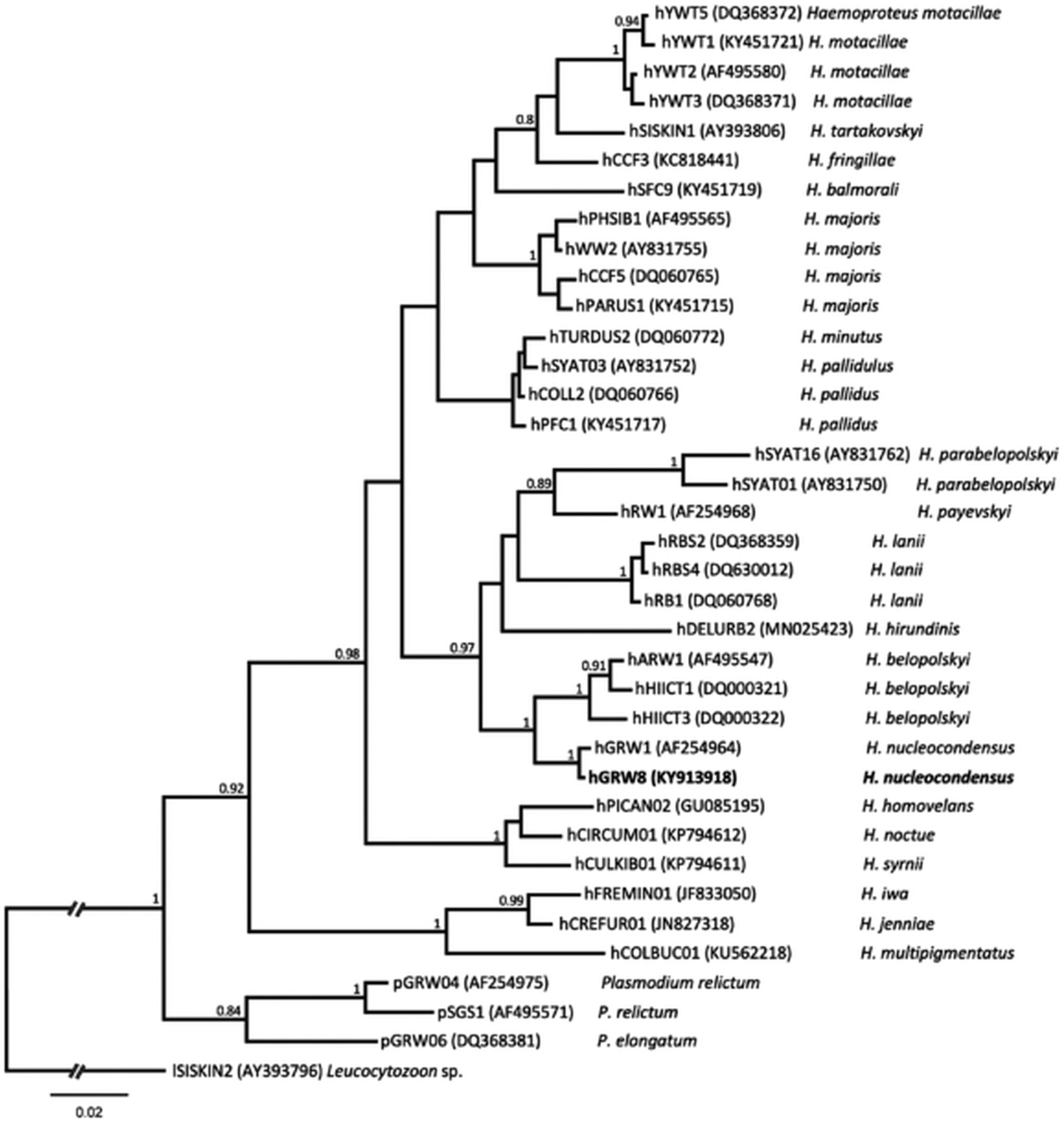
Bayesian analysis revealed that the investigated parasite lineage hGRW8 is phylogenetically closely related to another H. nucleocondensus genetic lineage hGRW1. Both lineages cluster together in the phylogenetic tree (Fig. 3). The difference only in one nucleotide of the mitochondrial cytb gene fragment also indicates that both lineages belong to the same species. The genetic divergence between them is 0.02%. This result is in accord with the morphological data, which showed marked morphological similarity of the blood stages of H. nucleocondensus lineages hGRW1 and hGRW8 (Križanauskienė et al., Reference Križanauskienė, Iezhova, Palinauskas, Chernetov and Valkiūnas2012; this study Fig. 1).
Bayesian phylogenetic tree of cyt b gene sequences (379 nucleotides) of 33 Haemoproteus lineages and three Plasmodium lineages. One Leucocytozoon sp. sequence was used as outgroup. Nodal support values indicate Bayesian posterior probabilities. The scale bar shows the expected substitutions per site. Lineage codes are given according to MalAvi database (Bensch et al., Reference Bensch, Hellgren and Perez-Tris2009), followed by GenBank accession number in parentheses and parasite species names. Haemoproteus nucleocondensus (hGRW8) parasite is shown in bold.

Based on the MalAvi database, H. nucleocondensus has been recorded in Bulgaria, France, Germany, Israel, Latvia, Lithuania, Nigeria, Romania, Russia (Kaliningrad Region), the Seychelles, Spain, Sweden, Turkey and Zambia.
Discussion
This study supports the results of former studies (Waldenström et al., Reference Waldenström, Bensh, Kibol, Hasselquist and Ottosson2002; Hasselquist et al., Reference Hasselquist, Östman, Waldenström and Bensch2007; Rojo et al., Reference Rojo, Hernandez, Campos, Santamaria, Dias and Casanueva2015) that showed that great reed warblers get infected by Haemoproteus (Parahaemoproteus) nucleocondensus outside of the Northern European breeding areas. The lineage hGRW1 of this parasite was first reported in this avian host in Sweden (Bensch et al., Reference Bensch, Stjenman, Hasselquist, Ostman, Hansson, Westerdahl and Pinheiro2000) and then described as a new species in Lithuania (Križanauskienė et al., Reference Križanauskienė, Iezhova, Palinauskas, Chernetov and Valkiūnas2012). The blood stages of H. nucleocondensus morphologically are similar to Haemoproteus payevskyi, particularly due to the condensed nuclear material in microgametocytes, explaining why the newly described species was named using this feature by Križanauskienė et al. (Reference Križanauskienė, Iezhova, Palinauskas, Chernetov and Valkiūnas2012). However, these two agents of haemoproteosis are readily distinguishable genetically (Fig. 3) and can be readily distinguished by the size of their mature gametocytes. In the 2012 study by Križanauskienė et al., the analysis that combined morphological features of blood stages with genetic and phylogenetic data, as well as information about the vertebrate host, was used to characterize H. nucleocondensus infection. However, the vectors of the lineage hGRW1 remained unidentified.
During this study, the new lineage hGRW8 of H. nucleocondensus was detected. Our analysis revealed that the blood stages of these two parasite lineages are morphologically indistinguishable, and the parasites are genetically similar, so certainly belong to the morphospecies H. nucleocondensus. In addition, sporogonic development of H. nucleocondensus (hGRW8) was examined for the first time (Fig. 2).
According to the available data (Waldenström et al., Reference Waldenström, Bensh, Kibol, Hasselquist and Ottosson2002; Križanauskienė et al., Reference Križanauskienė, Iezhova, Palinauskas, Chernetov and Valkiūnas2012; Rojo et al., Reference Rojo, Hernandez, Campos, Santamaria, Dias and Casanueva2015; Hammers et al., Reference Hammers, Komdeur, Kingma, Hutchings, Fairfield, Gilroy and Richardson2016), birds are infected with H. nucleocondensus in Southern Europe (Spain) or outside Europe in the wintering areas in tropical and subtropical regions. Haemoproteus nucleocondensus hGRW1 has been reported in birds from African Islands (Seychelles), sub-Saharan Africa and the Iberian Peninsula (Waldenström et al., Reference Waldenström, Bensh, Kibol, Hasselquist and Ottosson2002; Rojo et al., Reference Rojo, Hernandez, Campos, Santamaria, Dias and Casanueva2015; Hammers et al., Reference Hammers, Komdeur, Kingma, Hutchings, Fairfield, Gilroy and Richardson2016) where transmission occurs. However, the only record of hGRW1 in a non-Sylviidae bird, specifically in a white-throated dipper Cinclus cinclus (Rojo et al., Reference Rojo, Hernandez, Campos, Santamaria, Dias and Casanueva2015) in South Europe, should be taken with caution. This parasite is also present in adult birds in South East, Central and Northern Europe (Bensch et al., Reference Bensch, Stjenman, Hasselquist, Ostman, Hansson, Westerdahl and Pinheiro2000; Hasselquist et al., Reference Hasselquist, Östman, Waldenström and Bensch2007; Dimitrov et al., Reference Dimitrov, Zehtindjiev and Bensch2010; Križanauskienė et al., Reference Križanauskienė, Iezhova, Palinauskas, Chernetov and Valkiūnas2012). However, juvenile or resident non-migrant birds infected with H. nucleocondensus have only been reported in Spain and African regions, where the transmission of this infection occurs (Rojo et al., Reference Rojo, Hernandez, Campos, Santamaria, Dias and Casanueva2015; Hammers et al., Reference Hammers, Komdeur, Kingma, Hutchings, Fairfield, Gilroy and Richardson2016). This infection has not been found in juvenile great reed warblers before their seasonal migration in Central or Northern Europe (Hasselquist et al., Reference Hasselquist, Östman, Waldenström and Bensch2007; Križanauskienė et al., Reference Križanauskienė, Iezhova, Palinauskas, Chernetov and Valkiūnas2012; this study). It was unclear whether susceptible vectors occur in the temperate zones of Europe. This study shows that sporogonic development of H. nucleocondensus is completed in C. impunctatus at average air temperatures ranging from 14°C (at night) and 24°C (during the day) and, thus, theoretically this insect could be a natural vector in Northern Europe, where it is widespread, abundant and readily takes bird blood meals (Žiegytė et al., Reference Žiegytė, Palinauskas, Bernotienė, Iezhova and Valkiūnas2014). Sporogonic development was completed in this insect at temperatures close to natural during the summer months, indicating that both availability of vector and average air temperature probably are not the main factors restricting the transmission of this parasite at our study site. A recent study by Chagas et al. (Reference Chagas, Bukauskaitė, Ilgūnas, Bernotienė, Iezhova and Valkiūnas2019) revealed that the closely related lineage hGRW1 of H. nucleocondensus completes sporogony in laboratory-reared Culicoides nubeculosus at 24°C, which is widespread in Eurasia (Mathieu et al., Reference Mathieu, Cêtre-Sossah, Garros, Chavernac, Balenghien, Carpenter, Setier-Rio, Vignes-Lebbe, Ung, Candolfi and Delécolle2012) and is also a suitable vector for many avian haemoproteids.
It is worth noting that these data are in accord with observations by Valkiūnas et al. (Reference Valkiūnas, Žiegytė, Palinauskas, Bernotienė, Bukauskaitė, Ilgūnas, Dimitrov and Iezhova2015), who confirmed that malaria parasite P. relictum (pGRW4), which is transmitted mainly in Africa and other tropical regions, but not in temperate zone of Europe, completes sporogony and could be transmitted by C. pipiens mosquitoes at relatively low average temperature (19°C). This mosquito is widespread and abundant in the Central and Northern Europe, but the transmission of P. relictum (pGRW4) does not take place in these regions. In other words, there are no restrictions for the spreading of this parasite in European birds from the point of view of vector availability and air temperature necessary for successful sporogony, as is the case with H. nucleocondensus. These results indicate some general patterns in avian haemosporidian parasite transmission, but further studies on different pathogens are needed. Available research (Valkiūnas et al., Reference Valkiūnas, Žiegytė, Palinauskas, Bernotienė, Bukauskaitė, Ilgūnas, Dimitrov and Iezhova2015) and our observations on sporogony of H. nucleocondensus (Fig. 2) call for a more precise investigation of the ecological factors that might restrict the transmission of these haemosporidian parasites in Europe. Among them, the following factors are worth considering: (1) time-differences of maximum abundance of susceptible vectors and the presence of offspring of corresponding bird species at the breeding sites (temporary isolation); (2) non-overlapping biotopes of vectors and avian hosts (biotope and other spatial isolations); (3) mortality of infected juvenile birds (if transmission happens) and (4) long prepatent period of parasite development in the vertebrate host, resulting in the absence of detectable parasitaemia in juveniles before autumnal migration. Defensive behaviour by the host might play a role in avoiding bites and thus infection with vector-borne diseases. Walker and Edman (Reference Walker and Edman1985) showed that blood-sucking insects limit their attack on defensive birds, reducing the chances of becoming infected. To our knowledge, there are few studies addressing these issues in research on the transmission of avian haemosporidian parasites. We should consider these issues in more detail.
It is interesting that the period of the maximum abundance and activity of C. impunctatus does not coincide with the period when the offspring of the great reed warblers are present at our study site, and this might decrease the possibility of juvenile bird infection. Maximum activity and the greatest abundance of C. impunctatus biting midges are reported in mid-June, and the density of this insect sharply decreases in July at our study site (Liutkevičius, Reference Liutkevičius2000). The maximum hatching and fledging period of great reed warblers occurs in July. Thus, vector abundance does not coincide with the presence of juvenile great reed warblers at our study site. This discordance in timing might prevent the transmission of some infections from adult birds to juveniles. However, this situation may change with changes in arrival times of migrant birds due to climate change (Fuller et al., Reference Fuller, Bensch, Müller, Novembre, Pérez-Tris, Ricklefs, Smith and Waldenström2012), and transmission might be established. In some cases, successful infection of adult birds may be reduced by non-overlapping diurnal cycles of peak activity of vector and host, which cause temporal isolation (Atkinson, Reference Atkinson1991).
It is also possible that the breeding sites of great reed warblers (reeds located in open air water bodies and resulting windy conditions) are not attractive to Culicoides biting midges at our study site. The highest abundance of Culicoides vectors is observed in the moist coniferous forest (Glukhova and Valkiūnas, Reference Glukhova and Valkiūnas1993), localities that great reed warblers do not breed in and avoid visiting. Culicoides impunctatus midges usually fly within 75 m of their breeding areas (Kettle, Reference Kettle1951), and this might minimize contact rates as well as transmission opportunities. Other spatial factors, such as vertical stratification, may also be a barrier for vectors seeking hosts. It was shown by Garvin and Greiner (Reference Garvin and Greiner2003) that the abundance of some blood-sucking insects may decrease with each 2 m down starting from 12 m above the ground.
European birds gain many Haemoproteus parasites at the breeding sites (Valkiūnas, Reference Valkiūnas1993, Reference Valkiūnas2005), but precise information on the epizootiology of these infections remains insufficiently investigated. Numerous recent studies indicate that Haemoproteus parasites might be dangerous and even deadly, especially for non-adapted birds (Atkinson et al., Reference Atkinson, Thomas and Hunter2008; Donovan et al., Reference Donovan, Schrenzel, Tucker, Pessier and Stalis2008; Cannell et al., Reference Cannell, Krasnec, Campbell, Jones, Miller and Stephens2013; Jia et al., Reference Jia, Valkiūnas, Yang, Zheng, Pu, Zhang, Dong, Suo and Zhang2018; Kelly et al., Reference Kelly, Baldwi, Frame, Childress and Wellehan2018; Ortiz-Catedral et al., Reference Ortiz-Catedral, Brunton, Stidworth, Elsheikha, Pennycott, Schulze, Braun, Wink, Gerlach, Pendl, Gruber, Ewen, Perez-Tris, Valkiūnas and Olias2019; Groff et al., Reference Groff, Lorenz, Crespoc, Iezhova, Valkiūnas and Sehgal2019). The possibility that H. nucleocondensus infection could be virulent and even kill non-adapted juvenile birds cannot be ruled out due to the lack of experimental research or other observations on the virulence of this infection. According to the study on Seychelles warblers, Acrocephalus sechellensis by Hammers et al. (Reference Hammers, Komdeur, Kingma, Hutchings, Fairfield, Gilroy and Richardson2016), the probability of juveniles infected with H. nucleocondensus (hGRW1) surviving up to 1-year-old does not differ from those not infected with the haemoproteids. However, it is difficult to rule out that juvenile birds in that study had already survived primary infection when the most dangerous acute parasitaemia is present, thus the survival analysis between juveniles and 1-year-old birds was based on individuals containing relatively benign chronic infections. Numerous observations indicate that haemosporidian infections are more virulent in young birds, but adults of the same species might tolerate the disease or even be resistant (Valkiūnas, Reference Valkiūnas2005).
A possible explanation for the lack of reports of H. nucleocondensus in juvenile great reed warblers in Europe also might be due to undetectable parasitaemia resulting from the possible long-lasting prepatent period. In the Hammers et al. (Reference Hammers, Komdeur, Kingma, Hutchings, Fairfield, Gilroy and Richardson2016) study, which was based solely on PCR-based diagnostics, the prepatent period of H. nucleocondensus (hGRW1) in Seychelles warblers might be speculated to be short because this haemoproteid was detected both in nestlings (5% prevalence) and fledglings (prevalence 58%). However, it was not proved that the positive PCR signal was related to the erythrocytic stage (gametocyte), which is essential for the development of a true prepatent period in haemoproteids. PCR amplifies DNA of sporozoites or circulating merozoites from tissue meronts (Valkiūnas et al., Reference Valkiūnas, Iezhova, Loiseau and Sehgal2009; Palinauskas et al., Reference Palinauskas, Žiegytė, Iezhova, Ilgūnas, Bernotienė and Valkiūnas2016), thus is a highly sensitive tool for reports of parasite DNA in blood, but is insufficiently sensitive when used alone for determining the length of the prepatent period. These results reveal that unclear H. nucleocondensus (hGRW1) life cycle stages appear in the blood of A. sechellensis within 2 weeks of infection, but the prepatent period remains unclear. It is also unclear whether a long prepatent period is characteristic for H. nucleocondensus in great reed warblers. Thus, experimental studies using C. impunctatus vectors and juvenile great reed warblers are needed to clarify this.
Our study reveals that C. impunctatus is a suitable vector for the transmission of H. nucleocondensus (hGRW8) at the ambient temperatures of Northern Europe. Other factors important for the successful transmission of haemoproteids should be addressed in future studies combining field and experimental approaches, because answering these questions is important for better understanding disease epizootiology. Investigations into the development of parasites in birds and vectors, as well as precise consideration of ecological factors in disease epizootiology, remain insufficiently addressed in wildlife haemosporidian research, but this knowledge is crucial for the development of measures aimed at improving bird health.
Acknowledgments
We would like to thank the staff of the Biological Station ‘Rybachy’ for assistance in the field. The director of the Biological Station ‘Rybachy’ Nikita Chernetsov is acknowledged for generously providing facilities for the experimental research. We are grateful to Jos Stratford and Prof Ravinder Sehgal for correcting the English and comments on the manuscript. The experiments described herein comply with the current laws of the Republic of Lithuania and the Russian Federation.
Financial support
This research was funded by the European Social Fund (Project No 09.3.3-LMT-K-712-02-0047) and also supported by the Open Access to research infrastructure of the Nature Research Centre under Lithuanian open access network initiative.
Conflict of interest
None.
Ethical statement
Experimental procedures of this study were approved by the International Research Cooperation Agreement between the Zoological Institute of the Russian Academy of Sciences and the Nature Research Centre (1-12-2015–30-11-2020). All efforts were made to minimize handling time and potential suffering of animals.