Introduction
Fasciola hepatica, the trematode that causes fasciolosis, mainly affects domestic and wild herbivores and humans. Fasciola hepatica infection reduced dairy production by 20% and meat production by 8% in the Cuban bovine livestock industry, causing losses of US$200,000–800,000 (González et al., Reference González, Pérez and Brito2007). Inappropriate use of chemicals to control fasciolosis in the 1990s caused development of a strain resistant to triclabendazole in Scotland, Spain, Ireland, UK, Netherlands, Australia and, most recently, in Peru (Mitchell et al., Reference Mitchell, Maris and Bonniwell1998; Álvarez-Sánchez et al., Reference Álvarez-Sánchez, Mainar-Jaime, Pérez-García and Rojo-Vázquez2006; Brennan et al., Reference Brennan, Fairweather, Trudgett, Hoey, McCoy, McConville, Meaney, Robinson, McFerran, Ryan, Lanusse, Mottier, Alvarez, Solana, Virkel and Brophy2007; Brockwell et al., Reference Brockwell, Elliott, Anderson, Stanton, Spithill and Sangster2013; Ortiz et al., Reference Ortiz, Scarcella, Cerna, Rosales, Cabrera, Guzmán, Lamenza and Solana2013).
A protective vaccine for domestic animals is an important potential strategy for long-term control of fasciolosis, and excretion–secretion antigens, including glutathione S-transferase (Zafra et al., Reference Zafra, Pérez-Ecija, Buffoni, Mendes, Martínez-Moreno, Martínez-Moreno, Galisteo and Pérez2010), fatty acid binding proteins (FABPs) (López-Abán et al., Reference López-Abán, Nogal-Ruiz, Vicente, Morrondo, Diez-Baños, Hillyer, Martínez-Fernández, Feliciano and Muro2008), paramyosine (Cancela et al., Reference Cancela, Carmona, Rossi, Frangione, Goñi and Berasain2004), peroxiredoxin (Mendes et al., Reference Mendes, Pérez-Ecija, Zafra, Buffoni, Martínez-Moreno, Dalton, Mulcahy and Pérez2010), cathepsin L1 (CL1) (Mulcahy et al., Reference Mulcahy, O'Connor, Clery, Hogan, Dowd, Andrews and Dalton1999; Golden et al., Reference Golden, Flynn, Read, Sekiya, Donnelly, Stack, Dalton and Mulcahy2010) and leucine aminopeptidase (LAP) (Acosta et al., Reference Acosta, Cancela, Piacenza, Roche, Carmona and Tort2008; Maggioli et al., Reference Maggioli, Acosta, Silveira, Rossi, Giacaman, Basika, Gayo, Rosadilla, Roche, Tort and Carmona2011), have been identified as vaccine candidates. To improve protection, cocktails containing CL1, cathepsin L2 (CL2) and haemoglobin (Hb) have been used. The most effective combination, providing 72.4% protection, included CL2 and Hb (Dalton et al., Reference Dalton, McGonigle, Rolph and Andrews1996). Another cocktail included cathepsin L5 (CL5), cathepsin LG (CLG) and cathepsin B (CB), and the best result, 83% protection, was obtained with the combination of CL5 and CB (Jayaraj et al, Reference Jayaraj, Piedrafita, Dynon, Grams, Spithill and Smooker2009). A cocktail containing CL1, CL2 and LAP was also tested, achieving 79% protection. However, simultaneous immunization with multiple proteins may create competition and unpredictable results, including reduced efficacy rather than additive effects (Willadsen, Reference Willadsen2008).
In the present study, we propose a strategy to combine the antigenic and immunogenic characteristics of two proteins into one, to avoid competition and achieve additive effects. The LAP and CL1 enzymes were selected because they have essential roles in parasite physiology. LAP is expressed in epithelial cells (Acosta et al., Reference Acosta, Cancela, Piacenza, Roche, Carmona and Tort2008) in adult and juvenile trematodes (Piacenza et al., Reference Piacenza, Acosta, Basmadjian, Dalton and Carmona1999), where it is involved in the final stage of catabolizing peptides generated by CL1, which breaks collagen (types I, III and IV) and fibronectin, permitting Fasciola to penetrate and migrate across the intestinal wall, hepatic capsule and parenchyma (Berasain et al., Reference Berasain, Goni, McGonigle, Dowd, Dalton, Frangione and Carmona1997). CL1 is also involved in proteolysis of host tissue to obtain nutrients (Tort et al., Reference Tort, Brindley, Knox, Wolfe and Dalton1999). In addition, CL1 digests immunoglobulins (Smith et al., Reference Smith, Dowd, McGonigle, Keegan, Brennan, Trudgett and Dalton1993), impeding eosinophil adhesion to juvenile Fasciola (Carmona et al., Reference Carmona, Dowd, Smith and Dalton1993). CL1 may also promote a non-protective T-helper 2 (Th2) response in the host immune system (Dalton et al., Reference Dalton, Neill, Stack, Collins, Walshe, Sekiya, Doyle, Mulcahy, Hoyle, Khaznadji, Moiré, Brennan, Mousley, Kreshchenko, Maule and Donnelly2003).
The aim of this study was to design and construct a protein containing the most antigenic regions of F. hepatica LAP and CL1. Based on bioinformatic analyses, this protein may be used as the basis for serological diagnoses or for vaccine development and protection from fasciolosis.
Materials and methods
Design of the rFhLAP-CL1 chimeric protein
The F. hepatica LAP (GenBank AAV59016.1) and CL1 (GenBank CAC12806.1) amino acid sequences were analysed using the Kolaskar & Tongaonkar (Reference Kolaskar and Tongaonkar1990) antigenic prediction method (http://imed.med.ucm.es/Tools/antigenic.pl), BCEPRED (Saha & Raghava, Reference Saha, Raghava, Nicosia, Cutello, Bentley and Timis2004; http://www.imtech.res.in/raghava/bcepred/), and ABCPred (Saha & Raghava, Reference Saha and Raghava2006; http://www.imtech.res.in/raghava/abcpred/) to select antigenic regions to incorporate into the chimeric protein. The enzymatic domains were located with Pfam (Finn et al., Reference Finn, Mistry, Tate, Coggill, Heger, Pollington, Gavin, Gunesekaran, Ceric, Forslund, Holm, Sonnhammer, Eddy and Bateman2010; http://pfam.sanger.ac.uk/) software, the PSIPRED (http://bioinf.cs.ucl.ac.uk/) program was used to analyse the secondary structures and hydrophobicity predictions were conducted with ProtScale (http://www.expasy.ch/tools/protscale.html).
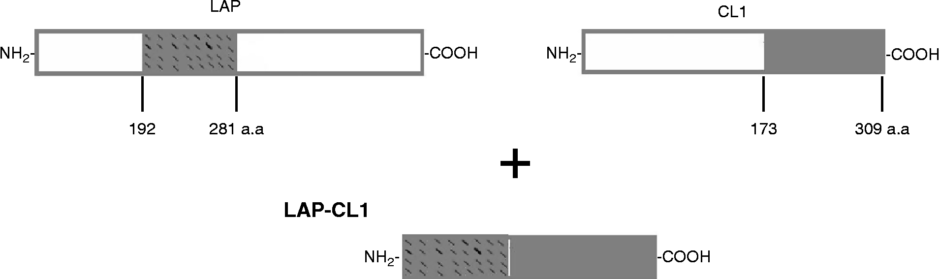
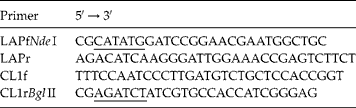
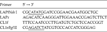
After selecting antigenic regions (fig. 1), primers were designed to amplify selected fragments of LAP and CL1. Restriction sites for NdeI and BglII were included in the LAP forward primer (LAPfNdeI) and the CL reverse primer (CL1rBglII), respectively. Complementary bases between the genes were added to the LAP reverse (LAPr) and CL forward (CL1f) primers to allow the individual polymerase chain reaction (PCR)-amplified fragments to bind in a modified overlap extension PCR (Grandori et al., Reference Grandori, Struck, Giovanielli and Carey1997; Wurch et al., Reference Wurch, Lestienne and Pauwels1998) (table 1).
Schematic representation of the protein chimeric rFhLAP-CL1 with the antigenic leucine aminopeptidase (LAP) and cathepsin L1 (CL1) regions used in the construction of this protein.

Leucine aminopeptidase and cathepsin L1 primers with restriction enzyme sites underlined.
Construction of the chimeric gene
Adult F. hepatica flukes were removed from liver bile ducts of infected cattle slaughtered at the municipal abattoir (Toluca, State of Mexico). Flukes were washed several times with chilled sterile phosphate-buffered saline (PBS; NaCl 137 mm, KCl 2.69 mm, Na2HPO4 10.2 mm, KH2PO4 1.77 mm, pH 7.4) until they were free of blood and bile, and later processed for isolation of total RNA for cDNA synthesis.
Isolation of total RNA from the liver flukes was carried out using TRIzol reagent (Invitrogen, Carlsbad, California, USA). Briefly, 100 mg of tissue from the trematodes was added to 1 ml of TRIzol. The subsequent steps followed the protocol recommended by the manufacturer.
Complementary DNA (cDNA) was synthesized by reverse transcription from 5 μg of total RNA, 1 μl (10 mm) of deoxynucleotide triphosphates (dNTP) mix, 1 μl (0.5 μg/μl) of oligo-dT18 primer, 1 μl (2 μm) of LAP/CL1 primer and water added to a total volume of 10 μl, following the protocol recommended by SuperScript (Invitrogen).
The cDNA was used as a template for PCR amplification; the LAP fragment was amplified in a 50-μl PCR reaction prepared with 2 μl of cDNA, 5 μl of PCR 10 × buffer, 15 mm MgCl2 (QIAGEN, Hilden, Germany), 1 μl of dNTPs (10 mm each), 1 μl of each primer (LAPfNdeI and LAPr; 20 pmol), and 0.1 U of Taq DNA polymerase (QIAGEN). Initial denaturation was done for 4 min at 95°C, followed by 25 cycles of denaturation for 1 min at 94°C, annealing for 45 s at 61°C, extension of 1 min at 72°C, and a final extension of 10 min at 72°C. The CL1 fragment was amplified with the CL1f and CL1rBglII primers (table 1) under the same conditions, except that the annealing temperature was 56°C. The PCR products were analysed by electrophoresis in 1% agarose gel, then purified using the QIAquick PCR Purification kit (QIAGEN); 40 μl of LAP and CL1 samples were purified using a microcentrifuge following the protocol recommended by the manufacturer.
Purified samples of LAP and CL1 were used as templates to generate the chimeric fragment in a new PCR reaction; 1 μl of LAP and 1 μl of CL1 PCR products were prepared in 50 μl as described above. The first five cycles were performed without primers at an annealing temperature of 56°C. At cycle 6, 1 μl of each primer, LAPfNdeI and CL1rBglII (20 pmol each; table 1) were added to the reaction. Amplification conditions were as follows: denaturation for 4 min at 95°C, 30 cycles of denaturation for 1 min at 94°C, annealing for 45 s at 64.9°C, an extension of 1 min at 72°C, and a final extension of 10 min at 72°C. The amplified chimeric product was evaluated in 1% agarose gel stained with ethidium bromide. The product from overlapping PCR was purified from 2% agarose gel with a 123-bp molecular marker (Sigma-Aldrich, St. Louis, Missouri, USA). Electrophoresis conditions were 80 V, 100 mA for 40 min in cold Tris–acetate–EDTA buffer. Purification was performed using the QIAquick® Gel Extraction kit following the protocol recommended by the manufacturer (QIAGEN).
The purified PCR chimeric product was cloned in pGEM®-T cloning vector (Promega Corporation, Madison, Wisconsin, USA) following the instructions of manufacturer. Recombinant colonies with the LAP-CL1 gene were confirmed by colony PCR and restriction digestion of the clones. The recombinant plasmid was transformed and amplified in Mach-1 Escherichia coli (Invitrogen). The nucleotide sequence of the cDNA clones was purified and sequenced at the Instituto de Biotecnología, Universidad Nacional Autónoma de México. Sequences were analysed using Sequencher® 4.2 software (Gene Codes Corporation, http://www.genecodes.com/). The sequence was confirmed by comparison with the theoretical sequence using ClustalW2 software (Thompson et al., Reference Thompson, Higgins and Gibson1994; http://www.ebi.ac.uk/Tools/msa/clustalw2/).
Expression and purification of rFhLAP-CL1
The chimeric gene was directionally sub-cloned at NdeI and BglII restriction sites of expression vector pET15b (Novagen, Gibbstown, New Jersey, USA) in a frame with a histidine tag; the plasmid was transformed into Rosetta (DE3) E. coli. Three positive clones were grown in overnight culture in Luria–Bertani medium with 100 μg/ml ampicillin (Sigma-Aldrich) at 37°C with shaking at 185 rpm. The recombinant strain culture was used to subculture 10 ml into 1 litre of Terrific broth; the recombinant gene was induced for protein expression by 0.25 mm isopropyl-β-d-1-thiogalactopyranoside (IPTG; Sigma) after 4 h of growth. Fifteen hours after induction, the culture was centrifuged at 12,490 × g for 25 min at 4°C, and the pellet was washed twice with PBS pH 7.4 and resuspended in 3 ml of PBS. This suspension was sonicated (Virsonic 550, Virtis Co., Gardiner, New York, USA) once for 45 s at 4°C with 1 mm phenylmethylsulphonyl fluoride (PMSF; Sigma). The sonicated suspension was centrifuged at 12,490 × g for 10 min at 4°C. The recombinant protein was purified under non-denaturing conditions with Ni-NTA affinity chromatography (QIAGEN) following the recommendations of the manufacturer. The eluted protein was dialysed overnight at 4°C against PBS (pH 7.2) with a step gradient of 6 m to 2 m urea.
The chimeric protein was evaluated by sodium dodecyl sulphate polyacrylamide gel electrophoresis (SDS PAGE) and Western blot analyses. The eluted protein was resuspended in PBS (pH 7.4) and samples were incubated at 90°C for 10 min with sample buffer (10% SDS, 10 mm 2β-mercaptoethanol, 20% glycerol, 0.2 m Tris–HCl pH 6.8, and 0.05% bromophenol blue) and resolved on a 15% SDS-PAGE gel (Laemmli, Reference Laemmli1970). Electrophoresis conditions were 60 V in the concentrator gel and 120 V in the separator gel. The protein was transferred to a 0.45 μm nitrocellulose membrane (BioRad, Hercules, California, USA; Towbin et al., Reference Towbin, Staehelin and Gordon1979) at 350 mA for 1.5 h, and the membrane was blocked for 30 min with 2% bovine serum albumin fraction V (BSA; Sigma) in PBS-T (0.05% Tween-20; Sigma). The membrane was then incubated with a peroxidase-conjugated mouse monoclonal anti-histidine tag antibody (Roche Applied Science, Penzberg, Germany) diluted 1:500 in PBS-T with 2% BSA for 16 h at 4°C. After incubation with the antibody, the membrane was washed three times in PBS-T and developed with 0.1 g of 3.3′-diaminobenzidine 99% (Sigma) and 10 μl of 30% hydrogen peroxide (J.T. Baker, Phillipsburg, New Jersey, USA) in 10 ml PBS (pH 7.4).
Functionality of the chimeric protein rFhLAP-CL1
Protein antigenicity was tested in 12 serum samples collected from grazing cattle in Chiquimitio Michoacán, México, that tested positive for F. hepatica by coprology, and 8 serum samples collected from feedlot cattle in Tizimin, Yucatan, that tested negative by coprology. The recombinant protein was subjected to SDS-PAGE and electro-transferred on to a polyvinylidene difluoride membrane (PVDF; BioRad) as described above. After blocking with 2% BSA in PBS-T, the membrane was incubated with a 1:200 dilution of the bovine sera in PBS-T (pH 7.4) with 2% BSA for 4 h at 23°C. Antibodies bound to the chimeric protein were detected after 1 h of incubation at 23°C with peroxidase-conjugated protein G diluted 1:2000 in PBS-T (pH 7.4) with 2% BSA.
For the immunogenicity experiment, two New Zealand rabbits were inoculated intradermally with 100 μg of purified recombinant rFhLAP-CL1 emulsified in incomplete Freud's adjuvant. Three boosters of the same dose were administered at 4-week intervals. Blood samples were collected on day 0 and 1 month after each inoculation. The blood samples were allowed to clot for 1 h at room temperature, centrifuged at 1000 × g for 25 min at 4°C to separate the sera, and stored at 20°C until use. Samples were evaluated by Western blot as described previously.
Results
Design, construction and expression of chimeric rFhLAP-CL1
A 90-amino acid fragment (192–281 aa) from the amino-terminal region of LAP and a 137-amino acid fragment (173–309 aa) from the carboxyl-terminal region of CL1, which included four and six antigenic determinants, respectively, were selected for inclusion in the chimeric protein. Complete catalytic domains were excluded to avoid producing a chimeric protein with enzymatic activity. Based on Pfam analyses, LAP belongs to the M17 peptidase family and CL1 belongs to the C1 peptidase family. Secondary structure analyses showed 3 alpha-helices, 8 beta-folds and 11 twists, whereas hydrophobic regions were not found.
Reverse transcription-polymerase chain reaction (RT-PCR) carried out using cDNA as the template resulted in amplification products of 270 bp LAP, 411 bp CL1 and 681 bp chimeric gene. The LAP-CL1 nucleotide sequence obtained was compared with the theoretical sequence (LAP AY644459.1 and CL1 AJ279092.1), which revealed 97% similarity. The sequence of amino acids (227 aa) was 96% similar to the theoretical sequence.
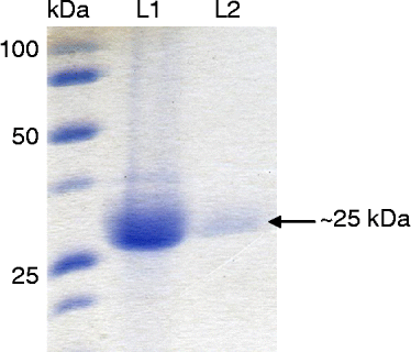
The LAP DNA sequence amplified in this study showed 100% identity with the selected LAP fragment (AAV59016.1). In contrast, the CL1 amplified sequence had 91% identity to the selected fragment of CL1 (CAC12806.1). Sixteen mutations (8 transitions and 8 transversions) were observed, 6 of which were synonymous and 10 non-synonymous. Comparison of the CL1 fragment with different sequences of F. hepatica CL1 reported in GenBank showed 91–98% identity. Identity with other L cathepsins (L2, L3, L4, L6) was below 80%. Three epitopes were consistently identified by the antigenic prediction software: DGRVVHLKY at position 53–61 corresponds to LAP, while the epitopes VTGYYTVHSGSEVELKNLV at position 118–136 and YQSQTCLPF at position 160–168 belong to CL1. The recombinant chimeric gene was expressed in E. coli. Western blot analysis showed expression of the ~25 kDa recombinant protein rFhLAP-CL1 (fig. 2). A time-course study for optimizing the level of protein expression showed that 15 h of growth of bacteria post-induction was sufficient for optimal expression. Likewise, 0.25 mm IPTG induced optimal protein expression, when concentrations of 0.25, 0.5 and 1 mM were checked for protein induction.
Expression of the rFhLAP-CL1 chimeric protein in E. coli strain Rosetta (DE3). Lanes: 1, protein marker (kDa); 2, rFhLAP-CL1 induced with 0.25 mm IPTG in culture for 16 h; 3, no induction.
Functionality of the rFhLAP-CL1 protein
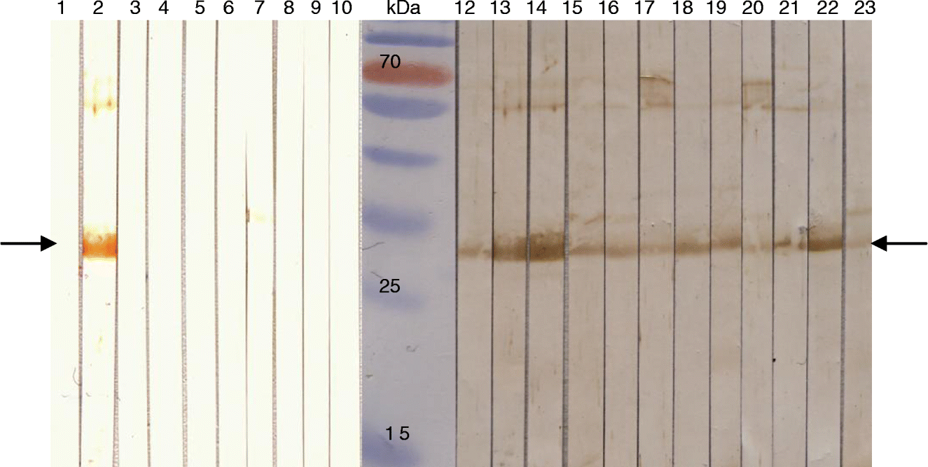
Recombinant rFhLAP-CL1 detected F. hepatica field infection in grazing livestock by Western blotting. Twelve cattle sera samples were collected in Chiquimitio, Michoacán. All positive samples recognized the chimeric protein, whereas negative samples did not recognize the ~25 kDa protein, indicating 100% sensitivity (fig. 3).
Recognition of rFhLAP-CL1 (arrowed at 25 kDa) in sera of infected cattle. Lanes: 1, negative control sera; 2, positive control; 3–10, bovine serum negative for F. hepatica; 11, protein molecular weight marker; 12–23, bovine serum positive for F. hepatica.
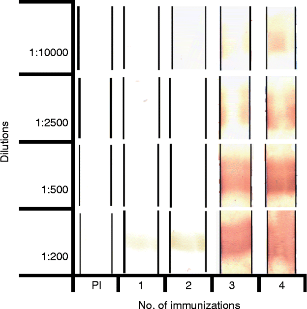
Rabbit pre-immune sera showed no reactivity to the rFhLAP-CL1 protein in Western blot assays. After the first immunization, specific antibodies against rFhLAP-CL1 were detected at a dilution of 1:200. The highest titre, 1:10,000 was reached after the fourth immunization. These results showed that the chimeric protein was highly immunogenic and coincided with the theoretical design. It may therefore have protective effects. Additional studies are needed to further define the immunogenicity of the chimeric protein (fig. 4).
Protein recognition in sera of rabbits immunized with 100 μg of purified antigen using Western blot; pre-immune sera (PI).
Discussion
Chimeric proteins are a new generation of recombinant products designed to replace native antigens. They may have several immunoreactive epitopes from different antigens, conferring enhanced immunogenicity (Holec-Gasior et al., Reference Holec-Gasior, Ferra and Drapala2012). Chimeric proteins can be designed to include protein epitopes from different stages of the parasite life cycle. Other designs may include fragments of proteins that are candidates for subunit vaccines, which may have greater protective effects in combination.
For diagnostics, the major advantage of using chimeric antigens rather than existing commercial assays would be to have a more ‘standardized’ antigen with high specificity and sensitivity for detection of antibodies (Holec-Gasior et al., Reference Holec-Gasior, Ferra and Drapala2012). In this study, the LAP and CL1 enzymes of F. hepatica were selected due to their protective effects. After in silico analyses of these proteins, the most antigenic regions were selected and incorporated into the recombinant chimeric protein. To include the most antigenic regions, three consensus epitopes (one from LAP and two from CL1) were included in the chimeric protein.
In the chimeric rFhLAP-CL1 sequence, no differences were found between the partial sequence of LAP and the sequence reported by Acosta et al. (Reference Acosta, Cancela, Piacenza, Roche, Carmona and Tort2008). However, differences were observed between the partial sequence of CL1 and the sequence reported by Cornelissen et al. (Reference Cornelissen, Gaasenbeek, Borgsteede, Holland, Harmsen and Boersma2001). These differences may represent a mechanism by which the parasite evades the host immune system or helps the trematode invade new hosts, as suggested by Sajid & McKerrow (Reference Sajid and McKerrow2002), Irving et al. (Reference Irving, Spithill, Pike, Whisstock and Smooker2003) and Robinson et al. (Reference Robinson, Tort, Lowther, Donnelly, Wong, Xu, Stack, Padula, Hebert and Dalton2008).
Antigenicity of the chimeric protein rFhLAP-CL1 was confirmed by Western blot. Indeed, antibodies present in sera from trematode-positive cattle recognized the chimeric protein. Of the two enzymes, CL1 has been the most fully evaluated in diagnostic techniques. O'Neill et al. (Reference O'Neill, Parkinson, Strauss, Angles and Dalton1998) developed a serological diagnosis (enzyme-linked immunosorbent assay (ELISA)) in humans, using the native enzyme to detect antibodies to Fasciola; the test detected 21% of sera with fasciolosis. A diagnostic ELISA with recombinant CL detected antibodies against F. hepatica in 100% and 99.1% of samples of sera from cattle and sheep, respectively (Cornelissen et al., Reference Cornelissen, Gaasenbeek, Borgsteede, Holland, Harmsen and Boersma2001). In the present study, the chimeric protein was able to differentiate between positive and negative sera. Therefore, this protein may be used as a tool for diagnosing fasciolosis.
We also determined that in rabbits the rFhLAP-CL1 protein was immunogenic due to the ability to induce a strong humoral immune response when high IgG titres were observed. These results agreed with those of Golden et al. (Reference Golden, Flynn, Read, Sekiya, Donnelly, Stack, Dalton and Mulcahy2010), who observed high titres of IgG in cattle immunized with recombinant CL1. By selecting the most antigenic regions of both enzymes, immunogenicity may be increased relative to either protein alone.
The F. hepatica rFhLAP-CL1 protein was constructed to increase the detection of each antigen (CL1 and LAP) for diagnosis and to provide greater stimuli for the immune system by concentrating antigenic regions in one protein. Construction of chimeric proteins is a viable and effective strategy for capitalizing on the antigenic and immunogenic properties of two or more proteins that are not bound in nature. Further assessment is needed to determine immunoprotective capabilities of the rFhLAP-CL1 protein in a natural host of the parasite. However, the F. hepatica rFhLAP-CL1 protein constructed in this study could be used as a tool for serological diagnosis of fasciolosis and serve as an immunogen to induce protection in natural hosts.
Acknowledgements
We would like to thank Dr Clara I. Espitia Pinzon and M.C. Cristina Parada Colin of the Departamento de Inmunología del Instituto de Investigaciones Biomédicas, FMVZ-UNAM, for their support.
Financial support
This study was co-financed by Dirección General Asuntos del Personal Académico-Programa de Apoyo a Proyectos de Investigación e Innovación Tecnológica (DGAPA-PAPIIT), Universidad Nacional Autónoma de México (UNAM) IN-219006, IN 219708, IT 230011, Consejo Nacional de Ciencia y Tecnología (CONACyT) and Programa de Apoyo para la Superación del Personal Académico-Universidad Nacional Autónoma de México (PASPA-UNAM).
Conflict of interest
None.
Ethical standards
The authors assert that all procedures contributing to this work comply with the ethical standards of the national and institutional guides on the care and use of laboratory animals (Reglamento para el uso del bioterio, Instituto de Investigaciones Biomédicas, Universidad Nacional Autónoma de México; Código ético para el Personal Académico del Instituto de Investigaciones Biomédicas, Norma Oficial Mexicana NOM-062-ZOO-1999).