Introduction
Reactive oxygen species (ROS) such as superoxide anion (1O2·−), hydrogen peroxide (H2O2), and hydroxyl radical (·OH) are responsible for many physiologic cellular disorders in humans.[Reference Veal, Day and Morgan1–Reference Tredwin, Naik, Lewis and Scully4] Of a particular concern is the interconvertibility[Reference Tredwin, Naik, Lewis and Scully4–Reference Alnuaimi, Rauf and Ashraf6] of the reactions that involve the production of these destructive chemical entities which also causes synergetic effects on the weights of damage inflicts on susceptible cellular targets. For example, superoxide anion converts to hydrogen peroxide readily in the presence of suitable hydrogen ion donors[Reference Veal, Day and Morgan1, Reference Ormond and Freeman7, Reference Huang, Chen, Dong, Luo, Yu, Moore and Bey8] while hydrogen peroxide transforms to reactive hydroxyl and hydro-peroxyl radicals in the presence of Fe2+ and Fe3+ ions, respectively, under Fenton reaction mechanism.[Reference Young, Fairley, Mohan and Jumeaux5, Reference Alnuaimi, Rauf and Ashraf6, Reference Huang, Chen, Dong, Luo, Yu, Moore and Bey8] However, among these ROS, H2O2 seems to have attracted considerable attention. This is attributed to the fact that, its presence could be linked to the existence of other ROS[Reference Veal, Day and Morgan1, Reference Tredwin, Naik, Lewis and Scully4, Reference Huang, Chen, Dong, Luo, Yu, Moore and Bey8, Reference Yang, Zhao and Liu9] or some chemical agents (e.g., glucose or cholesterol)[Reference Gu, Wang, Schultz and Camden10–Reference Chen, Cai, Ren, Wen and Zhao13] responsible for the promotion of certain physiologic diseases. In addition, H2O2 seems to be the only species out of all the ROS that has the capability to diffuse[Reference Veal, Day and Morgan1, Reference Marinho, Real, Cyme, Soares and Antunes14–Reference Naik, Tedwin and Scully16] relatively large distances within cells or across membranes, creating a conducive atmosphere for outburst of in situ oxidative stress in adjacent cells and tissues.[Reference Marinho, Real, Cyme, Soares and Antunes14, Reference Kaunig, Wang, Pu and Zhou15, Reference Franco, Schoneveld, Georgakilas and Panayiotidis17, Reference Kryston, Georgiev, Pissis and Georgakilas18] This has resulted in several cellular dysfunctions such as malignant revolutions,[Reference Kaunig, Wang, Pu and Zhou15, Reference Franco, Schoneveld, Georgakilas and Panayiotidis17–Reference Ziech, Franco, Pappa and Panayiotidis19] cardiovascular diseases, neurodegenerative disorders,[Reference Veal, Day and Morgan1] and aging.[Reference Marinho, Real, Cyme, Soares and Antunes14]
Possible sources of exposure to H2O2’s contamination include occupational activities emanating from textile processing, wine distillation, and sewage treatment environments[Reference Tredwin, Naik, Lewis and Scully4, Reference Naik, Tedwin and Scully16] as well as handling of hydrogen peroxide-containing pharmaceutical products (HPCPs), e.g., mouth rinses, disinfectant solutions, and teeth whitening kits such as carbamide peroxide gel and hydrogen peroxide strips, especially for oral health and aesthetic values.[Reference Naik, Tedwin and Scully16, Reference Consolaro20, Reference Sulieman, Addy, Macdonald and Rees21] However, among these sources, utility of HPCPs is of higher risks as many of these products are commercially available as self-administering drugs which are handled by many non-professional consumers.[Reference Tredwin, Naik, Lewis and Scully4, Reference Consolaro20] For example, 10% carbamide peroxide (about 3–5% H2O2) and 30–35% hydrogen peroxide solutions have been reported to exhibit effective teeth whitening functions[Reference Naik, Tedwin and Scully16, Reference Sulieman, Addy, Macdonald and Rees21] in association with adverse consequences such as tooth sensitivity and gingival irritation.[Reference Tredwin, Naik, Lewis and Scully4, Reference Naik, Tedwin and Scully16] In addition, 3% H2O2 solutions used as disinfectant to treat wound was reported to cause increase injury to damaged tissues, thus delaying healing.[Reference Naik, Tedwin and Scully16] Although the pharmacologic levels of H2O2 in these products are perceived to be safe for human's use under suitable management conditions, reports have shown that these materials have devastating effects on health, especially over persistent prolong usage[Reference Tredwin, Naik, Lewis and Scully4, Reference Naik, Tedwin and Scully16] as well as after accidental discharge of H2O2 on tissues.[Reference Tredwin, Naik, Lewis and Scully4] The major factor responsible for the utility of drugs over many cycles is incomplete therapies due to administration of lower doses.[Reference Sulieman, Addy, Macdonald and Rees21] Many HPCPs are susceptible to degradation under different light[Reference Alnuaimi, Rauf and Ashraf6] and heat[Reference Young, Fairley, Mohan and Jumeaux5] conditions, which further lower their required concentrations for effective therapeutic functions[Reference Young, Fairley, Mohan and Jumeaux5] and thus subsequently promote their extensive usage over time. As a result, it is essential to monitor the levels of H2O2 in commercially-available HPCPs to safeguard against overexposure to H2O2 contamination and thus possible cellular oxidative stress.
In the past years, various methods employed to detect and quantify H2O2 in various environments include electrochemical sensing, luminescence spectroscopy,[Reference Yang, Zhao and Liu9, Reference Huan, Fei, Shan, Wang, Xu and Feng22, Reference Chen, Fang, He, Zhang and Yao23] and plasmonic-surface-enhanced Raman spectroscopy.[Reference Gu, Wang, Schultz and Camden10] However, many of these techniques are expensive to manage,[Reference Chen, Cai, Ren, Wen and Zhao13] requires frequent calibrations for optimal performances, exhibit prolong operational times,[Reference Chen, Cai, Ren, Wen and Zhao13] and necessitate specialized skills to operate. As a result, low-cost, rapid, and sensitive alternative techniques are urgently needed for the detection and monitoring of H2O2 in aqueous environments.
Many iron-core porphyrin systems (e.g., heme proteins such as catalase, horseradish peroxidase, cytochrome c, and hemoglobin) exist in nature as excellent biosensors of hydrogen peroxide and have been reported to exhibit great potentials for enhancing the electron transfer kinetics of electrochemical sensing of H2O2 mechanism owing to their great redox capabilities.[Reference Chen, Cai, Ren, Wen and Zhao13] Also, the use of methylene blue-sodium dodecylsulphate-multiwalled carbon nanotube has been reported for detecting H2O2 with excellent sensing capability.[Reference Tiwari and Singh11] However, to the best of our knowledge, none of these reports has featured combined iron (III) meso-tetrakis(4-hydroxyphenyl)porphyrin-methylene blue hybrid complex for the detection and sensing of H2O2 using advanced colorimetry stripping technique. In this study, we present a low-cost, rapid visual colorimetric detection and advanced color stripping quantification of H2O2 in aqueous solution based on interaction of iron (III) complex of meso-tetrakis(4-hydroxyphenyl)porphyrin-methylene blue hybrid (Porphyrin-Blue) with H2O2. The complex was immobilized on environmental-friendly disposable paper materials to make color evolution (indicator) strips, making the technique relatively low-cost in nature. The generation of ROS’ radicals (HOO· and HO·) from the interaction of Fe3+ of the complex with H2O2, which subsequently interact with the background dye system to produce variant color evolutions, forms the basis of the present H2O2 detection and quantification mechanism. Furthermore, the present approach was used to quantify the true level of H2O2 in a self-administering 30%-labeled commercially-provisioned hydrogen peroxide disinfectant solution to demonstrate its practical applicability. The stripping method involves utilization of the red, green, and blue (RGB) color mode evolution pattern of a common free image editing software (GIMP 2.8.20) to detect and quantify various reference concentrations of H2O2 from 0% to 30%, while corresponding calibrated pixels counts based on the different color evolutions were used as sources of quantification in the real pharmaceutical product sample. To the best of our knowledge, no such technology exists in the literature, making this approach a new low-cost technique for the detection and quantification of H2O2 in aqueous and pharmaceutical solutions.
Experimental
Materials
All chemicals (Sigma Aldrich, South Africa) were analytical grades and used without further purification. The meso-tetrakis(4-hydroxyphenyl)porphyrin was synthesized following our previously reported method.[Reference Fakayode, Songca and Oluwafemi24] Deionized water was used for all aqueous solution preparations.
Development of iron (III) porphyrin-methylene blue dye strips
Solutions of iron (III) chloride (0.0202 g/5 mL water), porphyrin (5 mL of 0.0237 g/100 mL), and methylene blue (5 mL of 0.0082 g/250 mL ethanol) were added together in a 250 mL Pyrex beaker to obtain a green dye hybrid solution. This was followed by immersion of a Whatman No. 1 filter paper (90 mm) into the dye solution for 1 h 30 min. Afterwards, the filter paper was removed and dried under ambient condition. Finally, the dried paper was cut into rectangular strips, followed by storage under ambient condition for further studies.
Characterization of the dye complex
The dye complex was characterized using ultraviolet-visible spectrophotometry [Perkin Elmer UV-V is Lambda 25 spectrometer (UK); 1 nm slit width] and Fourier transform infrared spectroscopy (FTIR) (Spectrum two UATR spectrometer, Perkin Elmer, UK).
H2O2 detection
Calibrating test strips
Each test strip was dipped into a 5 mL test solution containing a given amount of reference H2O2 concentration (between 0% and 30%) and allowed to react for 10 min, followed by drying under ambient condition (room temperature 19.5 °C) for 15 min. At this time, the image of the test strip was captured using a camera. Deionized water was used as control (0% H2O2).
Detection of H2O2 in real pharmaceutical fluid sample
A test strip was dipped into a 5 mL pharmaceutical 30%-labeled H2O2 disinfectant solution and allowed to react under the same conditions as “Calibrating test strips” above.
Strip color analysis
The color of each paper strip was evaluated using the histogram of the RGB mode of image editor GIMP 2.8.20 for better color discrimination. Each strip was sampled from edge to edge using 84 × 102 (pixel × pixel) fixed size aspect ratio with characteristic 25,704 total pixel counts, 96 ppi resolution, and RGB color values ranging from 0 to 255.
Results and discussion
Synthesis and characterization of the complex
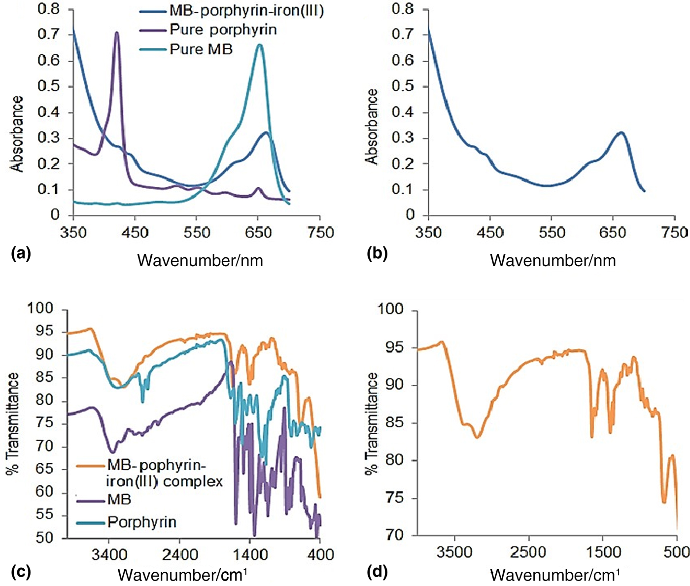
The complex evolved as a green solution after mixing the three precursor solutions. The UV-visible absorption spectra of the aqueous solution of the complex showed a typical iron(III)-porphyrin aggregate signal[Reference Huan, Fei, Shan, Wang, Xu and Feng22] with bathochromically shifted Soret band from 422 nm to bimodal peaks at 433 and 435 nm together with a broad Q-band between 470 and 535 nm, signifying molecular symmetry transformation from free base rectangular D 2h to metallo-structural square D 4h configurations,[Reference Hashimoto, Choe, Nakano and Hirao25] characteristic of S 0 → S 1 transitions, respectively.[Reference Huan, Fei, Shan, Wang, Xu and Feng22, Reference Hashimoto, Choe, Nakano and Hirao25]
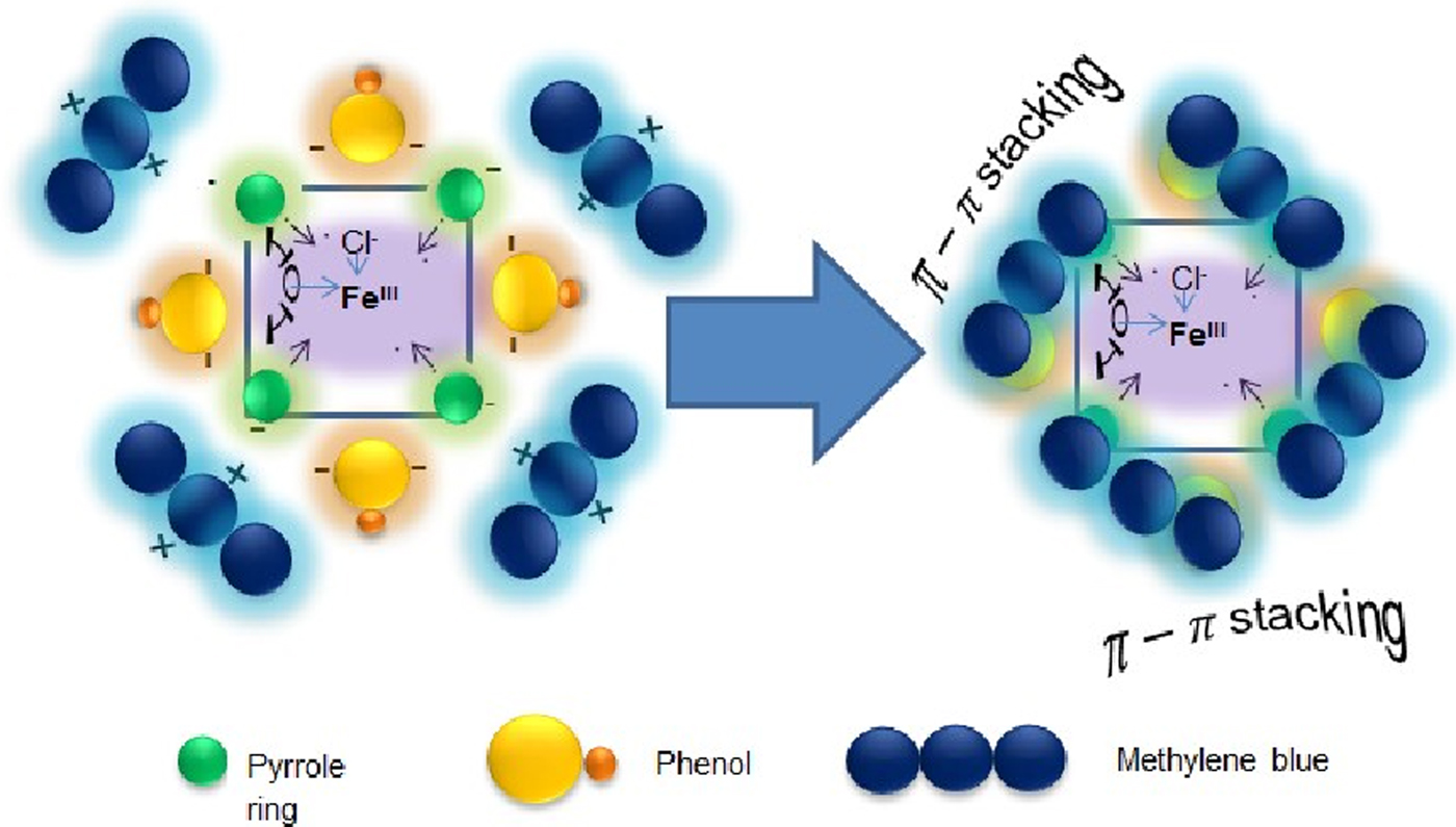
In addition, the methylene blue peak was bathochromically shifted from 652 to 663 nm [Figs. 1(a) and 1(b)]. The results from the FTIR analyses of the complex revealed major characteristic vibrational band peaks at 3423, 3261, 1654, 1414, and 692/cm corresponding to intermolecular hydrogen bonded phenolic hydroxyl group of the porphyrin, 2° ammonium ion (= N+–H) group of the protonated methylene blue, C = Cstr of porphyrin and methylene blue, O–Hbend of phenols and water (from aqua complex of FeIII) and Fe3+–Ostr of aqua complex of FeIII, respectively. This confirmed the possible association of the porphyrin, methylene blue, and FeIII within the complex. Typically, methylene blue and porphyrin may associate together via π − π electronic stacking, hydrogen bonding, and hydrophobic interactions[Reference Tiwari and Singh11, Reference Gao, Liu, Yin, Li, Bai and Zhang26] (Scheme 1). Furthermore, aromatic structures such as phenols and pyrroles may have negative charges on their ring structures due to resonance hybridization. These features readily attract the positively charged methylene blue via its ammonium ( = N+–H) or sulphonium ( = S+) ion site leading to the assembling of methylene blue molecules on porphyrin's structure (Scheme 1). These coupling interactions probably contributed to the red shiftings and reduction in the intensities of the O–Hstr and N–Hstr of the porphyrin and methylene blue, respectively, in the complex relative to their pristine state [Figs. 1(c) and 1(d)].
UV-visible absorption spectra of the aqueous solution of the complex and precursors: porphyrin in methanol, methylene blue (MB) in ethanol (a), UV-visible absorption spectra of the aqueous solution of the complex (b), FTIR spectra of the complex and precursors (c), and FTIR spectra of the complex (d).

Model demonstrating π–π aromatic stacking between the methylene blue molecules and phenolic and pyloric systems of the porphyrinoid iron (III) complex.

Detection of H2O2
Calibration of test strips
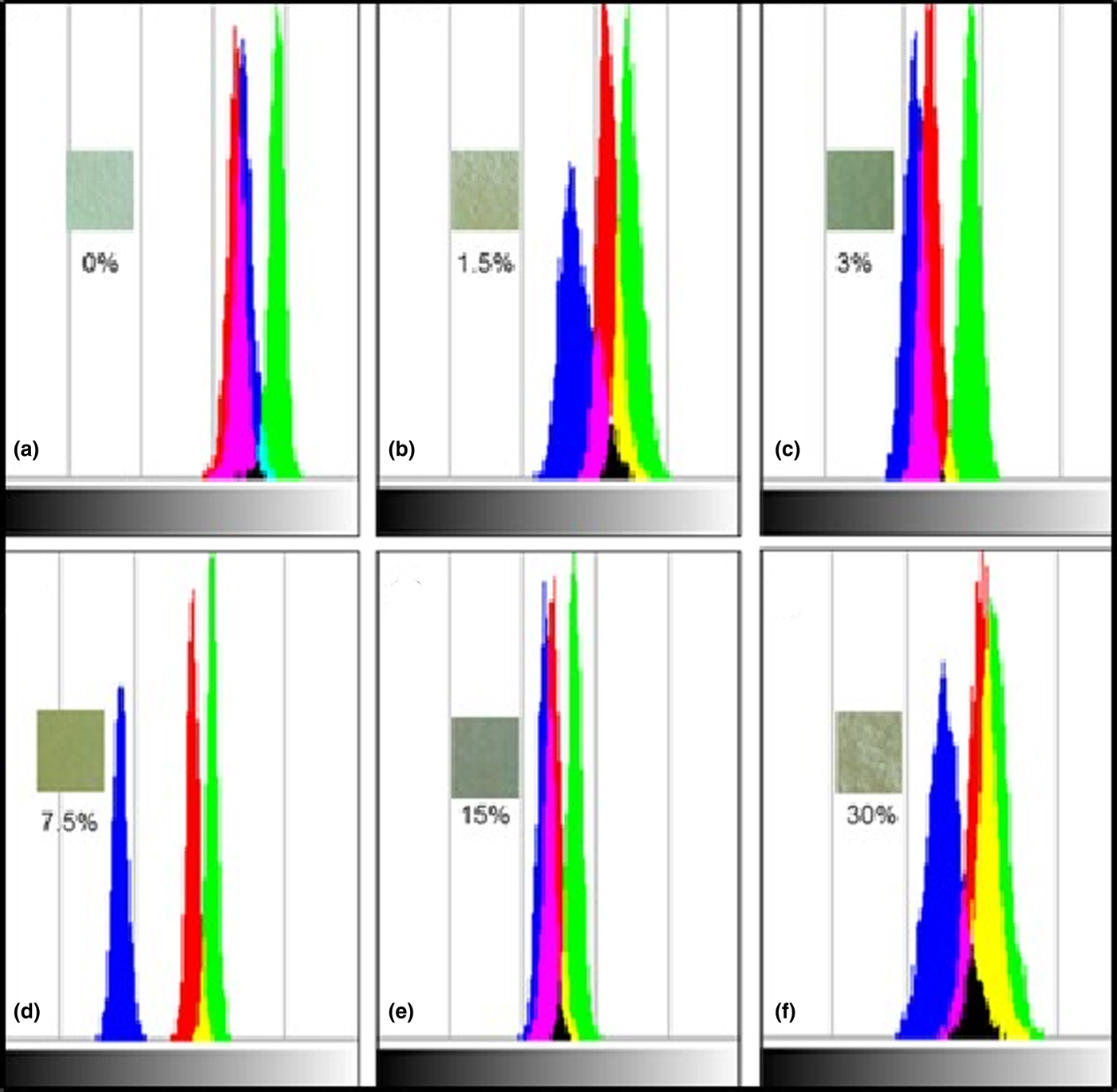
The results of the interaction of the test strips with different H2O2 concentrations are shown in Fig. 2. According to Fig. 2, each strip displayed different RGB colors with characteristic different orientations, patterns, and intensities, based on the different concentrations of H2O2.
Color discriminations with respect to different concentrations of H2O2. Insets: Test strips with different color evolutions at different H2O2 concentration of (a) 0%, (b) 1.5%, (c) 3%, (d) 7.5%, (e) 15%, and (f) 30%.
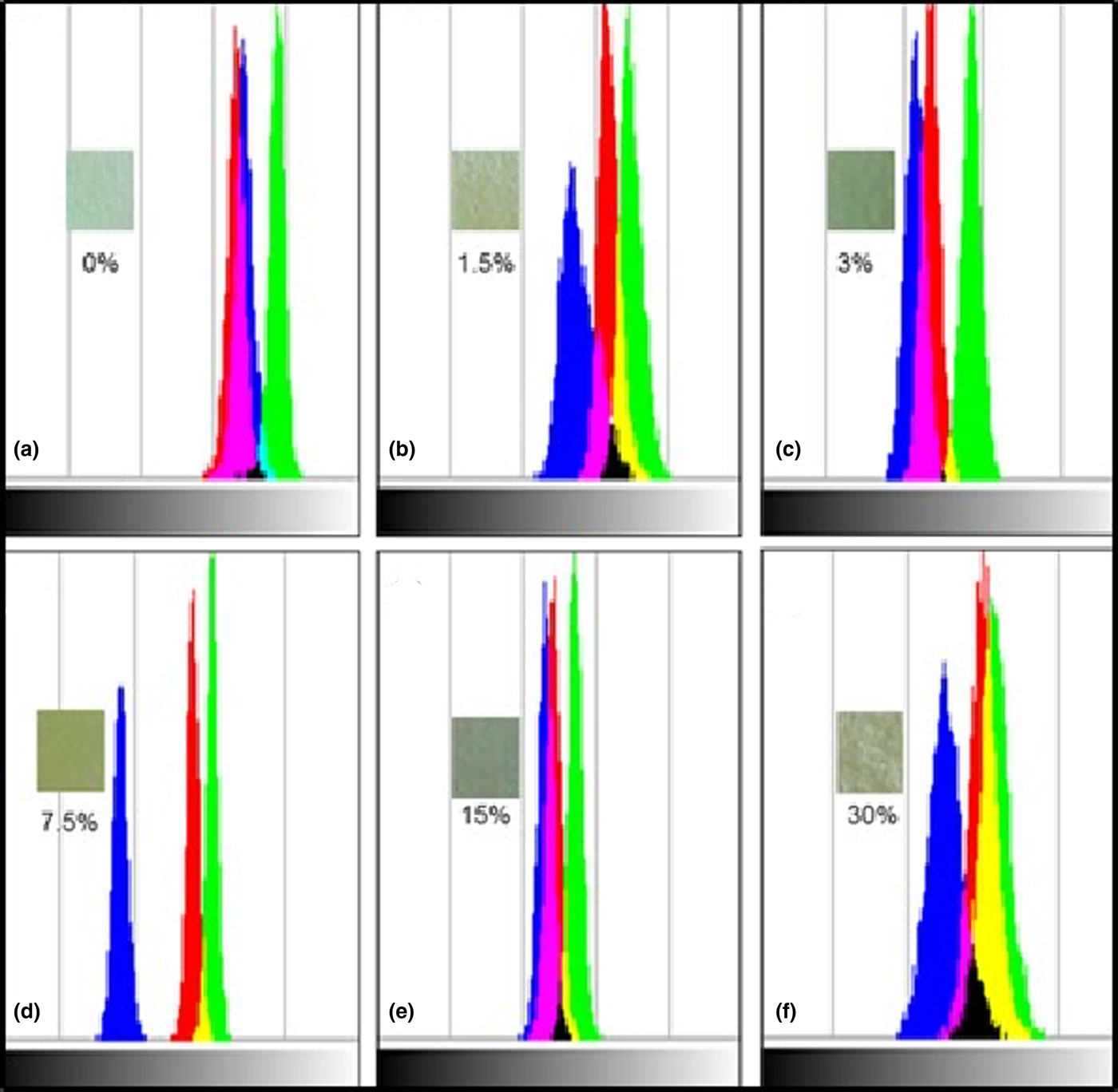
In addition, some strips such as 1.5%, 3%, 15%, and 30% displayed magenta, yellow, and black, while 7.5%-strip and the control (0%-strip) displayed yellow and black and cyan colors, respectively, in addition to the RGB colors. Collectively, these features enabled easy and reliable discrimination between the different concentrations of the H2O2.
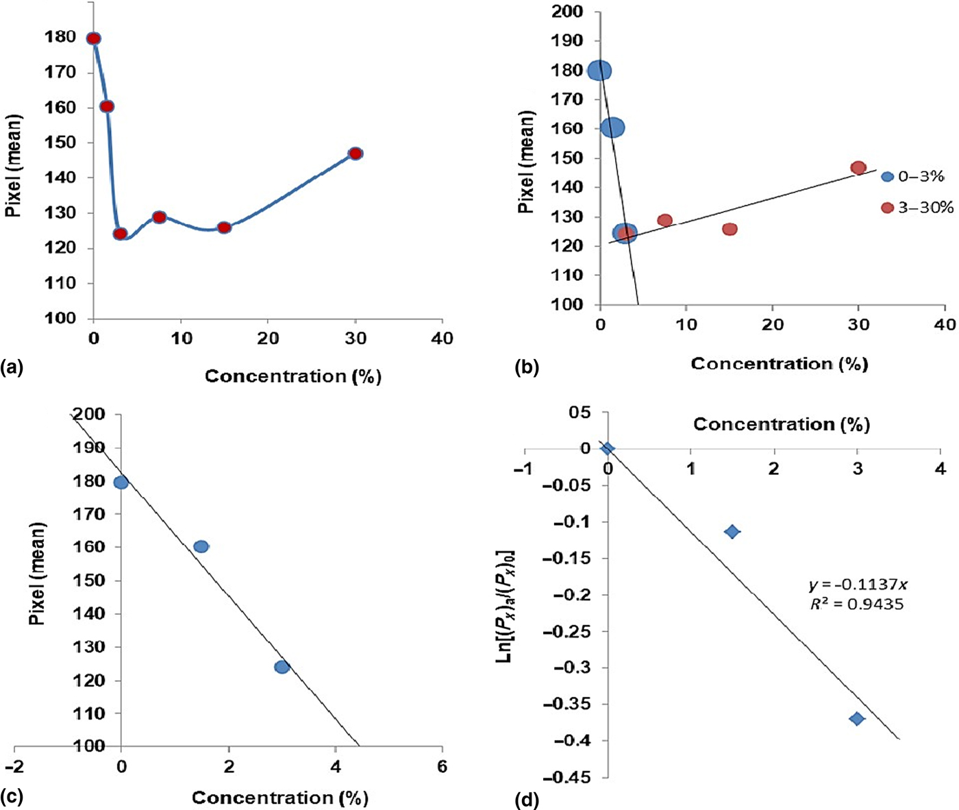
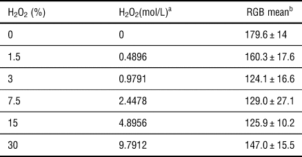
The plot of mean pixels counts versus concentration of the H2O2 solutions is shown in Figs. 3(a)–3(c). According to Fig. 3(a), the color of the pixel values of the strips decreased with increasing concentration of the H2O2 between 0% and 3% (0–1 M) and then increased beyond this point up to 30% (9.7912 M) with a slight deviation at the 15% (4.8956 M). This curve resembled a typical amperometric titration curve with two linear ranges between 0% and 3% (0–1 M) and 3% and 30% (1–10 M). However, 0–3% showed better linearity (R 2 = 0.97) than the 3–30% range (R 2 = 0.8274) with a detection limit (LOD) of 2.27% (0.7409 M). The latter was estimated using the expression 3σ/s (where σ = standard deviation of the blank and s = slope of the calibration graph).[Reference Gu, Wang, Schultz and Camden10, Reference Tiwari and Singh11] Furthermore, the 0–3% linear range could be described using first-order exponential expression as shown in Eq. (1) and Fig. 3(d):
 $$(P_x)_a/(P_x)_0 = e^{-kc},$$
which linearized to give Eqs. (2) and (3)
 $$\ln (P_x)_a - \ln (P_x)_0 = -kc$$
or
 $$\ln [(P_x)_a/(P_x)_0] = -kc,$$
where (P x)a = mean pixel counts for a given H2O2 concentration, (P x)0 = mean pixel count before decay, k = first-order decay rate constant, and c = concentration of H2O2 in mol/L (Table I).
Exponential relationship between Pixel mean and H2O2 concentration (a, b), calibration curve for 0–3% linear region (c), and first-order linear plot for the 0–3% range (d).

Data for the plots of the exponential decay and calibration curve.

a Density = 1.11 g/cm3; molar mass = 34.01 g/mol.
b Pixel counts = 25,704 for 84 × 104 sampling.
A similar observation has been reported by Sulieman et al. which observed that the number of applications to achieve uniform tooth whitening effect decreased exponentially with increasing H2O2 concentration.[Reference Sulieman, Addy, Macdonald and Rees21] The 0–3% linear range in this study indicates that this approach has great potential to be used for the quantification of hydrogen peroxide in food drinks and biologic fluids which usually accommodate low H2O2 concentrations.[Reference Gu, Wang, Schultz and Camden10, Reference Tiwari and Singh11]
H2O2 quantification in pharmaceutical disinfectant sample
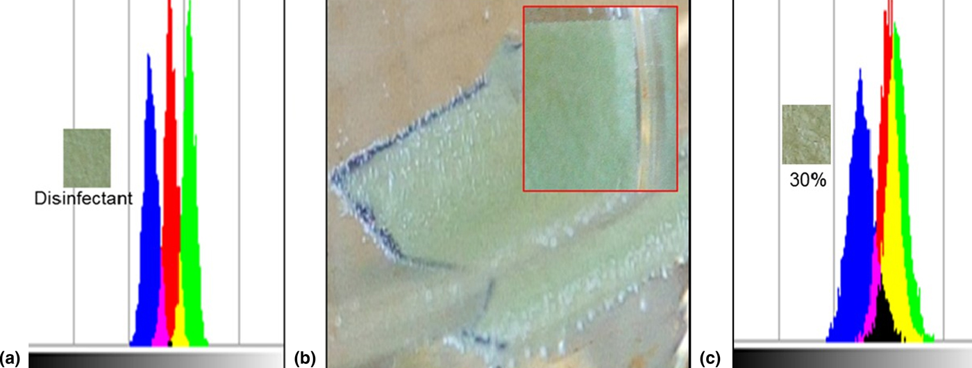
The result of the evaluation of H2O2 level in a pharmaceutical 30%-labeled H2O2 disinfectant solution is shown in Fig. 4(a). According to Fig. 4(a), the 30% H2O2-labeled pharmaceutical fluid showed RGB color pattern, which fall between 1.5% and 3.0% reference H2O2 patterns [see Figs. 2(b) and 2(c)]. This observation agreed with the quantification value of 0.7466 mol/L (i.e., 2.29%, Pixel mean = 140.1 ± 15.5) estimated from the calibration curve [Fig. 3(c)]. The lower concentration detected in this sample (about 92.37% deviation from the label) may be due to the exposure of the disinfectant solution to heat and light within the commercial environment in which it was purchased which have the potential to degrade H2O2 in solutions.[Reference Young, Fairley, Mohan and Jumeaux5] One proof that showed that the commercial product had deviated from its labeled concentration was the inability of the solution to generate gas bubbles after being in contact with the test strip [inset, Fig. 4(b)] whereas the 30%––reference H2O2 solution exhibited visible evolution of gas [Fig. 4(b)]. In addition, the RGB color pattern [Fig. 4(a)] of the pharmaceutical fluid was totally different from that of the reference [Fig. 4(c)].
RGB color discrimination of 30% H2O2-labeled pharmaceutical disinfectant (a), digital image showing gas evolution with 30% H2O2 reference solution (b), inset: digital image showing absence of gas evolution in the 30% H2O2-labeled pharmaceutical disinfectant solution, and (c) RGB color discrimination of 30% H2O2 reference solution.

Thus, this study shows that constant routine monitoring of hydrogen peroxide levels in HPCPs such as disinfectant solutions and teeth whitening products is highly essential to ensure reliability of the product's concentration as deviation from these labels may have occurred over exposure of these products to ambient heat and light conditions. This consequentially may enforce the applicability of these materials over many cycles of time which may lead to toxicity consequences.[Reference Tredwin, Naik, Lewis and Scully4, Reference Naik, Tedwin and Scully16] In addition, it is essential that, such products should be stored under refrigeration in the dark to avoid accidental degradation.
Proposed detection mechanism
The proposed mechanism of the detection of H2O2 using the current approach is given in Eqs. (4)–(8).
 $$\eqalign{&{\rm PO}{\rm P}^{\rm a}-{\rm M}{\rm B}^{\rm b}-{\rm F}{\rm e}^{{\rm III}} + {\rm HO}-{\rm OH} \cr & \qquad = {\rm POP}-{\rm MB}-{\rm F}{\rm e}^{{\rm II}} + {\rm HOO}\cdot + {\rm H}^{+}}$$
 $$\eqalign{{\rm POP}-{\rm MB}-{\rm F}{\rm e}^{{\rm III}} + {\rm HOO}\cdot & = {\rm POP}-{\rm MB}-{\rm F}{\rm e}^{{\rm II}} + {\rm O}_{\rm 2} \cr & \quad + {\rm H}^{+}}$$
 $$\eqalign{&{\rm POP}-{\rm MB}-{\rm F}{\rm e}^{{\rm II}} + {\rm HO}-{\rm OH} \cr & \qquad = {\rm POP}-{\rm MB}-{\rm F}{\rm e}^{{\rm III}} + {\rm HO}\cdot + {\rm H}{\rm O}^{-}}$$
 $$\eqalign{&{\rm POP}-{\rm MB}-{\rm F}{\rm e}^{{\rm III}} + {\rm HOO}\cdot /{\rm HO}\cdot \cr & \qquad = {\rm POO}{\rm P}^{\rm c} + {\rm MOO}{\rm B}^{\rm d} + {\rm F}{\rm e}^{{\rm III}}}$$
 $${\rm F}{\rm e}^{{\rm III}} + {\rm H}{\rm O}^- = {\rm Fe(HO}{\rm )}_{\rm 3}.$$
Note:
aPOP––porphyrin; bMB––methylene blue; cPOOP––oxidized porphyrin; dMOOB––oxidized methylene blue. The evidence of this proposed mechanism is further shown in Charts 1(a) and 1(b).
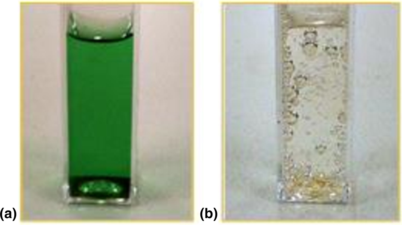
Green solution of the iron (III) meso-tetrakis(4-hydroxyphenyl)porphyrin-methylene blue complex (a) and the resulting solution after addition of a few drops of 30% H2O2 solution (b).

According to Eqs. (4)–(8), ferric ion oxidized H2O2 to hydro-peroxyl radical[Reference Young, Fairley, Mohan and Jumeaux5] which in turn further reacted with the ferric ion to generate oxygen gas[Reference Young, Fairley, Mohan and Jumeaux5, Reference Alnuaimi, Rauf and Ashraf6] [Figs. 4(b); Charts 1(a) and 1(b)]. The ferrous ion generated as a result of this reaction reacted with more H2O2 to produce hydroxyl radical[Reference Young, Fairley, Mohan and Jumeaux5, Reference Alnuaimi, Rauf and Ashraf6, Reference Alnuaimi, Sauf and Ashraf27] which together with hydro-peroxyl radical degraded the color of the porphyrin-methylene blue on the test strips. Finally, the FeIII remaining in the solution precipitated as yellow-orange Fe(OH)3 as shown in Chart 1. Since the extent of color degradation depends on the concentration of H2O2 in the test solution, a feasible quantification procedure was established.
Conclusion
Ferric chloride solution of meso-tetrakis(4-hydroxyphenyl)porphyrin-methylene blue was immobilized on a paper material to make indicator test strips for colorimetric discrimination of different concentration of H2O2 in aqueous solution. The UV-visible absorption and FTIR spectra confirmed the presence of metallated porphyrin stacked with methylene blue. This configuration enabled the feasible interaction between the core FeIII ion and H2O2 to produce reactive oxygen––radicals which degraded the color of the porphyrinoid–– methylene blue dye. This current approach was extended to determine the true concentration of H2O2 in a commercial 30%-labeled H2O2 disinfectant solution. The result showed about 92.37% deviation from the labeled quantity which showed the need for regular monitoring of the H2O2 level in H2O2-containing pharmaceutical fluids. The current technique may be extended for the detection and quantification of H2O2 in biologic fluids and for monitoring the levels of H2O2 in environmental waters, foods, drinks, biologic media, and self-administering pharmaceutical and industrial-based H2O2-containing solutions.
Acknowledgments
The authors are grateful to the National Research Foundation (NRF), South Africa under the Nanotechnology Flagship Programme (Grant no: 97983) and Competitive Programme for Rated Researchers (Grant no: 106060) for financial support.