

Milk secretion rate in various mammals is tightly associated with the fluctuating nutritional demands of offspring, and mammary gland (MG) development stages are closely related to the nurse's reproduction cycles (Oftedal, Reference Oftedal1984; Wilde & Peaker, Reference Wilde and Peaker1990). Regulation of day-to-day variation in milk secretion (Wilde & Peaker, Reference Wilde and Peaker1990; Daly et al. Reference Daly, Owens and Hartmann1993) and induction of MG involution (Silanikove et al. Reference Silanikove, Merin and Leitner2006) are ruled by a milk-borne negative feedback (MBNF) system. The plasminogen activator (PA)-plasminogen (PG)-plasmin (PL) enzymic system is ubiquitously expressed in the milk of humans (Heegaard et al. Reference Heegaard, Larsen, Rasmussen, Hojberg, Petersen and Andersen1997), rodents (Tonner et al. Reference Tonner, Allan and Flint2000) and ruminants (Silanikove et al. Reference Silanikove, Merin and Leitner2006), and was found to be associated with regulation of milk secretion in both cows and goats (Silanikove et al. Reference Silanikove, Shamay, Shinder and Moran2000, Reference Silanikove, Shapiro and Shinder2009) and activation of involution in rodents (Tonner et al. Reference Tonner, Allan and Flint2000), goats (Shamay et al. Reference Shamay, Shapiro, Mabjeesh and Silanikove2002) and cows (Politis, Reference Politis1996; Shamay et al. Reference Shamay, Shapiro, Leitner and Silanikove2003; Silanikove et al. Reference Silanikove, Shapiro, Shamay and Leitner2005b).
As in other body tissues, PL is presented in milk mainly in its inactive zymogen form PG, whose conversion to PL is modulated by PAs (Politis, Reference Politis1996). The two types of PAs that exist in mammals systemic fluids, urokinase-type PA (u-PA) and tissue-type PA (t-PA) are also presented in milk (Heegaard et al. Reference Heegaard, Rasmussen and Andresen1994b; White et al. Reference White, Zavizion, O'Harea, Gilmore, Guoa, Kindstedt and Politis1995; Politis, Reference Politis1996; Tonner et al. Reference Tonner, Allan and Flint2000). In milk, PL, PG and t-PA are closely associated with the casein micelles, whereas u-PA is associated with neutrophils in close association with its specific receptor (Heegaard et al. Reference Heegaard, Rasmussen and Andresen1994b; Politis, Reference Politis1996).
The MBNF system associated with regulation of milk secretion was shown to comprise the PA-PG-PL system that specifically forms a β-casein (CN) fragment (f) (1–28) from β-CN, which in return serves as the negative control signal by closing potassium channels on the apical membrane of the epithelial cells of the MG (Silanikove et al. Reference Silanikove, Shamay, Shinder and Moran2000, Reference Silanikove, Shapiro and Shinder2009). Down-regulation of these channels induces undefined inwardly directed cellular signals that inhibit milk secretion. Interestingly, a further activation of the PA-PG-PL system, which was coupled to more extensive degradation of casein induced involution of the MG in lactating goats and cows and forcefully activated the innate immune system (Shamay et al. Reference Shamay, Shapiro, Mabjeesh and Silanikove2002, Reference Shamay, Shapiro, Leitner and Silanikove2003; Silanikove et al. Reference Silanikove, Shapiro, Shamay and Leitner2005b). Based on these findings, a casein hydrolysate (CNH) preparation was developed to reduce the suffering from MG engorgement associated with abrupt cessation of milking (the conventional procedure to induce involution in modern dairy cows) (Leitner et al. Reference Leitner, Jacoby, Maltz and Silanikove2007) and to treat and prevent common clinical and subclinical infections of the udder in dairy cows (Silanikove et al. Reference Silanikove, Iscovich, Leitner and Hogeveen2005a; Leitner et al. Reference Leitner, Jacoby and Silanikove2011a, Reference Leitner, Koren, Jacoby, Merin and Silanikove2012). It was also found that CN-derived peptides induced by PL activity inhibit milk clotting (Merin et al. Reference Merin, Fleminger, Komanovsky, Silanikove, Bernstein and Leitner2008; Fleminger et al. Reference Fleminger, Heftsi, Merin, Silanikove and Leitner2011, Reference Fleminger, Heftsi, Merin, Silanikove and Leitner2012), which is important during mastitis and milk stasis in preventing uncontrolled inflammation (Leitner et al. Reference Leitner, Merin and Silanikove2011b).
There is convincing evidence that under physiological situations, t-PA is the main factor involved in casein hydrolysis (Heegaard et al. Reference Heegaard, Christensen, Rasmussen, Benfeldt, Jensen, Sejrsen, Petersen and Andersen1994a; Politis, Reference Politis1996; Tonner et al. Reference Tonner, Allan and Flint2000). However, the picture remains unclear because: firstly, there are lots of data showing that u-PA is a major factor involved in CN hydrolysis in stored milk (Ismail & Nielsen, Reference Ismail and Nielsen2010); and secondly, the increase of u-PA secretion by MG epithelial cells under inflammation is claimed to be responsible for CN degradation in goats’ milk under mastitis (Heegaard et al. Reference Heegaard, Christensen, Rasmussen, Benfeldt, Jensen, Sejrsen, Petersen and Andersen1994a; Politis, Reference Politis1996; Weng et al. Reference Weng, Chang, Chen, Chou, Peh, Huang, Chen and Nagahata2006).
The association of t-PA with the CN micelles suggests that it constitutes a pivotal component of the MBNF system; however, as noted above, evidence for the involvement of u-PA confronts this assumption. The ability of CNH to up-regulate the PL system activity in a manner that imitates in an accelerated manner the events associated with MG involution stage I (Shamay et al. Reference Shamay, Shapiro, Mabjeesh and Silanikove2002; Silanikove et al. Reference Silanikove, Shapiro, Shamay and Leitner2005b) represent an opportunity to test the assumption regarding the pivotal role of t-PA in CN degradation under relevant physiological situations.
The aims of the present study were: firstly, to test the assumption that t-PA and PG embedded within casein micelles form a functional complex that rules casein hydrolysis, by analysing the distribution of the PL system in different fractions of bovine milk in control and in response to CNH treatment; secondly, to test the assumption that t-PA, PG and CN micelle are secreted by MG cells as functional units by verifying the presence of t-PA and PLG in CN micelles in freshly secreted milk; and thirdly, to exclude the role of u-PA in CN hydrolysis under the experimental conditions.
Materials and methods
H-D-Norleucyl-hexahydrotyrosol-lysine-pnitroanilide diacetate [Spectrozyme PL (SpecPL)], bovine PG, and cyanogens bromide fibrinogen digest (FIBGN) were purchased from American Diagnostica (Greenwich CT, USA). N-methylsulphonyl-D-Phe-Gly-Arg-4-nitroanilide acetate substrate was obtained from Boehringer Mannheim (Chromozym t-PA; UK, East Sussex, UK). Polyclonal rabbit anti-human t-PA IgG was obtained from Oxford Biomedical Research (Oxford, UK) and plasminogen activator inhibitor-1 (PAI-1), was from Calbiochem (USA). Other mentioned chemicals were purchased from Sigma (Rheovot, Israel).
Ethical considerations
All protocols were approved by the Institutional Animal Care Committee of the Agricultural Research Organization, which is the legitimate body for such authorizations in Israel.
Experiment 1
Six Israeli Holstein heifers with low leucocyte content, as indicated by low somatic cell count (<70 000 cells/ml) and no bacterial finding according to preliminary analysis (Leitner et al. Reference Leitner, Jacoby and Silanikove2011a), milk yield ∼36 litre/d, in their second to third lactation were used. Two MGs, one front and one rear quarter, were infused with sterile saline solution while the other two counter glands were infused with casein hydrolysate (CNH).
The experiment was carried out during November under natural lighting regimen, with typical noon temperatures of 24 °C and night temperatures of 12 °C, which is within the thermoneutral zone of cows (Silanikove, Reference Silanikove2000). The cows were milked thrice daily (5·30, 12·30 and 21·30) and milk yield and exact milking times were individually recorded automatically (Leitner et al. Reference Leitner, Koren, Jacoby, Merin and Silanikove2012).
All experimental procedures were carried out during the noon milking. Milk samples (100 ml) were taken from every gland of each cow at −24, 0, +24 h relative to treatment with saline solution and CNH, where 0 h refers to day of infusion. Milk samples on day 0 were taken prior to the infusion. On day 0, a dose of 10 ml of CNH (Leitner et al. Reference Leitner, Jacoby and Silanikove2011a), with a peptide concentration of ∼7 mg/ml was infused into each treated gland with a special applicator following careful sterile cleaning of the teat. The control glands of the cows in the first group were infused with 10 ml of sterile saline. Milk was discarded for 3 d following the infusion.
Analytical procedures
One set of samples (10 ml) was sent to the central laboratory for the determination of total protein, fat, lactose and somatic cell count (Silanikove et al. Reference Silanikove, Shapiro and Shinder2009). A second set of milk samples (2×10 ml) were defatted at 4 °C (Silanikove & Shapiro, Reference Silanikove and Shapiro2007) and analysed according to previously described procedures for: concentration of lactose, protein, fat, casein, whey protein, proteose peptones, lactoferrin, albumin, Na+ and K+ and the activity of xanthine oxidase, lactoperoxidase, and the concentration of nitrite (by the DAN reagent), nitrate (by the Griess reaction) and uric acid (Silanikove et al. Reference Silanikove, Shapiro, Shamay and Leitner2005b). A sub-set of skim milk was ultracentrifuged and clear milk serum (whey) devoid of membranous particles and casein micelle pellet were separated (Silanikove & Shapiro, Reference Silanikove and Shapiro2007).
A third set of samples (70 ml) was used to isolate somatic cells from milk. The samples were centrifuged at 2000 g at 4 °C for 30 min; then the fatty fraction and supernatant were removed. Cells from the bottom layer were suspended in 500 μl PBS (pH 7·4) containing 0·02% NaN3 and centrifuged twice (400 g at 4 °C for 15 min) to concentrate cells. After separation, the cells were lysed by at least 3 freeze–thaw cycles.
Activities of PL, PG, PA, t-PA and u-PA were determined in the whey and in the re-dissolved casein micelle pellets and isolated somatic cells (Ismail et al. Reference Ismail, Choi, Were and Nielsen2006). Whey, casein micelle pellets (1 mg/ml), cell lysates and other reagents were dissolved and diluted in 0·05 modified Tris buffer (MTB) composed of 0·05 m-Tris, 0·1 m-NaCl, 0·01% Tween 80, pH 7·6. The actual amount of proteins in the reaction mixtures was determined by the Bradford method.
The types of PA present in the CN micelles were further established as follows: (i) PA activity was determined in the presence of fibrinogen fractions (FIBGN; 16·2 μg/ml), with and without 4 mm of amiloride (Heegaard et al. Reference Heegaard, Rasmussen and Andresen1994b) (ii) Polyclonal rabbit anti-human t-PA IgG was included to a final concentration of 0·1 mg/ml in the reaction mixture for direct determination of t-PA activity, and (iii) the effects of PAI-1 at a final concentration of 500 ng/ml was determined in the CN micelles under the conditions of the direct t-PA analysis (Sorrell et al. Reference Sorrell, Shand, Tonner, Gamberoni, Accorsi, Beattie, Allan and Flint2006).
Experiment 2
Milk (∼50 ml per cow) was obtained from udders of six Israeli-Holstein cows of milk yield ∼40 litre/d. The sample from each cow was taken from a mixed yield of a bacteria-free (see Experiment 1) single udder, and was designated as mature milk. Fresh milk samples (milk secreted within 10 min after being secreted) were obtained with the use of oxytocin injections (Silanikove et al. Reference Silanikove, Shapiro and Leitner2007) and transported to a nearby laboratory, where they arrived at a temperature of 6–10 °C, and were analysed within less than 20 min for their content of xanthine+hypoxanthine (Silanikove et al. Reference Silanikove, Shapiro and Leitner2007). Fresh milk was fractionated to obtained whey, CN micelle and somatic cells and these fractions were analysed for the content of t-PA, u-PA, PG and PL as described for Experiment 1.
Statistical analysis
Results of Experiment 1 were analysed by using repeated-measures analysis as described previously for the effects of treatment, day, and treatment×day interactions (Shamay et al. Reference Shamay, Shapiro, Leitner and Silanikove2003). Effects of parity and of days in milk were not significant (P>0·25) and therefore were not included in the analyses presented here.
Results
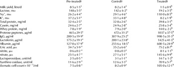
Treating MG with CNH induced dramatic changes in the secretion and composition of milk in the treated glands, whereas no significant changes were recorded in the control glands (Table 1). Milk yield fell 5·5-fold in comparison with pre-treated or control levels, and lactose concentration drooped 7·7-fold, so that lactose secretion was reduced by ∼42-fold. Na+ concentration increased ∼4-fold whereas K+ concentration decreased ∼4-fold, so that their level in milk of treated glands resembled the expected level of Na+ and K+ in blood plasma. Protein concentration increased in the skim milk of treated glands by ∼22%, which could be related to increase of 78% in whey protein concentration. This was particularly associated with a dramatic increase in the concentration of proteose peptones (CN hydrolysis peptides), and soluble components of the acquired (IgG), and innate (lactoferrin, albumin, lactoperoxidase, xanthine oxidase) immune system and related metabolites (uric acid, nitrite and nitrate). CNH treatment was also associated with ∼14-fold increase in the treated glands in the count of somatic cells, which were composed mainly of leucocytes.
Effect of treatment with casein hydrolysate (CNH) on milk yield (on a single gland level) and skim milk composition of protein, casein degradation products (proteose peptones), Na, K and components of the immune system in pre-treated, control and treated glands (mean±sd)

† Results from Experiment 1
a, b Values marked by different superscript are significantly different, at least P<0·01
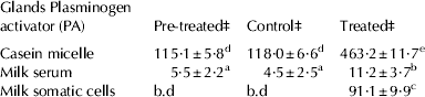
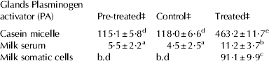
Most of the PA activity in pre-treated and control glands was associated with the CN micelles and only a minority was found in milk serum and somatic cells (Table 2). CNH treatment induced increase in PA activity in CN micelle and somatic cells. However, the activity of PA in CN micelles per ml milk was 4-fold higher than in somatic cells. No PG activity could be detected in milk serum or in the somatic cells (data not shown in a table).
Effect of casein hydrolysate (CNH) treatments on the distribution of plasminogen activator (PA) activity (unit/ml†) in milk fractions (mean±sd)
† Unit/ml: using dilution and protein content, activity of PA was calculated to be based per ml of reconstituted raw milk
‡ Results from Experiment 1
b.d, below detection level
a,b,c,d,e Values marked by different superscript are significantly different, at least P < 0·01
Activity of t-PA in CN micelles: pre-treated, control and treated glands accounted for the vast majority of PA activity (Table 3). CNH treatment induced dramatic reduction in PG activity without change in total PG+PL activity. Thus, the large increase in PL activity in the CNH-treated glands can be related to conversion of PG to PL and was associated with dramatic reduction in PG to PL ratio (Table 3).
Effect of casein hydrolysate (CNH) treatments on the activities of the plasminogen activator (PA)-plasminogen (PG)-plasmin (PL) (unit/ml†) in isolated casein micelles in pre-treated, control, freshly secreted milk and treated gland (mean±sd)

† Unit/ml: using dilution and protein content, activities of PA, PLG and PL were calculated to be based per ml of reconstituted raw milk
‡ Results from Experiment 1
§ Results from Experiment 2
a,b Values marked by different superscript are significantly different, at least P < 0·01
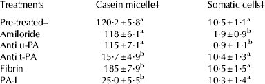
PA activity was measured with the following additions in CN micelles and somatic cells (Table 4). Amiloride and anti-u-PA antibody did not affect PA activity in CN micelles, but reduced it in somatic cells. PAI-1 and anti t-PA antibody dramatically reduced PA activity in CN micelles, but not in somatic cells. Addition of fibrin increased PA activity in CN micelles, but not on somatic cells.
Effect of amiloride, anti-U-PA, PA-I and fibrin on plasminogen activator (PA) activity (unit/ml†) in isolated casein micelles and isolated somatic cells (mean±sd)
† Unit/ml: using dilution and protein content, activity of PA was calculated to be based per ml of reconstituted raw milk
‡ Milk from pre-treated gland (6 cows) was used to isolate casein micelles and somatic cells
a,b Values marked by different superscript are significantly different from the pre-treated values by paired t test analysis, at least P < 0·01
Concentration of xanthine+hypoxanthine in milk defined as freshly secreted was 38·5±5 μm. On the other hand, no xanthine+hypoxanthine could be detected in milk defined as mature milk, or in freshly secreted milk stored at 37 °C for 30 min (data not shown in a table). We can conclude that freshly secreted milk represent milk that was sampled within 10 min or less after being secreted (Silanikove et al. Reference Silanikove, Shapiro and Leitner2007). The content of t-PA, PG and PL in freshly secreted milk was found to be similar to those found in milk from pre-treated and control glands (Table 4).
Discussion
It is well established that amiloride affects u-PA but not t-PA that PA-1 affects t-PA but not u-PA and that fibrin accelerates t-PA activity but not u-PA activity (Heegaard et al. Reference Heegaard, Rasmussen and Andresen1994b; White et al. Reference White, Zavizion, O'Harea, Gilmore, Guoa, Kindstedt and Politis1995; Politis, Reference Politis1996; Sorrell et al. Reference Sorrell, Shand, Tonner, Gamberoni, Accorsi, Beattie, Allan and Flint2006; Ismail & Nielsen, Reference Ismail and Nielsen2010). Based on that, it can be concluded that under physiological conditions, t-PA in milk is vastly associated with the CN micelles, whereas, u-PA cannot be traced in the micelles. This conclusion is further supported by the interaction with respective antibodies to t-PA and u-PA. As mentioned, this conclusion is also consistent with some previous reports (Heegaard et al. Reference Heegaard, Christensen, Rasmussen, Benfeldt, Jensen, Sejrsen, Petersen and Andersen1994a; Politis, Reference Politis1996; Tonner et al. Reference Tonner, Allan and Flint2000). However, what is unique to the present study is that the large increase in t-PA activity was induced by CNH. In previous studies, it was shown that CNH induces accelerated MG involution, which was associated with intense activation of the MG immune system (Shamay et al. Reference Shamay, Shapiro, Mabjeesh and Silanikove2002; Silanikove et al. Reference Silanikove, Shapiro, Shamay and Leitner2005b). This conclusion is supported by the present results, which demonstrate that the treatment with CNH precipitously reduced mammary secretion, disrupted the tight junction (increase in Na+ and decrease in K+ concentrations), induced hydrolysis of CN, and activated various elements of the innate and acquired immune system. These aspects were considered in detail in previous publications (Shamay et al. Reference Shamay, Shapiro, Mabjeesh and Silanikove2002; Silanikove et al. Reference Silanikove, Shapiro, Shamay and Leitner2005b, Reference Silanikove, Merin and Leitner2006) and therefore were not considered here in detail. However, the data are consistent with the theory that the PL system plays a key role in inducing MG involution by degradation of CN micelles and liberating or inducing the formation of active components that in turn affect MG epithelial cells to commit involution. In the present study, we have identified t-PA as the principal PA, which is responsible for the conversion of PG to PL.
In accordance with previous studies, PG was found to be closely associated with the casein micelles (Politis, Reference Politis1996; Weng et al. Reference Weng, Chang, Chen, Chou, Peh, Huang, Chen and Nagahata2006). However, perhaps the most novel finding of this study, namely, the presence of t-PA and PG in the CN micelles of freshly secreted milk provides new insight into the setting of PL-casein interactions. It was already demonstrated that t-PA is produced and secreted by MG cells (Politis, Reference Politis1996; Zavizion et al. Reference Zavizion, Heegard, White, Cheli and Politis1996). Thus, the close presence of t-PA and CN micelles should not be surprising as they both share the same excretory pathway through secretory vesicles released from the Golgi apparatus and because t-PA has high affinity to the CN micelles (Silanikove et al. Reference Silanikove, Iscovich, Leitner and Hogeveen2005a; Ismail & Nielsen, Reference Ismail and Nielsen2010). Following early suggestion (Politis, Reference Politis1996), PG is generally considered to leak to milk from blood plasma. Though, to the best of our knowledge, there is no evidence that supports this notion. The presence of PG in the CN micelles of fresh milk and its lack of presence in the whey of fresh milk suggest that it is secreted into milk through the secretory vesicles route embedded within the CN micelles along with its activator, t-PA, and based on the results of Sorrel et al. (Reference Sorrell, Shand, Tonner, Gamberoni, Accorsi, Beattie, Allan and Flint2006), most likely along with PA-1 because it secreted by MG cells (Zavizion et al. Reference Zavizion, Heegard, White, Cheli and Politis1996). In a close remark, we would like to relate to the following example as further support for this assumption. The presence of substances such as albumin and PG in milk in animals whose mammary gland epithelial tight junction is intact is a conundrum in that they are able to prevent the passage of ions, such as Na+ and K+. It has been shown that albumin, which also conventionally has been considered to derive in milk from blood plasma, is produced and secreted by mammary gland cells of bovine as a component of the innate immune system (Shamay et al. Reference Shamay, Homans, Fuerman, Levin, Barash, Silanikove and Mabjeesh2004). In view of the impressive synthesizing repertoire of the mammary epithelial cells, it should not be surprising to find that they are capable of synthesizing PG. However, obviously, further research applying molecular and cell biology techniques to mammary epithelial cells is needed to support this assumption.
The close association between PL, t-PA, PA-1 and CN suggests that these components serve as functional complexes that regulate the liberation of active components from the CN micelle. Such an arrangement allows effective fine tuning of the CN micelle degradation process: (i) it allows the complex to function as a time machine; milk stasis will result in longest exposure and thus higher degradation, and (ii) it allows fast responsive reaction to relevant systemic hormonal effects, which either attenuate or stimulate CN hydrolysis (Silanikove et al. Reference Silanikove, Merin and Leitner2006).
Localization of u-PA with somatic cells and lack of u-PA activity in milk serum is consistent with previous reports (Heegaard et al. Reference Heegaard, Christensen, Rasmussen, Benfeldt, Jensen, Sejrsen, Petersen and Andersen1994a, Reference Heegaard, Rasmussen and Andresenb; Politis, Reference Politis1996). In more detailed studies, it was demonstrated that milk u-PA is bound mostly to u-PA receptors on polymorphonuclear cells. Recently it was shown that MG cells respond to lipopolysacharide challenge (pro-inflammatory stress) by increasing the expression of u-PA. The physiological role of u-PA was attributed to its ability to induce basal membrane degradation that helps to induce inflow of polymorphonuclear cells to injured or infected tissue (Theodorou et al. Reference Theodorou, Bizelis, Rogdakis and Politis2009; Baldi et al. Reference Baldi, Pecorini, Rebucci, Saccone, Cheli, Miranda-Ribera, Lecchi and Ceciliani2012).
According to the present findings, the reported correlation between increased u-PA activity and CN hydrolysis during mastitis (Weng et al. Reference Weng, Chang, Chen, Chou, Peh, Huang, Chen and Nagahata2006), merely reflects the fact that they respond similarly to the inflammatory stress. Our results and others (Heegaard et al. Reference Heegaard, Christensen, Rasmussen, Benfeldt, Jensen, Sejrsen, Petersen and Andersen1994a, Reference Heegaard, Rasmussen and Andresenb; White et al. Reference White, Zavizion, O'Harea, Gilmore, Guoa, Kindstedt and Politis1995; Politis, Reference Politis1996) clearly indicate that there is no direct relation between u-PA activity and CN hydrolysis in raw milk during inflammation. The substantial evidence for the major role of u-PA in CN degradation in stored milk may be explained by the dissociation of u-PA from its receptor on leucocytes and the tendency of u-PA to form interaction with CN. There are many reports that show that heat treatment, such as that applied to milk during pasteurization, inactivates PAI-1 and increase the association between u-PA and CN (Ismail & Nielsen, Reference Ismail and Nielsen2010).