INTRODUCTION
Schistosomiasis is the most important human helminth infection when it comes to morbidity and mortality (McManus and Loukas, Reference McManus and Loukas2008). About 800 million people live in risk areas in 76 countries worldwide, of which 207 million people are infected and 20 million develop the severe disability form of the disease (Steinmann et al. Reference Steinmann, Keiser, Bos, Tanner and Utzinger2006; WHO, 2010). It is estimated that 15 000 people die per year, however, mortality rate could be as high as 280 000 per year in Africa (van der Werf et al. Reference van der Werf, de Vlas, Brooker, Looman, Nagelkerke, Habbema and Engels2003; Lustigman et al. Reference Lustigman, Prichard, Gazzinelli, Grant, Boatin, McCarthy and Basanez2012). Many countries, especially in sub-Saharan Africa, invest in intervention strategies based on short-term control programmes using mass drug administration together with supply of safe water, improvement in sanitation and snail control (McManus and Loukas, Reference McManus and Loukas2008; Prichard et al. Reference Prichard, Basanez, Boatin, McCarthy, Garcia, Yang, Sripa and Lustigman2012). The main goal in schistosomiasis treatment is to achieve the reduction of disease transmission and morbidity. Praziquantel (PZQ) is the main single drug used in schistosomiasis treatment, but it does not kill the larval stage form of schistosome and does not prevent reinfection. Besides, some cases of drug-resistant parasites should also be considered (Gray et al. Reference Gray, McManus, Li, Williams, Bergquist and Ross2010; McManus, Reference McManus2012). Since PZQ mass treatment has so many limitations, many believe that an integrative measure is to combine the traditional PZQ treatment with an effective vaccine (Bergquist et al. Reference Bergquist, Leonardo and Mitchell2005). The most promising targets for vaccine development are membrane proteins present in the parasite's outer surface or tegument identified through bioinformatics analysis of Schistosoma mansoni sequence databases, such as transcriptomes, genome and proteomics (Pinheiro et al. Reference Pinheiro, Martins, Assis, Figueiredo, Morais, Azevedo and Oliveira2011).
Proteomic analysis of S. mansoni tegument (Braschi and Wilson, Reference Braschi and Wilson2006; Braschi et al. Reference Braschi, Curwen, Ashton, Verjovski-Almeida and Wilson2006) provided the schistosome vaccine field with promising candidates for vaccine design, such as Sm29 (Cardoso et al. Reference Cardoso, Macedo, Gava, Kitten, Mati, de Melo, Caliari, Almeida, Venancio, Verjovski-Almeida and Oliveira2008) and TSP-2 (Tran et al. Reference Tran, Pearson, Bethony, Smyth, Jones, Duke, Don, McManus, Correa-Oliveira and Loukas2006). A recent study on the abundance of S. mansoni tegument surface proteins (Castro-Borges et al. Reference Castro-Borges, Simpson, Dowle, Curwen, Thomas-Oates, Beynon and Wilson2011) indicates that aquaporin is the most abundant transmembrane protein. The S. mansoni aquaporin (SmAQP – GenBank: EU780065.1) is one small tegumental membrane protein that transports water and other small solutes such as glycerol (Faghiri and Skelly, Reference Faghiri and Skelly2009). In addition to being very abundant in the tegument, this protein is expressed in all developmental stages of the parasite and plays a crucial role in osmoregulation, nutrient transport and drug uptake (Faghiri et al. Reference Faghiri, Camargo, Huggel, Forster, Ndegwa, Verrey and Skelly2010). SmAQP is vital for schistosome survival, since the inability to control water movement affects the parasite's biochemistry, leading to increased mortality in vitro (Faghiri and Skelly, Reference Faghiri and Skelly2009).
Taking into account the importance of SmAQP in parasite survival, the goal of the present study was to test this protein as a vaccine candidate in the murine model. In order to characterize the host immune responses and evaluate the prophylactic potential of this protein, we formulated a vaccine with a chimeric form of SmAQP in Freund's adjuvant and assessed the protection after challenge. As described below, vaccination with cSmAQP did not reduce worm burden in mice nor ameliorate pathology in the liver.
MATERIALS AND METHODS
Mice and parasites
Six- to 8-week-old female C57BL/6, TLR4 KO and Swiss mice were purchased from the Federal University of Minas Gerais (UFMG) animal facility. All animal experiments were conducted in accordance with the Brazilian Federal Law no. 11.794, which regulates the scientific use of animals, and IACUC guidelines. The protocols involving animals used in this study were approved by the Federal University of Minas Gerais Ethics Committee on animal experimentation (CETEA no. 179/2010). Cercariae of S. mansoni (LE strain) were maintained routinely in Biomphalaria glabrata snails at Rene Rachou Research Center (CPqRR, Fiocruz, Brazil) and prepared by exposing infected snails to light for 2 h to induce shedding of parasites. Cercariae numbers and viability were determined using a light microscope prior to infection.
Antigen preparation
The plasmid pJ414 containing the cDNA sequence for cSmAQP (pJ414::cAQUA) was manufactured by DNA 2.0, Inc. USA (https://www.dna20.com) using DNA 2.0 optimization algorithms for expression in Escherichia coli. This plasmid was transformed into E. coli Rosetta-gami™ (Merck KGaA, Darmstadt, Germany) competent cells. Transformants harbouring the designed plasmid were screened on LB agar plates containing ampicillin (50 μg mL−1) and chloramphenicol (34 μg mL−1) and the selected transformant was designated as Aqua-Rosetta. One litre of Aqua-Rosetta was cultured in a 3 L Erlenmeyer flask on a rotary shaker at 200 rpm at 37 °C to an optical density at 600 nm of approximately 0·5–0·8 and gene expression was induced by using 1 mm isopropylthiogalactoside (IPTG). After 5 h of induction, the bacterial cells were harvested by centrifugation at 4000 g for 20 min. Using gentle vortexing or pipetting, the pellet was resuspended in 50 mL of 10 mm Na2HPO4, 10 mm NaH2PO4, 0·5 m NaCl and 20 mm imidazole. Subsequently, the cells were submitted to three cycles of sonication lasting 30 s each and centrifuged at 5400 g for 20 min. The cSmAQP was recovered solubilized in the supernatant and purified by affinity chromatography on a Ni-Sepharose column (Hitrap chelating 5 mL) using an AKTA explorer chromatography system (GE Healthcare, São Paulo, Brazil). After protein binding to the Ni-Sepharose column, washes with 50 mm imidazole were performed and the protein was eluted with 500 mm imidazole. Fractions containing the protein were determined through Bradford's method (Coomassie Protein Assay Kit, Pierce) and also SDS/PAGE-12% and dialysed against PBS pH 7·0. The dialysis was carried out at 4 °C using a Spectra/Por2 membrane (MWCO 6 to 8 kDa; Spectrum Medical Industries, Inc., Laguna Hills, CA). The recombinant protein was quantified using the Bradford's method and used as antigen for vaccination and immunological experiments. All reagents were purchased from Sigma-Aldrich, CO (St. Louis, MO, USA) unless otherwise specified.
Mice immunization, challenge infection and worm burden recovery
Two groups of 10 mice each (female C57BL/6 aged 6–8 weeks) were subcutaneously injected in the nape of the neck with 25 μg of cSmAQP or PBS, as a control, at days 7, 22 and 37. Both preparations were formulated with Complete Freund's Adjuvant (CFA) for the first immunization and Incomplete Freund's Adjuvant (IFA) for the last two immunizations. All reagents were purchased from Sigma-Aldrich, CO (St. Louis, MO, USA). Fifteen days after the last immunization, the mice were challenged through percutaneous exposure of abdominal skin to water containing 100 cercariae (LE strain) for 1 h. Forty-five days after the challenge, adult worms were perfused from the portal veins of each animal, as previously described (Fonseca et al. Reference Fonseca, Brito, Alves and Oliveira2004). The immunization and challenge protocol is demonstrated in supplementary Fig. 1A. Three independent experiments were performed to determine the vaccine protection level. Protection was calculated by comparing the number of worms recovered from the group vaccine with cSmAQP and the control group, as follows: PL = [(WRCG–WREG)/WRCG]×100, where PL = protection level, WRCG = worms recovered from control group and WREG = worms recovered from experimental group.
Measurement of anti-cSmAQP specific antibodies
Following immunizations, sera of 10 vaccinated mice from both groups (cSmAQP or PBS) were collected at days 0, 15, 30, 45, 60, 75 and 90. Measurements of specific antibodies were performed using indirect ELISA. Maxisorp 96-well microtitre plates (Nunc, Denmark) were coated with 10 μg mL−1 of cSmAQP in carbonate-bicarbonate buffer, pH 9·6 for 16 h at 4 °C, then blocked for 2 h at room temperature with 200 μL well−1 PBST (phosphate buffered saline, pH 7·2 with 0·05% Tween-20) plus 10% FBS (foetal bovine sera). For each serum, 20 dilutions were evaluated (starting at 1 : 20 and following a two-fold serial dilution); 100 μL of each dilution was added per well and incubated for 1 h at room temperature. Plate-bound antibody was detected after 1-h incubation with peroxidase-conjugated anti-mouse IgG, IgG1 and IgG2a diluted in PBST 1 : 5000, 1 : 10 000 and 1 : 2000, respectively. Colour reaction was developed by addition of 100 μL per well of 200 pmol OPD (o-phenylenediamine) in citrate buffer, pH 5·0 plus 0·04% H2O2 for 10 min and stopped with 50 μL of 5% sulphuric acid per well. The plates were read at 495 nm in an ELISA plate reader (BioRad, Hercules, CA). All reagents were purchased from Sigma-Aldrich, CO (St. Louis, MO, USA) unless otherwise specified.
Purification of anti-cSmAQP specific antibodies
For the purification of anti-cSmAQP antibodies, serum from the fourth bleeding was utilized (day 45 – after the third immunization and before challenge). Briefly, 500 μg of the recombinant protein was adsorbed to a nitrocellulose membrane. The membrane was then blocked with TBST (tris buffered saline, pH 7·2 with 0·05% Tween-20) containing 5% non-fat dry milk for 2 h at room temperature. After three washes using TBST, the membrane was incubated with 2 mL of pooled serum from mice immunized with cSmAQP for 16 h at 4 °C. After another set of three washes with TBST, the antibodies were eluted with 400 μL of triethylamine 0·014% for 5 min at room temperature and then neutralized with 100 μL of 10× TBS. The eluate was dialysed against TBS pH 7·0. The dialysis was carried out at 4 °C using a Spectra/Por2 membrane (MWCO 6 to 8 kDa; Spectrum Medical Industries, Inc., Laguna Hills, CA, USA). All reagents were purchased from Sigma-Aldrich, CO (St. Louis, MO, USA) unless otherwise specified.
Oogram
To evaluate the effect of immunization with cSmAQP in granuloma formation, following perfusion for the recovery of the schistosomes, livers from eight mice per group were collected. Liver fragments from each animal were separated and the smallest part was weighed and digested with 10% KOH overnight at 37 °C. The eggs were obtained by centrifugation at 900 g for 10 min and resuspended in 1 mL of saline. Egg number was counted using a light microscope. Quantitative oograms were obtained calculating the number of eggs per gram of liver tissue. The liver samples removed from the central part of the left lateral lobe were fixed with 10% buffered formaldehyde in PBS (phosphate buffered saline, pH 7·2). All reagents were purchased from Sigma-Aldrich, CO (St. Louis, MO, USA).
Liver histopathological analysis
Histological sections were performed using a microtome at 6 μm and stained on a slide with picrosirius-haematoxylin-eosin (PSHE). The count of granulomas was performed at a microscope with 10× objective lens. Each liver section was scanned for calculating its whole area (mm2) using the ImageJ software (http://rsbweb.nih.gov/ij/index.html). For measurement of the total area of granulomas, a microscope with 10× objective lens was used; images were obtained through a JVC TK-1270/RBG microcamera attached to the microscope. Twenty granulomas with a single well-defined egg were randomly selected, in each liver section, and the granuloma area was measured using the ImageJ software. All reagents were purchased from Sigma-Aldrich, CO (St. Louis, MO, USA).
Mice immunization and cytokine analysis
Cytokine experiments were performed using splenocyte cultures from individual mice (C57BL/6 or TLR4 KO) immunized three times with 25 μg of cSmAQP or PBS (control) both formulated with Complete/Incomplete Freund's Adjuvant (n = 5 for each group). The cytokine protocol is demonstrated in supplementary Fig. 1B. Ten days after the last immunization, splenocytes were isolated from macerated spleen of individual mice and washed twice with sterile PBS. After washing, the splenocytes were adjusted to 1×10⁶ cells per well in RPMI 1640 medium (Invitrogen, Carlsbad, CA, USA) supplemented with 10% FBS, 100 U mL−1 of penicillin G sodium and 100 μg mL−1 of streptomycin sulphate, for IL-4, IL-5, IL-10, IL-17, IFN-γ and TNF-α assays. Splenocytes were maintained in culture with medium alone or stimulated with the cSmAQP (25 μg mL−1) or with concanavalin A (ConA) (5 μg mL−1), or LPS (1 μg mL−1), as positive controls. The 96-well plates (Nunc, Denmark) were maintained in an incubator at 37 °C with 5% CO2 (Fonseca et al. Reference Fonseca, Pacifico, Barsante, Rassi, Cassali and Oliveira2006; Pacifico et al. Reference Pacifico, Fonseca, Chiari and Oliveira2006). For C57/BL6 mice cells, polymyxin B (30 μg mL−1) was added to the cultures since this treatment completely abrogates the cytokine response to LPS as previously described (Cardoso et al. Reference Cardoso, Araujo, Goes, Pacifico, Oliveira and Oliveira2007). Culture supernatants were collected after 24 h for IL-4 and IL-5, after 48 h for TNF-α and after 72 h for IL-17 and IFN-γ. The assays for the measurements of all cytokines were performed using the Duoset ELISA kit (R&D Diagnostic, Minneapolis, MN, USA) according to the manufacturer's directions. All reagents were purchased from Sigma-Aldrich, CO (St. Louis, MO, USA) unless otherwise specified.
SDS-PAGE and immunoblotting
SDS-PAGE was conducted on 15% polyacrylamide gels prepared as previously described (Laemmli, Reference Laemmli1970). Protein samples of cSmAQP or SWAP (soluble worm antigen preparation), obtained from adult worms recovered from perfused infected Swiss mice, were run at 120 V for 2–3 h. The gel was electroblotted onto nitrocellulose membrane using a wet system (Towbin et al. Reference Towbin, Staehelin and Gordon1979). The membrane was then blocked with TBST (tris buffered saline, pH 7·2 with 0·05% Tween-20) containing 5% non-fat dry milk for 16 h at room temperature. Then, the membrane was incubated in a 1 : 2000 dilution of anti-HIS antibodies (GE Healthcare) or in 1 : 1000 anti-cSmAQP murine polyclonal antibodies or in 1∶1000 naive mice serum in TBST for 1 h at room temperature. After three washes using TBST, the membrane was incubated in 1 : 2000 mouse IgG conjugated with alkaline phosphatase (AP) treated with AP reaction developing buffer containing nitroblue tetrazolium (NBT) and 5-bromo-4-chloro-3-indolyl-1-phosphate (BCIP). After the reaction was developed, the membrane was washed using distilled water and dried on filter paper. All reagents were purchased from Sigma-Aldrich, CO (St. Louis, MO, USA) unless otherwise specified.
Immunolocalization
Adult male worms used in fluorescence microscopy studies were recovered from perfused infected Swiss mice. Parasites were fixed in Omnifix II (Ancon Genetics, St Petersburg, FL, USA) and sectioned in 7 μm slices, and were then deparaffinized with xylol series. Parasites were blocked with 1% BSA in PBST (phosphate buffered saline, pH 7·2 with 0·05% Tween-20) for 1 h and incubated with anti-cSmAQP serum diluted 1 : 20 in blocking buffer. Serum from non-immunized mice was used as a negative control. Samples were washed three times with PBST and incubated with anti-mouse IgG antibody conjugated to FITC diluted 1 : 100 in blocking buffer containing rhodamine phalloidin to stain actin microfilaments. The samples were washed four times and mounted in anti-fade reagent with DAPI. The parasites were visualized using immersion 40× objective in a Nikon fluorescence microscope at the Microscopy Center of Biological Sciences Institute (CEMEL), Federal University of Minas Gerais. All probes and anti-fade were purchased from Molecular Probes, Life Technologies (Grand Island, NY, USA).
Measurement of human anti-SmAQP IgG responses in schistosomiasis patients
Sera were obtained from individuals living in two different endemic areas for schistosomiasis (‘Melquiades’ and ‘Côrrego do Onça’, Minas Gerais, and ‘Conde’, Bahia, Brazil). These individuals were classified into two groups, regarding their infection status. Non-infected (NIN) individuals (n = 15) were healthy people without any parasite infection or contaminated water contact and infected (INF) individuals (n = 15) showed stool-positive examination and no treatment history. All patients were negative for other helminthic infections (for study population see supplementary Table 1). These patients or their legal guardians gave informed consent after explanation of the protocol that had been previously approved by the Ethical Committee of the Federal University of Minas Gerais, as previously described (Cardoso et al. Reference Cardoso, Pacifico, Mortara and Oliveira2006). Sera from these patients were used in an ELISA to measure the levels of total IgG to SmAQP (Brito et al. Reference Brito, Fonseca, Goes, Azevedo, Simpson and Oliveira2000). For this assay, 96-well flat-bottom microtitre plates (Nunc, Denmark) were coated overnight at 4 °C with 100 μL of cSmAQP protein or SWAP (soluble worm antigen preparation), obtained from adult worms recovered from perfused infected Swiss mice, at a concentration of 5 μg mL−1 in 0·1 m carbonate bicarbonate buffer (pH 9·6) per well. The plates were then blocked with 10% bovine foetal serum in PBS (pH 7·4) for 2 h at room temperature. Subsequently, the plates were washed three times with PBST. For total IgG, serum samples were diluted 1 : 50 in PBST (100 μL well−1) were added in duplicate and the plates incubated for 1 h at room temperature. Peroxidase-labelled anti-human IgG was added at dilutions of 1 : 10 000 (100 μL well−1). After 1 h at 37 °C, the plates were washed and orthophenyl-diaminobenzidine plus 0·05% hydrogen peroxide in phosphate citrate buffer (pH 5·0) was added (100 μL well−1). The plates were then incubated for 30 min at room temperature, and the reaction was stopped by addition of 5% H2SO4 (50 μL well−1). Absorbance was read at 492 nm using a microplate reader (Bio-Rad, Hercules, CA). All reagents were purchased from Sigma-Aldrich, CO (St. Louis, MO, USA) unless otherwise specified.
Epitope analysis
The amino acids sequence of SmAQP was obtained from SchistoDB (http://schistodb.net/schisto/). Linear B-cell epitopes were predicted based on the algorithm BepiPred, the score threshold parameter ranged from 0·15 to 0·90. We also performed a prediction with the cSmAQP sequence to evaluate which epitopes were maintained and which ones were lost in the construction of the chimera.
Statistical analysis
Statistical analysis was performed with the Student's t-test for comparison between two experimental groups using the software package GraphPad Prism (La Jolla, CA, USA). Bonferroni adjustments were included for multiple comparisons. P values obtained by these methods were considered significant if they were <0·05.
RESULTS
Design and production of recombinant SmAQP
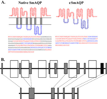
The S. mansoni aquaporin (SmAQP) is a large transmembrane insoluble protein which has six transmembrane helices. In order to overcome problems with protein size and solubility and to improve its expression in E. coli, we developed a chimeric protein in which the transmembrane domains are absent. So, both the intracellular and the extracellular hydrophilic portions were fused together, generating what we denominated chimeric aquaporin (cSmAQP). Chimeric SmAQP was designed through the fusion of cDNA sequences of predicted non-membranous SmAQP portions with a six-histidine tag in its C-terminal. Bioinformatic analysis of the resultant sequence revealed a soluble 169-aa protein with a predicted molecular mass of 18 705 Da and a predicted pI of 8·59 (Fig. 1A). The native aquaporin is a 304-aa protein, with 32 893 Da and pI of 8·67 (Faghiri and Skelly, Reference Faghiri and Skelly2009). Besides, since this chimeric protein was used in vaccine studies, we also performed linear B-cell epitope analysis of both cSmAQP and the native protein sequences. The native aquaporin has 10 predicted epitopes, nine of them in its hydrophilic portions (see supplementary Table 2 for epitopes’ sequence). cSmAQP has seven predicted epitopes, all of them with a similar sequence to the native molecule (Fig. 1B).
Schistosoma mansoni aquaporin schematic representation. (A) Schistosoma mansoni native aquaporin is represented on the left. The hydrophilic portions used in the chimera construction are represented as: intracellular (red), extracellular (blue); the transmembrane portions (grey) are not present in the designed chimeric recombinant aquaporin (cSmAQP), represented on the right. (B) The native aquaporin is represented on the top row with empty rectangles representing the transmembrane regions and grey rectangles representing the predicted B-cell linear epitopes. The cSmAQP, represented on the bottom row, maintained most of the original protein's epitopes.

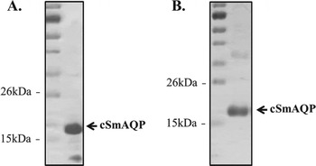
The cSmAQP was expressed in E. coli and purified by affinity chromatography. The expression and purity of cSmAQP as a 6×His-tag fusion protein were checked by SDS-PAGE and western blotting analysis with anti-HIS antibody, which revealed a protein with approximately 18 kDa corresponding to predicted mass of cSmAQP (Fig. 2).
Heterologous expression and purification of cSmAQP. (A) Western blot analysis of cSmAQP in the bacterial lysate (10 μg) probed with monoclonal mouse anti-His tag antibodies. (B) 15% SDS-PAGE stained with Coomassie brilliant blue of the purified cSmAQP (5 μg). The molecular weight protein standard (first lane) is a broad range pre-stained ladder from BioRad.

Antibody profile following mice immunization
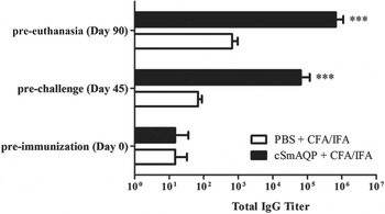
To evaluate the levels of total IgG, IgG1 and IgG2a antibodies to cSmAQP, sera from 10 vaccinated animals of each group were tested by ELISA. All mice vaccinated with cSmAQP produced significant levels of specific IgG antibodies compared with the control group vaccinated with PBS at day 45, pre-challenge with cercariae, and at day 90, before euthanasia (Fig. 3). The measurement of IgG isotypes levels revealed that the cSmAQP-vaccinated group had increased levels of IgG1 and IgG2a when compared with the PBS-administered group. Moreover, the IgG1/IgG2a ratio was increased at day 45. After challenge infection with schistosome cercariae, this elevated IgG1/IgG2a ratio was maintained (see supplementary Table 3).
Kinetics of specific anti-cSmAQP IgG induced in mice immunized with recombinant cSmAQP. Sera of immunized mice were collected at days 0, 45 and 90 and assayed by ELISA. Results are presented as the mean of the antibody titres for 10 mice in each group and error bars indicate s.d. The results shown are representative of three independent experiments. Statistically significant differences of recombinant cSmAQP vaccinated mice compared with PBS control group is denoted by three asterisks for P<0·001.

Antibodies against cSmAQP recognize native aquaporin in S. mansoni
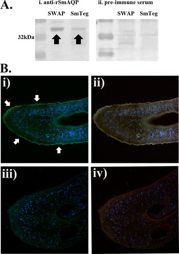
In order to confirm whether polyclonal antibodies raised against cSmAQP were able to recognize native aquaporin, we performed western blot analysis to detect aquaporin in skin-stage schistosomula tegument (SmTeg, kindly provided by Dr Cristina T. Fonseca) or SWAP. Sera from naïve mice were used as a control. In both protein extract samples, the anti-cSmAQP (1 : 1000) recognized a single band with approximately 33 kDa, which is the predicted size for aquaporin (Fig. 4A). This result indicates that anti-cSmAQP were able to recognize the native S. mansoni aquaporin. Additionally, we confirmed that anti-cSmAQP can recognize native aquaporin in fluorescence microscopy experiments. The anti-cSmAQP were used in immunolocalization of male worms and they recognized native aquaporin predominantly on the worm tegument (Fig. 4B), as demonstrated by Faghiri et al. (Reference Faghiri, Camargo, Huggel, Forster, Ndegwa, Verrey and Skelly2010).
Antibodies anti-cSmAQP recognized native aquaporin through western blot and immunolocalization. (A) Twenty micrograms of SWAP or 10 μg of SmTeg were applied onto 15% SDS-PAGE and transferred to a nitrocellulose membrane. (i) The probing with polyclonal antibodies raised against cSmAQP protein demonstrated the presence of one band with the predicted size of native aquaporin, around 33 kDa, in both samples (black arrows). These data show that antibodies against cSmAQP are able to recognize native aquaporin. (ii) As control, serum from naïve mice was used. (B) Fluorescence microscopy images of male adult worm of S. mansoni are shown. Polyclonal anti-cSmAQP and secondary antibody coupled to FITC (green) were used for fluorescence detection of aquaporin on male adult worm sections (i and ii). Serum from naïve mice was used as negative control (iii and iv). Rhodamine phalloidin (red) was used for the actin localization. DAPI (blue) was used for nucleus staining. (i) The aquaporin localization is represented in green. Arrows indicate the worm tegument. (ii) aquaporin and actin staining merged. (iii) The absence of green staining in the negative control. (iv) FITC and actin staining merged.

Cytokine profile
To determine the cytokine profile induced by vaccination with cSmAQP, we measured the production of IFN-γ, TNF-α, IL-4, IL-5, IL-10 and IL-17 in spleen cells of two mice strains, C57BL/6 and TLR4 KO immunized mice 10 days after the third immunization. TLR4 is the major receptor for macrophage activation by bacterial LPS (Medzhitov, Reference Medzhitov2007). Considering that the antigen utilized in this experiment was expressed and purified from bacterial cultures, the bacterial LPS contamination could have affected the cytokine production. Therefore, by using TLR4 KO we eliminated any possible effect of LPS in cytokine synthesis. This mouse strain, together with the samples treated with polymyxin B eliminated any possible LPS contamination of the antigen.
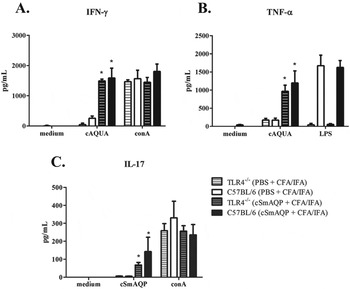
As a result of antigen stimulation, we detected significant levels of IFN-γ in supernatant of both cells from immunized TLR4 KO (1493·7±61·1 pg mL−1) and C57BL/6 (1589·7±322·9 pg mL−1) when compared with the PBS immunized control group (Fig. 5). We also detected high levels of TNF-α in cell supernatants of vaccinated mice stimulated with cSmAQP compared with PBS immunized groups (1196·0±335·2 pg mL−1 for C57BL/6 mice and 968·0±168·1 pg mL−1 for TLR4 KO mice). The Th2 type of cytokines, IL-4 and IL-5 and the regulatory cytokine IL-10 were produced in very low levels with no statistical significance when compared with control groups (data not shown). Upon stimulation, spleen cells of the cSmAQP vaccinated group were also able to produce IL-17 (147·4±56·9 pg mL−1 for C57BL/6 mice and 67·9±14·6 pg mL−1 for TLR4 KO mice), which characterizes a Th17 response. These results indicate that immunization with cSmAQP formulated with Freund's adjuvant induced a Th1/Th17 type of immune response characterized by the production of IFN-γ, TNF-α and IL-17 and the absence of Th2 cytokines.
Cytokine profile of mice immunized with cSmAQP. Ten days after the final immunization with cSmAQP or PBS, splenocytes from five mice C57BL/6 or TLR4 KO were isolated and assayed for the determination of cytokine profile. (A) IFN-γ, (B) TNF-α and C. IL-17 production in response to cSmAQP (25 μg) were measured in the supernatants of spleen cells. The results are presented as the mean±s.d. for each group. The results shown are representative of three independent experiments. Significant differences from mice immunized with cSmAQP and their respective control group (either C57BL/6 or TLR4 KO) are denoted by one asterisk for P<0·05.

Worm burden recovery
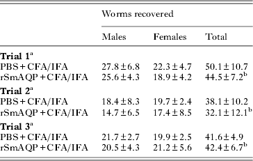
Protective immunity induced by vaccination with cSmAQP was evaluated 45 days after challenge with 100 S. mansoni cercariae. Mice vaccinated with cSmAQP showed no statistically significant reduction in worm burden recovery compared with control animals (Table 1). We performed three independent trials of cercarial challenge after immunization.
Protective immune response induced in mice by vaccination with cSmAQP
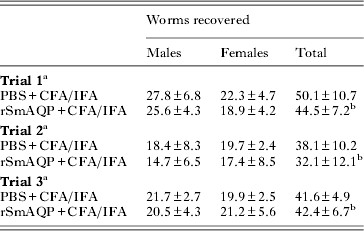
a 8–10 animals per group, values are mean number of recovered worms ±s.d.
b There was no statistically significant difference of recombinant cSmAQP vaccinated mice compared with PBS control group.
Liver pathology
Histological analysis by digital morphometry of PSHE-stained sections obtained from liver of mice immunized with cSmAQP showed no effect in granuloma volume and number compared with mice that received only PBS. Additionally, no difference in the number of eggs in the liver was observed in both groups. In general, these findings suggest that immunization with cSmAQP did not alter the hepatic conditions of mice infected with S. mansoni, as shown in Supplementary Fig. 2.
Human IgG antibody response to cSmAQP
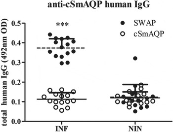
We also tested cSmAQP as a potential tool for diagnosing schistosomiasis patients. Before testing human antibodies response to cSmAQP, we tested murine polyclonal anti-SmTeg (kindly provided by Dr Cristina T. Fonseca) against a purified sample of cSmAQP in western blot assay. The antibodies (1 : 1000) recognized cSmAQP (data not shown), suggesting that both recombinant and native aquaporin probably have similar epitopes to B-cells. Then, considering that anti-SmAQP might be able to recognize the chimeric form of the protein, ELISA was performed to investigate the presence of IgG antibodies anti-cSmAQP in sera of infected S. mansoni individuals. Total IgG levels in sera of schistosomiasis patients and non-infected individuals were evaluated. The schistosomiasis patients studied had significantly higher levels of IgG to SWAP and no significant levels of IgG to cSmAQP compared with the non-infected group (Fig. 6). These results demonstrate that cSmAQP is not a good antigen to discriminate infected from healthy patients, showing no potential as an antigen for diagnostic purposes.
Levels of IgG anti-SWAP or anti-cSmAQP in sera of schistosomiasis patients. Analysis of human IgG antibody responses in sera of infected patients (INF) or non-infected individuals (NIN). The results are presented as the mean±s.d. for each group (n = 15). The results shown are representative of three independent experiments. Statistically significant difference of infected compared with non-infected patients is denoted by one asterisk for P<0·001.

DISCUSSION
Tegument proteins are of great importance in the development of schistosome vaccine because they are the major host–parasite interface (Loukas et al. Reference Loukas, Tran and Pearson2007). Recently, Castro-Borges et al. (Reference Castro-Borges, Simpson, Dowle, Curwen, Thomas-Oates, Beynon and Wilson2011) identified aquaporin as the most abundant transmembrane protein in the tegument of S. mansoni. Schistosoma mansoni aquaporin (SmAQP) is a well-known protein that plays crucial roles in parasite survival, such as osmoregulation, nutrient transport and drug uptake. The vital importance of SmAQP was demonstrated through RNAi experiments; the suppressed parasites exhibit lower viability in culture relative to controls (Faghiri et al. Reference Faghiri, Camargo, Huggel, Forster, Ndegwa, Verrey and Skelly2010).
Considering that SmAQP plays a vital role in parasite survival in culture, our group decided to evaluate its potential as vaccine candidate in prophylactic treatment against S. mansoni infection using the murine model. One of the main difficulties in working with SmAQP is that it has six transmembrane domains, making its expression in bacteria complicated. To overcome this problem, we designed a chimera protein by fusion of S. mansoni aquaporin's soluble portions. The use of chimeras is a strategy to optimize the immunological response against schistosome proteins (Romeih et al. Reference Romeih, Hassan, Shousha and Saber2008; Pearson et al. Reference Pearson, Pickering, McSorley, Bethony, Tribolet, Dougall, Hotez and Loukas2012).
In the mouse model, immunization with recombinant aquaporin (cSmAQP) formulated with Freund's adjuvant induced high levels of anti-cSmAQP IgG compared with PBS, also formulated with Freund, as control group. Through western blot analysis and immunolocalization, we demonstrated that these polyclonal antibodies were able to recognize both cSmAQP and native aquaporin, suggesting that the recombinant chimera induces an immune response similar to the native protein. We also evaluated IgG isotypes and found high levels of IgG1 and IgG2a in the cSmAQP vaccinated group compared with the control group. Previous studies have shown that high levels of IgG1 are associated with the induction of schistosomula death by antibody-dependent cell-mediated cytotoxicity, and the activation of complement (Capron et al. Reference Capron, Bazin, Dessaint and Capron1975; Khalife et al. Reference Khalife, Dunne, Richardson, Mazza, Thorne, Capron and Butterworth1989). Moreover, the stimulation of splenocytes with cSmAQP resulted in increased levels of IFN-γ, TNF-α and IL-17 in both C57/BL6 and TLR4 KO mice, the latter used as control due to its absence of response to LPS, assuring that the cytokine production was triggered by the antigen itself. IFN-γ is important on the protective immunity against S. mansoni, the production of this cytokine probably is stimulated by the larval schistosomulum when it passes through the lungs. This involves the recruitment of macrophages and lymphocytes around the worms, hindering their movement in the lungs and moving them to the airways, with the consequent elimination of these parasites (Wilson et al. Reference Wilson, Coulson, Betts, Dowling and Smythies1996; Jankovic et al. Reference Jankovic, Wynn, Kullberg, Hieny, Caspar, James, Cheever and Sher1999). Another described mechanism mediated by IFN-γ is the activation of macrophages to kill worms inducing the production of nitric oxide (Jankovic et al. Reference Jankovic, Wynn, Kullberg, Hieny, Caspar, James, Cheever and Sher1999; Pearce and MacDonald, Reference Pearce and MacDonald2002). TNF-α is another cytokine related to a protective immune response against S. mansoni, it probably operates in granuloma formation and, together with IFN-γ, increases the levels of nitric oxide for recruited macrophages (Amiri et al. Reference Amiri, Locksley, Parslow, Sadick, Rector, Ritter and McKerrow1992; Cheever et al. Reference Cheever, Poindexter and Wynn1999; Pearce and MacDonald, Reference Pearce and MacDonald2002). The role of IL-17 in schistosoma infection has recently been described. This cytokine, which is the marker of a Th17 response, is important in egg-induced inflammation. The mouse strain knock-out for both IL-17 and IFN-γ generated smaller liver granulomas when compared with wild-type mice (Rutitzky and Stadecker, Reference Rutitzky and Stadecker2011). In a study of schistosomiasis co-infection with the nematode helminth Heligmosomoides polygyrus, the switch from a Th1/Th17-polarized response to a Th2-polarized response, accompanied by a decrease in IL-17, IFN-γ and TNF-α and an increase in IL-4, IL-5 and IL-10, significantly reduced immunopathology in mice with severe pathology (Bazzone et al. Reference Bazzone, Smith, Rutitzky, Shainheit, Urban, Setiawan, Blum, Weinstock and Stadecker2008). In this study, the absence of production of IL-4, IL-5 and IL-10 seems to have affected cytokine polarization, which maintained high levels of IL-17, IFN-γ and TNF-α and resulted in no amelioration of the immunopathology of the group immunized with cSmAQP when compared with the control group.
Despite high antibody titres and the induction of cytokine production, vaccination of mice with cSmAQP plus Freund's adjuvant failed to reduce the parasite burden. Therefore, there was no apparent correlation between the antibodies generated and protective efficacy. Concerning the antibodies’ interactions with the parasite, two main reasons might justify the lack of effective protection: either the antibodies are not binding to the native SmAQP in live parasites or the antibody binding does not provoke protein function impairment, which would not affect the transport of water and excretion products.
When we tested the recognition of cSmAQP by human sera from schistosomiasis patients, the sera were unable to detect significant levels of IgG when compared with healthy subjects. The IgG molecules are important in S. mansoni infection resistance since they have functional activity of opsonization and cell-dependent cytotoxicity, and activate the classical complement pathway (Delgado and McLaren, Reference Delgado and McLaren1990). The absence of anti-cSmAQP IgG in infected schistosomiasis patients might be an indication that aquaporin is not important during the human immune response to S. mansoni. However, even considering that the two molecules share some of the predicted epitopes for B cells, and anti-cSmAQP can recognize the native aquaporin, no evidence is presented on whether schistosomiasis patients’ antibodies bind native aquaporin. At this point, what we do know is that the chimeric protein designed in this study cannot distinguish infected patients from healthy ones and thus it is not a good antigen for schistosomiasis diagnosis.
Aquaporins occur as tetramers in plasma membranes, each monomer serving as a separate pore with six membrane-spanning helices connected by five inter-helix loops, three external to the plasma membrane (Gonen and Walz, Reference Gonen and Walz2006). Recent proteomic study of S. mansoni indicated that aquaporin represents 17% of the total tegument proteins (Castro-Borges et al. Reference Castro-Borges, Simpson, Dowle, Curwen, Thomas-Oates, Beynon and Wilson2011). Besides its abundance in the tegument, this protein is almost completely inserted in the plasma membrane, which might impair the recognition of SmAQP by the immune system, protecting this essential protein against host cleavage and presentation to T cells. This is the first study evaluating SmAQP as a vaccine target; other protein portions or even peptides should be tested, as well as other adjuvants. Many methods, such as innovative vaccine delivery procedures, can be tried to make SmAQP immunogenic. Conversely, minor schistosome plasma membrane components could be better targets because fewer molecules would need to be blocked by the host immune system. The major protective antigens in the tegument, TSP-2 and Sm29, are present at very low rates (2·43 and 0·31%, respectively) (Castro-Borges et al. Reference Castro-Borges, Simpson, Dowle, Curwen, Thomas-Oates, Beynon and Wilson2011).
Our study demonstrated that a vaccine formulated with a chimeric form of aquaporin was not effective against schistosomiasis in mice. The use of this protein as a vaccine target requires more research and use of new vaccine technologies to make it immunogenic. Despite the results of this study, many researchers maintain hope in aquaporin as a vaccine antigen. However, future studies are needed to determine whether the native protein is immunogenic or not. Besides vaccine studies, the development of drugs which impair water transportation in the parasite could also provide an effective alternative to S. mansoni elimination.
SUPPLEMENTARY MATERIAL
For supplementary material accompanying this paper, visit http://dx.doi.org/10.1017/S0031182014000468.
ACKNOWLEDGEMENTS
The authors thank Dr Cristina T. Fonseca for kindly providing skin-stage schistosomula tegument (SmTeg) and antibodies raised against this preparation (anti-SmTeg).
FINANCIAL SUPPORT
This study was supported by Conselho Nacional de Desenvolvimento Científico e Tecnológico (CNPq), Fundação de Amparo à Pesquisa do estado de Minas Gerais (FAPEMIG), Coordenação de Aperfeiçoamento de Pessoal de Nível Superior (CAPES), INCT-Doenças Tropicais.