Wolff–Parkinson–White syndrome is characterised by the presence of one or more atrioventricular accessory pathways that predispose patients to recurrent bouts of paroxysmal supraventricular tachycardia, termed orthodromic re-entrant tachycardia, and less frequently atrial fibrillation. Inherent in symptomatic patients with Wolff–Parkinson–White syndrome is a small but finite risk of cardiac arrest or sudden cardiac death. The mechanism of such a catastrophic event results from an ectopic beat initiating orthodromic re-entrant tachycardia, which is supervened by atrial fibrillation and with a rapid ventricular response of the accessory pathway leading to ventricular fibrillation.Reference Dreifus, Haiat, Watanabe, Arriaga and Reitman 1 , Reference Olen, Baysa, Rossi, Kanter and Fishberger 2 Of great concern is the observation that asymptomatic patients whose surface electrocardiogram (ECG) demonstrates manifest ventricular pre-excitation may present with a life-threatening dysrhythmia. Before embarking on strategies and/or clinical pathways to best identify the “at-risk” patient with ventricular pre-excitation, however, it is worthwhile understanding the evaluative historical lessons surrounding these accessory pathways learnt over the past century.
History
The constellation of clinical findings we now refer to as Wolff–Parkinson–White syndrome was first described in 1930.Reference Wolff and White 3 Given the prevailing electroanatomical model of cardiac conduction at that time,Reference Scheinman 4 it is not surprising that the authors concluded that “available evidence points to vagal influence as the controlling factor” of the clinical and electrocardiac features manifested by their patients. In 1952, two decades after Drs Wolff, Parkinson, and White published their landmark paper, Holtzmann and Scherf were the first to describe pre-excitation as being due to antegrade conduction over an accessory pathway. Shortly thereafter, Pick, Langendorf, and Katz revealed that the arrhythmias exhibited by patients with Wolff–Parkinson–White syndrome were related to key differences in the electrophysiological properties between the atrioventricular node and the accessory pathway.Reference Pick and Katz 5 In 1967, Drs Durrer and Wellens definitively showed that the characteristic re-entrant tachycardia with normal QRS duration could be initiated by premature cardiac stimulation with antegrade conduction over the atrioventricular node and retrograde conduction via the accessory pathway.Reference Durrer, Schoo, Schuilenburg and Wellens 6 Understanding the pathophysiological contribution of the manifest accessory pathway to the re-entrant circuit opened the door for a therapeutic approach. In 1967, Burchell et alReference Burchell, Frye, Anderson and McGoon 7 used epicardial mapping to localise a right-sided accessory pathway and achieved transit conduction block by myocardial procaine injection. In 1968, Sealy and a team at Duke University performed the first successful surgical ablation of an accessory pathway. Sealy and the team at Duke University, including Gallagher, Madison Spach, and Klein, became the epicentre of Wolff–Parkinson–White syndrome.Reference Cobb, Blumenschein, Sealy, Boineau, Wagner and Wallace 8 , Reference Klein, Bashore, Sellers, Pritchett, Smith and Gallagher 9 Although the 1980s witnessed the explosion of catheter-based ablation for Wolff–Parkinson–White syndrome by Scheinman, Morady, Jackman, and Calkins, it was the initial experiences and observations at Duke University in the 1970s that profoundly shaped our awareness and legitimate concerns regarding the potentially life-threatening arrhythmias in previously healthy children with ventricular pre-excitation.
Early in the experience with Wolff–Parkinson–White syndrome, the appreciation of risk for sudden cardiac death in ostensibly healthy individuals became a major concern. In 1979, Klein et al compared electrophysiological findings in 25 predominantly adult patients with Wolff–Parkinson–White syndrome and documented ventricular fibrillation with 73 patients without ventricular fibrillation. They found that the shortest R-R interval during induced atrial fibrillation (SPERRI) was the most useful predictor in distinguishing patients with ventricular fibrillation from those without.Reference Klein, Bashore, Sellers, Pritchett, Smith and Gallagher 9 All patients with ventricular fibrillation had a SPERRI<250 ms. The antegrade effective refractory period of the accessory pathway showed similar overlap between the two groups and was believed at this early stage to be a poor predictor of risk.Reference Klein, Bashore, Sellers, Pritchett, Smith and Gallagher 9 Almost as an aside, a single sentence in the result section commented that “ventricular fibrillation was the sole presenting manifestation of the WPW syndrome in 3 young patients in the ventricular fibrillation group: a nine year-old girl, a sixteen year-old boy, and an eight year-old boy”.Reference Klein, Bashore, Sellers, Pritchett, Smith and Gallagher 9 It became clear at this juncture that asymptomatic children with ventricular pre-excitation are potentially at risk for a life-threatening arrhythmia. What then should be the proscriptive recommendations for the child incidentally identified with a delta wave on the ECG in the absence of dizziness, syncope, or palpitations? Similar retrospective studies have shown that in children with Wolff–Parkinson–White syndrome who have had a cardiac arrest, 10–48% were previously asymptomatic, and the single most statistically robust association remains a SPERRI in atrial fibrillation<250 ms;in fact, most are in the range of 140–220 ms.Reference Klein, Bashore, Sellers, Pritchett, Smith and Gallagher 9 – Reference Timmermans, Smeets, Rodriguez, Vrouchos, von den Dool and Wellens 11 The overall risk of sudden cardiac death in individuals with asymptomatic ventricular pre-excitation, however, is estimated to be quite low at 0.05–0.2% per annum.Reference Obeyesekere, Leong-Sit and Massel 12
There is little dispute that catheter ablation is highly successful for accessory pathways; however, it does alter our ability to perform large-scale longitudinal natural history studies. Hence, at first glance, catheter ablation for all patients having pre-excitation would seem to be reasonable. Ablation is not without risk of morbidity and very rare mortality. Thus, adjudicating “an ablation for all strategy” is not only costly but also may not truly be for everyone. The risk of sudden cardiac death with Wolff–Parkinson–White syndrome is low and, likewise, the risks of a complication for ablation are equally low. Herein lies the conundrum: the low risk of sudden death is a limitation to identifying risk factors with robust positive predictive value.Reference Obeyesekere, Gula, Skanes, Leong-Sit and Klein 13 How then does the parent of an asymptomatic child with Wolff–Parkinson–White syndrome decide whether or not to consent to a procedure with low risk/high success knowing that there is also a low or even lower risk of sudden cardiac death? If the option of catheter ablation is not chosen, it is critical that if at any point in time the patient develops palpitations, syncope, or documented tachycardia, they are no longer “asymptomatic”. Although such a statement seems profoundly simple, it is important to re-iterate those comments to the parents and child as he/she transitions from adolescence and begins to care for him/herself. So then, how do we as paediatric electrophysiologists direct an asymptomatic patient towards or away from catheter ablation? What is the best contemporary information we can provide to the asymptomatic patient with Wolff–Parkinson–White syndrome and his/her family with regard to an ablation approach versus a “wait and see” approach? The next section addresses these questions.
Understanding the accessory pathway
Asymptomatic children and adolescents with a ventricular pre-excitation pattern may lose pre-excitation, remain asymptomatic, develop orthodromic re-entrant tachycardia, or present with rapidly conducting atrial fibrillation. In retrospective natural history studies that predated routine ablation and that predominantly involved adolescents and young adults, 8–30% either developed orthodromic re-entrant tachycardia or symptoms of palpitations.Reference Fitzsimmons, McWhirter, Peterson and Kruyer 14 – Reference Munger, Packer and Hammill 17 Documented tachycardias or palpitations render the previously asymptomatic patient symptomatic. The intent of risk stratification is less about identifying the patient with ventricular pre-excitation who will develop symptoms, but rather should be focussed on identifying the patient “at risk” for sudden cardiac death. The critical obligatory condition for that pathophysiological process is a short antegrade functional refractory period of the accessory pathway. It was this premise that led to a combined 2012 Pediatric and Congenital Electrophysiology Society and Heart Rhythm Society consensus statement providing recommendations regarding a management strategy for the asymptomatic child with isolated ventricular pre-excitation without associated CHD.Reference Cohen, Triedman and Cannon 18 The combined paediatric and adult electrophysiologists on the writing committee came from the United States of America, Canada, Australia, and Europe and brought a wealth of clinical experience captured in the document. The consensus guidelines recommended that in patients with persistent ventricular pre-excitation it is reasonable to begin risk stratification with a non-invasive exercise stress test, looking for clear and abrupt loss of the pre-excitation pattern. Sudden disappearance of the delta wave suggests block of the accessory pathway conduction, identifying an accessory pathway with a long antegrade effective refractory period. This is in contradistinction to gradual disappearance of the delta wave, which may be the result of preferential conduction through the atrioventricular node versus the pathway as sympathetic tone increases. This is most commonly seen in patients with a left-sided accessory pathway given the distance between the sinoatrial and atrioventricular nodes compared with the far left lateral portion of the mitral valve annulus. Exercise stress testing with persistent pre-excitation at the maximum heart rate predicted a SPERRI ⩽250 ms with a sensitivity of 96% but a specificity of only 17% (positive predictive value 40%, negative predictive value 88%).Reference Bershader 19 – Reference Daubert, Ollitrault, Descaves, Mabo, Ritter and Gouffault 22 The exercise stress test is most helpful in deciding who is not at risk as opposed to truly identifying the patient at risk for sudden cardiac death.
In patients where the exercise stress test does not convincingly affirm clear and abrupt loss of pre-excitation, the 2012 consensus statement recommends proceeding with an invasive – oesophageal or intra-cardiac – electrophysiology study to measure the SPERRI in atrial fibrillation, and if atrial fibrillation is not inducible use of a minimal cycle length of pre-excitation during incremental atrial pacing as a surrogate is recommended. The authors felt it reasonable that if the SPERRI or minimal cycle length of pre-excitation was ⩽250 ms to proceed with an ablation (Class IIA) or alternatively if inducible supraventricular tachycardia was present regardless of the SPERRI to consider an ablation (Class IIB). Although the guidelines were well received, they sparked considerable discussion and provided an impetus for ongoing clinical research. Since the guidelines were implemented, 31 publications have appeared. The next section addresses some of the questions that arose after publication of the document and served as a catalyst for clinical research, addressing whether the guidelines still hold or whether some sections should be modified.
Value of exercise stress testing in the asymptomatic patient
The sudden loss of ventricular pre-excitation during exercise suggests a low-risk accessory pathway; two recent studies have evaluated exercise testing in 152 children with pre-excitation. Only 27 (17.7%) patients had sudden loss of the delta wave and were classified as low risk. All 27 low-risk patients were found to have non-rapid conduction through the accessory pathway at baseline. Low-risk classification by exercise alone to identify patients with non-rapid conduction through the accessory pathway at follow-up exercise had a specificity and positive predictive value of 100%. Sudden and abrupt loss of ventricular pre-excitation during exercise in an asymptomatic patient appeared to hold as a reasonable non-invasive starting point in risk stratification; however, it is equally clear from previous studies as well as these two contemporary studies that only 15–18% of patients will be classified as low-risk on the basis of the results of an exercise stress test. As such, although exercise testing is a reasonable starting point in the risk stratification of asymptomatic children with Wolff–Parkinson–White syndrome, the likelihood that such testing will be the final arbiter is less than 1 in 5.Reference Wackel, Irving, Webber, Beerman and Arora 23 , Reference Spar, Silver, Hordof and Liberman 24
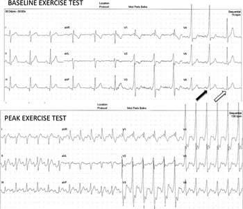
Unfortunately, the results of exercise testing is sometimes not binary – persistent pre-excitation or not – for example, consider the situation wherein the delta wave is only intermittently appreciated at rest but becomes persistent as exercise begins and remains manifest throughout the entirety of the stress test. An example of that is shown below.
A shown in Figure 1, the exercise stress test shows intermittent pre-excitation at rest (closed and open arrows). The second panel (same patient) shows maximal and persistent pre-excitation at peak exercise.
Exercise stress test showing intermittent pre-excitation at baseline (upper panel, closed and open arrows) and marked and persistent pre-excitation at peak exercise in the same patient (lower panel).

Approaching the asymptomatic patient with intermittent pre-excitation
Intermittent loss of the delta wave in patients with ventricular pre-excitation has long been considered a benign condition and unlikely to conduct rapidly if atrial fibrillations were to develop.Reference Klein and Gulamhusein 25 In fact, the 2012 PACES/HRS Consensus statement went so far as to recommend that in patients whose resting surface ECG demonstrated intermittent pre-excitation no further testing is required, except that if symptoms were to develop they should be re-evaluated as a “symptomatic patient with WPW”.Reference Cohen, Triedman and Cannon 18 It is important to remember that, although intermitted pre-excitation is believed to be a predictor of poor antegrade conduction via the accessory pathway, it does not preclude the possibility of having orthodromic reciprocating tachycardia. Fitzsimmons reported on 196 asymptomatic military aviators with pre-excitation. Over a mean follow-up of 21 years, 23% with constant pre-excitation developed tachycardia compared with only 8% with intermittent pre-excitation.Reference Fitzsimmons, McWhirter, Peterson and Kruyer 14 It is likely that the absence of inducible tachycardia in a patient with intermittent pre-excitation is equally important to confirm. On the basis of case reports of patients with Wolff–Parkinson–White syndrome developing ventricular fibrillation, it seems that the initial event is orthodromic re-entrant tachycardia degenerating to atrial fibrillation.Reference Dreifus, Haiat, Watanabe, Arriaga and Reitman 1 , Reference Olen, Baysa, Rossi, Kanter and Fishberger 2 In theory, if a patient with intermittent pre-excitation does not have inducible tachycardia, it is unlikely that they are at risk of sudden death. Over the last few years, there have been two publications specifically reviewing clinical outcomes data in symptomatic and asymptomatic children and adolescents with intermittent pre-excitation. Combining the data from Boston Children’s Hospital (2005–2011) and Children’s Hospitals Colorado (1996–2013), 623 patients with persistent pre-excitation were compared with 80 patients with intermittent pre-excitation.Reference Kiger, McCanta and Tong 26 , Reference Mah, Sherwin and Alexander 27 Although neither retrospective study was intended a priori to be an assessment of risk stratification, both groups used similar definitions of high risk: an accessory pathway effective refractory period or minimal pre-excited cycle length during incremental atrial pacing ⩽250 ms. From a purely electrophysiological standpoint, accessory pathway effective refractory period is different than a measured SPERRI or minimal pre-excited cycle length during atrial incremental pacing. For starters, there is clear evidence that the measured accessory pathway refractory period is not consistently reproducible from one measured time to another; in fact, the reproducibility is even worse on isoproterenol.Reference Oliver and Brembilla-Perrot 28 Choosing a combined accessory pathway effective refractory period and minimal pre-excited cycle length during atrial incremental pacing ⩽250 ms likely increases the specificity, but may reduce the sensitivity of detecting a high-risk accessory pathway. Furthermore, the accessory pathway effective refractory period as an isolated variable is of little prognostic value for the risk of syncope, atrial fibrillation, or atrioventricular reciprocating tachycardia and is less well correlated than the SPERRI in atrial fibrillation;Reference Klein, Bashore, Sellers, Pritchett, Smith and Gallagher 9 , Reference Lee, Hwang, Tai, Chiang, Yu and Chen 29 , Reference Paul, Guccione and Garson 30 nonetheless, the authors of these two studies showed that 5% of patients with intermittent pre-excitation may have a high-risk accessory pathway at the time of an invasive electrophysiology study, and if isoproterenol is utilised up to 11% may have pathways deemed high risk. Although none of the asymptomatic patients with intermittent pre-excitation had a high-risk accessory pathway, the numerator may be too small to detect a difference. Although the two studies supported the recommendations of the 2012 PACES/HRS Asymptomatic WPW Guidelines, larger-scale, multi-institutional studies are warranted to follow-up patients with intermittent pre-excitation. A question that remains for future discussions is “Are asymptomatic patients with intermittent pre-excitation who have inducible tachycardia at the time of EPS at greater risk than those who do not have inducible tachycardia?”.
Isoproterenol or no isoproterenol in assessing the asymptomatic patient with Wolff–Parkinson–White syndrome
Electrophysiologists continue to discuss the use of isoproterenol in risk stratification of patients with pre-excitation. Aside from some practices that use oesophageal pacing in risk stratification, most electrophysiological tests with catheters involve some sort of sedation and often general anaesthesia, especially in younger children. Isoproterenol has long been considered a means to counterbalance the effects of sedation and anaesthesia and mimic the real-life situations of exercise and stress. Isoproterenol decreases conduction times throughout the re-entrant circuit and may allow a patient who is non-inducible to have inducible tachycardia.Reference Cohen and Triedman 31 In a recent study from the Czech Republic, Kubus et alReference Kubus, Vit, Gebauer, Materna and Janousek 32 showed that among 44 asymptomatic patients with a Wolff–Parkinson–White pattern, an additional 36% met criteria for ablation, either because of inducible tachycardia or rapid antegrade conduction. Isoproterenol increases the sensitivity but decreases the specificity in assessing risk. The current role of using isoproterenol for risk stratification in asymptomatic patients remains unclear, and in the absence of long-term natural history studies it may be difficult to ever answer the question. Some experts consider it reasonable to use isoproterenol for risk assessment in the following ways: to ascertain whether or not the asymptomatic patient has inducible tachycardia, which itself is a risk factor; and to use a shorter SPERRI (⩽220 ms) as a categorical risk marker. All of these questions are reasonable to ask. How they will be answered remains the challenge.
Acknowledgements
None.
Financial Support
This research or review received no specific grant from any funding agency or from commercial or not-for-profit sectors.
Conflicts of Interest
None.
Ethical Standards
The authors assert that all referenced work contributing to this review complies with the ethical standards of biomedical or medicolegal investigation.