I. INTRODUCTION
There is increasing interest in the fabrication of hollow nanostructures, which find important functional applications in catalysis, drug delivery, energy storage, solar cells, absorption, photonics, chemical sensors, and reactors in confined space at nanoscale.Reference Caruso, Caruso and Mohwald1–Reference Deng and Lee5 Various fabrication methods have been developed for the preparation of hollow nanostructures, and new routes of fabrication are of continued interest. Noticeably, templating techniques are widely used to generate the hollow interiors. Both soft templates (e.g., emulsion droplets,Reference Dinsmore, Hsu, Nikolaides, Marquez, Bausch and Weitz6, Reference Strohm and Lobmann7 surfactantsReference Guo, Zhang, Li, Meng and Li8) and hard templates (e.g., silica particles, carbon spheres) as supports have been used for the fabrication of hollow nanomaterials. The materials of interest are first deposited or encapsulated on template supports, and subsequently the templates are removed by dissolving/etching/burning to achieve the desired interiors. To avoid complications arising from the templating or final removing process, it is also desirable to fabricate hollow nanostructures by one-step template-free routes.Reference Li and Zeng9–Reference Zheng, Meng, Tang, Zhang and Ren12 For example, hollow magnetite, titania, and tin dioxide nanostructures have been synthesized by hydrothermal synthesis.Reference Cheng, Tang, Qi, Sheng and Liu10, Reference Yin, Li, Zhang, Hao, Liu, Chen and Wang11, Reference Yang and Zeng13 The hydrothermal synthesis usually involves high temperature and high pressure with special sealed reactors. On the other hand, wet chemistry methods under mild reaction conditions (below 100 °C) using nontoxic solvents in conventional flasks for the fabrication of hollow structures is highly desirable, but less explored.Reference Lou, Deng, Lee, Feng and Archer14
In addition, it is preferred to obtain hollow structures with nanoporous shells to offer excellent surface permeability with the external environment. The nanoporous shells can provide effective and even controlled barriers for transfer of sensitive drugs, reactive molecules, ions, and other materials in and out of hollow voids.Reference Deng and Lee15 Such structures with increased surface area are also of interest to catalysis.Reference Li, Chen, Wang, Wang, Zhou and Shen16 Furthermore, it may be desirable to easily convert the chemical composition of the hollow nanoporous structures from one to another without any morphological compromise, i.e., in a topotactic manner.
In this study, we present a route to gram-scale synthesis of hollow porous nanospheres (HPNS) under mild reaction conditions. As a model example, a simple one-step wet chemical method was developed for the production of hydroxyl titanium oxalate HPNS having colloidal units forming the shells. Hydroxyl titanium oxalate could be of interest as an electrorheological material.Reference Liu, Xu, Wu, Cheng, Guo and Cui17, Reference Wen, Huang, Yang, Lu and Sheng18 To the best of our knowledge, hollow-structured nanospheres of hydroxyl titanium oxalate have not been reported before, although solid irregular particles of hydroxyl titanium oxalate of ~400-nm size have been obtained using titanium butoxide.Reference Liu, Xu, Wu, Cheng, Guo and Cui17 Barium–strontium titanate powders have also been obtained by oxalate coprecipitation.Reference Zhang, Su and Gao19 Furthermore, we show that the chemical composition of the hydroxyl titanium oxalate can be converted to anatase TiO2—an important semiconductor—with similar starting morphology of hollow nanospheres. TiO2 is widely used for applications in pigment, sunscreen, gas sensors, energy storage, water splitting, solar cells, environmental purification, and photocatalysis.Reference Chen and Mao20–Reference Peng, Richard-Plouet, Ke, Lee, Chiu, Marhic, Puzenat, Lemoigno and Brohan22 Templating and hydrothermal methods were used to fabricate hollow TiO2 spheres, but the diameter of the spheres were usually in mesoscale or in microscale.Reference Yang and Zeng13, Reference Deng and Lee15 It is still challenging to synthesize hollow TiO2 spheres with nanoscale diameter, especially so if the spheres to be formed by high-order aggregation of smaller crystalline nanounits of TiO2 nanoparticles. The nanospheres reported here are formed by high-order aggregation of single crystalline anatase TiO2 nanoparticles (anatase TiO2 usually more catalytically active). In this study, we focus on the synthesis and characterization of different kinds of nanospheres and understanding their formation mechanism.
II. EXPERIMENTAL SECTION
A. Synthesis of materials
In a typical synthesis sequence of HPNS of hydroxyl titanium oxalate, titanium tetraisopropoxide was injected to 45-mL ethanol in round bottom flask under vigorous stirring at room temperature to prepare 0.1 M solution. After stirring for 5 min, 45 mL an oxalic acid aqueous solution (0.1 M) was added drop by drop. The flask was put in an oil bath on a hotplate and maintained at 75 °C for 2 days. The precipitate was then collected and thoroughly rinsed with water and ethanol three times with centrifugation and dried in vacuum. In a typical synthesis of anatase TiO2 HPNS, the powder obtained was spread on a glass slide and calcinated in a tube furnace in air at 550 °C for 2 h (at ramp rate of 10 °C/min) and the furnace was cooled down naturally.
B. Characterization of materials
The nanospheres were characterized by high-resolution field emission scanning electron microscopy (FESEM; Supra55VP/Ultra55, Zeiss, Germany), energy dispersive x-ray spectroscopy (EDS; Supra55VP/Ultra55, Zeiss), transmission electron microscopy (TEM; JEM-2100 operating at 200 kV, JEOL, Peabody, MA) including selected area electron diffraction (SAED; JEM-2100 operating at 200 kV, JEOL), powder x-ray diffraction (XRD) on a Scintag XDS 2000 x-ray diffractometer using Cu Kα radiation, and Fourier transform infrared spectroscopy (FTIR) on a Nicolet 670 spectrometer.
III. RESULTS AND DISCUSSION
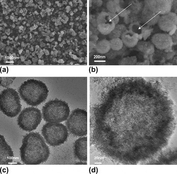
The morphology and internal structure of the hydroxyl titanium oxalate HPNS prepared by the wet chemistry route were characterized by scanning electron microscopy (SEM) and TEM. The overall morphology shown in the low-magnification FESEM image [Fig. 1(a)] reveals that the sample consisted of a large number of spheres of ∼200 nm in diameter. There were a few broken spheres with dark contrast in the center region, demonstrating their hollow structures. The hollow structure was further evidenced at high magnification, as shown in Fig. 1(b). The white arrows highlight two broken shells. This FESEM image further shows that the spheres had a rough surface, indicating that the shells were formed by smaller colloidal units. With the loose aggregation of colloidal units, the shells were nanoporous. This structure is different from that previously reported for irregularly shaped large solid nanoparticles (200–500 nm in size) of hydroxyl titanium oxalate.
Field emission scanning electron microscopy (FESEM) images of the hollow porous nanospheres (HPNS) of hydroxyl titanium oxalate: (a) low magnification showing the overall morphology and (b) high magnification view with white arrows highlight the broken hollow spheres. Transmission electron microscopy (TEM) images: (c) low magnification overall view and (d) high magnification view of a typical hollow sphere.

TEM characterization provided further insight into the structure, in particular validating that the spheres were hollow and nanoporous and formed by colloidal aggregation. The low-magnification TEM image [Fig. 1(c)] shows a few representative spheres. The dark contrast circles are the shells and the light contrast ones within the shells show the interior regions. Figure 1(c) also suggests that the internal and external surfaces of the shells were highly rough and porous. The shells were formed by the aggregation of small colloidal particles, consistent with the FESEM observation. Figure 1(d) is a magnified view of a representative hollow sphere. The shell is about 30 nm in thickness. No evident ring pattern was observed in electron diffraction, indicating their amorphous nature.
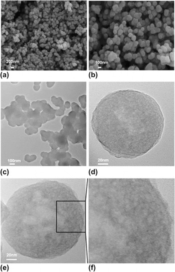
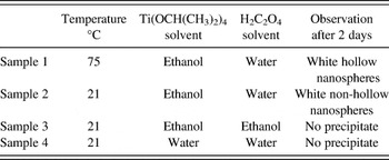
To understand the formation mechanism of the hollow nanospheres of hydroxyl titanium oxalate, additional experimental work was carried out and the conditions are listed in Table I. We note here that a large number of variables (e.g., time, ethanol/water ratio, temperature, concentration, solvents, addictives, and so forth) can affect the results for wet chemical reactions, and in this regard we carried out a limited number of experiments in the present work. Experimentally, it was found that reaction temperature could significantly affect the structure of the hydroxyl titanium oxalate. When the reaction was held at 21 °C instead of 75 °C, white precipitates were similarly obtained and characterized. At low temperature, only non-hollow nanospheres of hydroxyl titanium oxalate were obtained. The morphology is shown in low- and high-magnification FESEM images [Figs. 2(a) and 2(b)], and the size is about 200 nm. The SEM observation showed no obvious differences in overall morphology as compared to those hollow spheres. However, the low-magnification TEM image showing internal structure revealed that they were not hollow [Fig. 2(c)]. This finding indicates that temperature significantly affected the internal structure. Figures 2(d) and 2(e) show two typical hydroxyl titanium oxalate nanospheres, which revealed the existence of nanopores inside the sphere. A TEM image at higher magnification of a section of a typical particle clearly revealed the porous structure [Fig. 2(f)], in which the lower contrast region indicates nanopores. This suggests that the non-hollow spheres were formed through the aggregation of colloidal particles as well.
FESEM images of the non-hollow porous nanospheres of hydroxyl titanium oxalate (a) at low and (b) high magnification. TEM images: (c) low magnification showing a few nanospheres and (d,e) high magnification of two typical spheres revealing the structure in details; (f) higher magnification view of the section highlighted in (e) and the lower contrast region indicates nanopores.

Synthesis experiments carried out under different conditions.

When only ethanol (sample 3) or only water (sample 4) was used as the solvent, no precipitate was observed after 2 days of reaction. Only when both water and ethanol were used (samples 1 and 2), the solid precipitate was obtained.
In another experiment, the same concentration of oxalic acid dissolved in ethanol instead of water was added into the ethanol solution of titanium tetraisopropoxide, keeping other experimental conditions unchanged. However, no precipitate was formed after 2 days and the solution was colorless and transparent, although titanium oxalate is insoluble in ethanol.Reference Choi and Park23 This observation indicates that water plays a crucial role for the formation of hydroxyl titanium oxalate. Experimentally, it was also observed that when only water was used as the solvent, no precipitate formed after 2 days of reaction and the solution was colorless and transparent. The explanation is that hydroxyl titanium oxalate is soluble in strong acid environment.Reference Choy, Han and Kim24 Therefore, the formulated water and ethanol mixture could provide the right solvent conditions for the formation of these nanostructures.
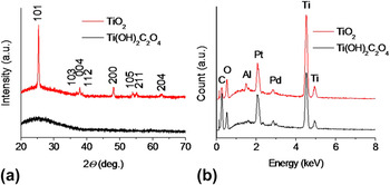
The amorphous nature of the hydroxyl titanium oxalate is also indicated by the absence of any peaks in the XRD pattern [Fig. 3(a), black pattern]. This is typically observed for hydroxyl titanium oxalate.Reference Liu, Xu, Wu, Cheng, Guo and Cui17 As expected, the EDS analysis [Fig. 3(b), black pattern] indicated that the hydroxyl titanium oxalate consisted mainly of elements of Ti, C, and O. The trace Al and Pt/Pd arose from the SEM sample holder as well as Pt/Pd sputtered during the SEM sample preparation. The atomic ratio of Ti:C measured from EDS is about ∼1:2, which is consistent with the initial concentration ratio of titanium tetraisopropoxide and oxalic acid [i.e., 0.1 M Ti(OCH(CH3)2)4 and 0.1 M H2C2O4, respectively].
(a) X-ray diffraction pattern and (b) energy dispersive spectroscopy spectrum of the hydroxyl titanium oxalate nanospheres obtained by wet chemistry (black patterns) and nanoporous anatase titanium dioxide obtained by calcination (red patterns).

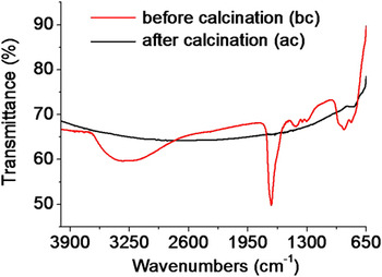
The chemical composition of hydroxyl titanium oxalate was further confirmed by FTIR analysis (Fig. 4, red pattern). The broadband centering around 3400 cm−1 was assigned to the symmetric and asymmetric vibration of O–H group. The sharp absorption band at 1695 cm−1 arose from the C=O stretching vibration. The other bands at 1419, 1349, and 1300 cm−1 were attributed to the O–C=O asymmetric and symmetric stretch vibrations as well as the C–O stretching vibration.Reference Liu, Xu, Wu, Cheng, Guo and Cui17, Reference Berger, Rodes and Gomez25 Bands between 650 and 900 cm−1 were assigned to C–C and Ti–O vibrations.Reference Liu, Xu, Wu, Cheng, Guo and Cui17, Reference Fang, Wang, Qian, Bao, Jiang and Huang26, Reference Thongtem and Thongtem27 The FTIR spectrum, therefore, showed all the characteristic absorption bands of oxalate and hydroxyl, confirming the formation of hydroxyl titanium oxalate. Based on the literature results and our experimental observation, the formation of hydroxyl titanium oxalate was consistent with the following chemical reactionReference Liu, Xu, Wu, Cheng, Guo and Cui17, Reference Choy, Han and Kim24:

Fourier transform infrared spectroscopy spectra of the hydroxyl titanium oxalate (red) and nanoporous anatase titanium dioxide obtained by calcination (black).

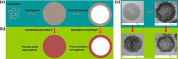
Based on these experimental observations, a model for the formation of hollow spheres of hydroxyl titanium oxalate is qualitatively proposed in Fig. 5(a). Once precipitation occurs, tiny colloids form first because of the low solubility of the hydroxyl titanium oxalate in the formulated ethanol/water solution.Reference Choi and Park23, Reference Choy, Han and Kim24 The colloidal particles have a tendency to aggregate to minimize surface energy and at the same time assemble into loosely packed nanospheres. Since no bubbles form under these experimental conditions, the mechanism of bubble template leading to the formation of hollow structures as proposed earlier likely may not play a significant role in this work.Reference Feng, Yang and Liu28, Reference Zhang, Liu, Liu, Zhang and Li29 Since the growth process is in a solvent, the formation of voids in the spheres is likely due to Ostwald ripening mechanism.Reference Li and Zeng9, Reference Cheng, Tang, Qi, Sheng and Liu10, Reference Yang and Zeng13 The initial high supersaturation causes faster nucleation and thereby the formation of primary particles of smaller sizes. The colloidal aggregation formed at the core is loosely packed with low density distribution.Reference Yu and Zhang30 Compared to those near the outer surface, the colloidal particles agglomerated in the inner core area have higher surface energy due to smaller primary spheres that have high curvature or high surface energy.Reference Yang and Zeng13 In the other words, the primary spheres located in the inner core area are more easily dissolved as compared to those on the external spheres. Ostwald ripening is strongly dependent on interfacial energy, crystal growth rate, and equilibrium solubility, and these parameters are in turn strongly influenced by temperature.Reference Yang and Zeng13, Reference Madras and McCoy31 During the ripening process, the low-density cores dissolve and are redistributed to the high-density outer surfaces to minimize energy.Reference Liu and Zeng32 Therefore, hollow structures are formed. We further demonstrate that the chemical composition and crystallinity can be tuned by topotactic conversion, as illustrated in Fig. 5(b).
(a) Scheme that illustrates the proposed mechanism in the formation of non-hollow and hollow nanospheres of hydroxyl titanium oxalate and (b) their subsequent conversion to solid and hollow nanoporous anatase TiO2 spheres. (c) The corresponding TEM images showing the morphology.

We next consider the transformation of these aggregates into titania. Topotactic conversion or transformation is a process of atomic displacements, which may include loss or gain of material, so that the initial and final structures are coherent. Topotactic conversion has been explored previously in the fabrication of metal oxides and nitride nanostructures.Reference Lou, Deng, Lee, Feng and Archer14, Reference Roberts and Markel33, Reference Kolmakov, Zhang and Moskovits34 Upon calcination in air at 550 °C, the hydroxyl titanium oxalate spheres can be converted to anatase TiO2 with the same morphological structure. This may be considered like a topotactic transformation process since it is a solid state conversion process that maintains the initial hollow structures. Several TiO2 nanostructures have been reported in literature, ranging from nanoparticles prepared by sol–gel method,Reference Liu, Gan, Liu, Zhang and Zeng35 nanotubes prepared from anodic aluminum oxide templateReference Liu, Qian, Li, Cui, Zhou, Guo and Ding36 and by anisotropic corrosion,Reference Liu and Aydil37 nanorods prepared by hydrothermal method,Reference Zhong, Yin, Gates and Xia38 and mesoscale hollow spheres of TiO2 prepared using polystyrene beads as templates.Reference Li and Zeng9, Reference Yang and Zeng13 It is desirable to find new routes for fabrication of anatase TiO2 (given its excellent photocatalytic activity) by simple procedures and this work could be relevant in this context.
The successful conversion of the hydroxyl titanium oxalate into anatase TiO2 is evidenced by XRD [Fig. 3(a), red pattern], EDS [Fig. 3(b), red pattern], and FTIR (Fig. 4, black pattern). The XRD peaks were assigned to crystalline TiO2 with the tetragonal anatase phase (JCPDS No. 21-1272). There were no other peaks observed, indicating that the transformation was complete at 550 °C and that anatase phase TiO2 was obtained. Detailed analysis of the peak broadening of the (101) reflection by Scherrer equation gave an average crystallite size ∼18 nm, indicating that the spheres were composed of nanocrystalline subunits. The EDS analysis shows that the C peak dramatically decreased as compared to its hydroxyl titanium oxalate precursor due to thermal decomposition of oxalate, and the trace amount of C could be from the carbon tape used in SEM sample preparation. FTIR analysis also showed that the vibration bands associated to hydroxyl and oxalate were not observed indicating successful conversion. The flat response observed for TiO2 indicating it was transparent from 650 to 3900 cm−1, which is consistent with literature reports.Reference Erkov, Devyatova, Molodstova, Malsteva and Yanovskii39, Reference Zhang, Boyd, O’Sullivan, Hurley, Kelly and Senateur40 According to literature, the characteristic infrared absorption peaks of TiO2 are typically below 610 cm−1.Reference Erkov, Devyatova, Molodstova, Malsteva and Yanovskii39, Reference Zhang, Boyd, O’Sullivan, Hurley, Kelly and Senateur40 The conversion was complete and the chemical reaction could be the following:

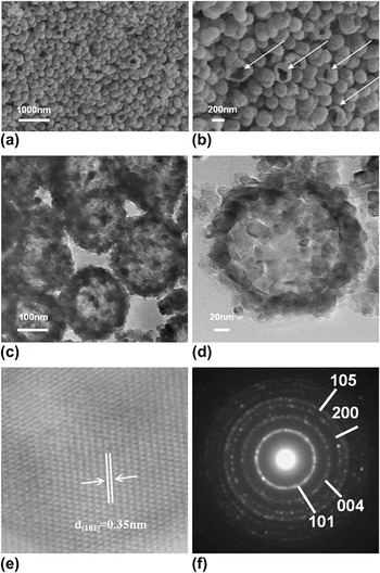
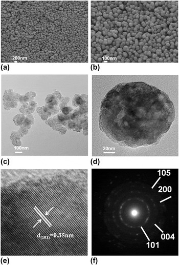
The morphology and structure of the anatase TiO2 nanospheres were characterized as well. The low- and high-magnification FESEM images [Figs. 6(a) and 6(b)] reveal that the calcination products were similar in morphology to their hydroxyl titanium oxalate precursors. The hollow structure was also maintained. The white arrows highlight a few spheres with broken shells in Fig. 6(b). It is worth noting that the shells were formed by crystalline TiO2 nanoparticle subunit aggregation or high-order assembly, leading to a rough surface. TEM characterization provided further insight into the structures and confirmed that the spheres were hollow and porous. The low-magnification TEM image [Fig. 6(c)] shows a few representative particles, and Fig. 6(d) shows a close-up view. The shell had a thickness of ∼20 nm and was made up of nanocrystals ranging from ∼10 to 20 nm as building subunits. The morphological integrity indicated a self-templated mechanism for the topotactic conversion of hydroxyl titanium oxalate precursors to anatase TiO2. High-resolution transmission electron microscopy (HRTEM) showed clear crystalline lattice fringes [Fig. 6(e)] indicating that the building units were crystalline. The SAED further confirmed its crystallinity, with all the diffraction rings being assigned to tetragonal anatase phase TiO2, in agreement with the XRD analysis. Similarly, anatase TiO2 nanospheres without hollow interiors were obtained by calcining its corresponding precursor of solid nanospheres of hydroxyl titanium oxalate (Fig. 7). The as-obtained solid anatase TiO2 spheres were also nanoporous, as revealed by low- and high-magnification TEM observation [Figs. 7(c) and 7(d)]. HRTEM observation and SAED analysis confirm that the converted TiO2 nanospheres are crystalline and in anatase phase [Figs. 7(e) and 7(f)].
Hollow nanoporous anatase TiO2 nanospheres obtained by calcination at 550 °C: FESEM images (a) at low and (b) high magnification. The white arrows highlight broken hollow nanospheres. TEM images (c) at low magnification of a few hollow particles and (d) at high magnification of a typical hollow nanosphere. (e) High-resolution transmission electron microscopy (HRTEM) image of the building unit and (f) selected area electron diffraction (SAED) patterns of the nanospheres.

Non-hollow nanoporous anatase TiO2: FESEM images (a) at low and (b) high magnification. TEM images (c) at low magnification of a few spheres and (d) high magnification of a typical sphere. (e) HRTEM and (f) SAED patterns of the spheres.

IV. CONCLUSIONS
A facile wet chemical route under mild experimental conditions was investigated for the large-scale fabrication of hollow, as well as non-hollow, porous nanospheres of hydroxyl titanium oxalate. Simple thermal treatment converted the spheres into anatase TiO2 while retaining morphological integrity. High-order assembly of subunit of anatase TiO2 nanocrystals into spherical aggregates about 200 nm in diameter was achieved. The nanostructured anatase titania reported herein could be of interest to several emerging energy and environmental applications. In principle, this synthesis route of wet chemistry and solid state reaction could be used for the fabrication of other nanostructured oxides.