Introduction
In vitro maturation of oocytes, both nuclear and cytoplasmic is an important step and preliminary requirement for embryo production in vitro. While nuclear maturation can be checked by staining techniques using Hoechst 33342 (Purse et al., Reference Purse, Wall, Rexroad, Hammer and Brinster1985) or Orcein (Liu et al., Reference Liu, Sun, Li, Jiao and Wang2003; Talukder et al., Reference Talukder, Shamsuddin, Rahman, Bari and Parrish2009; Wani, Reference Wani2008), cytoplasmic maturation can be checked by normal embryo development after fertilization or artificial activation of such oocytes (Eppig, Reference Eppig1996). Even glutathione has been reported as one of the key indicators of cytoplasmic maturation in goat oocytes (Abazari-Kia et al., Reference Abazari-Kia, Mohammadi-Sangcheshmeh, Dehghani-Mohammadabadi, Jamshidi-Adegani, Veshkini, Zhandi, Cinar and Salehi2014). For nuclear and cytoplasmic maturation of oocytes gonadotropins like follicle-stimulating hormone (FSH) and LH have been supplemented in the maturation medium in most animal species (Phillips and Dekel, Reference Phillips and Dekel1982; Zuelke and Brackett, Reference Zuelke and Brackett1990; Wani, Reference Wani2002; Junk et al., Reference Junk, Dharmarajan and Yovich2003; Wani and Nowshari, Reference Wani and Nowshari2005, Wani, Reference Wani2009) including goat (Younis et al., Reference Younis, Zuelke, Harper, Oliveira and Brackett1991; Ongeri et al., Reference Ongeri, Bormann, Butler, Melican, Gavin, Echelard, Krisher and Behboodi2001). Supplementation of cystine in goat oocyte maturation medium has been reported to increase the proportion of mature oocytes, however higher concentrations decrease blastocyst rates (Zhou et al., Reference Zhou, Wu, Li, Lan, Wang, Gao and Tan2008).
Activation of mature oocytes, which is an important step in the success of somatic cell nuclear transfer has been achieved by various chemical and physical treatments (Cuthbertson et al., Reference Cuthbertson, Whittingham and Cobbold1981; Nagai, Reference Nagai1987; Ozil, Reference Ozil1990; Jones, et al., Reference Jones, Carroll and Whittingham1995; Wani, Reference Wani2008). Chemicals such as ionomycin, ethanol, calcium ionophore, and strontium are generally used to induce the activation of oocytes in most animal species. Treatment with 6-dimethylaminopurine (6-DMAP) immediately after ionomycin or ethanol exposure has been used for activation of porcine (Kim et al., Reference Kim, Uhm, Ju, Lee and Chung1997) bovine (Rho et al., Reference Rho, Kawarsky, Johnson, Kochhar and Betteridge1998), buffalo (Gasparrini et al., Reference Gasparrini, Boccia, Rosa, Palo, Campanile and Zicarelli2004), camel (Wani et al., Reference Wani, Wernery, Hassan, Wernery and Skidmore2010) and goat (Ongeri et al., Reference Ongeri, Bormann, Butler, Melican, Gavin, Echelard, Krisher and Behboodi2001; Lan et al., Reference Lan, Han, Wu, Han, Ma, Liu, Chang and Tan2005) oocytes. To the best of our knowledge, there have been no studies on chemical activation of oocytes from prepubertal goats.
Culture medium plays a major role in the development of the embryo to the blastocyst stage in vitro. Several commercial complex culture media such as TCM-199, Hams-F10, Hams-F12 (Wani, Reference Wani2002) and simple formulation media such as synthetic oviductal fluid (SOF) (Tervit et al., Reference Tervit, Whittingham and Rowson1972), Charles Rosenkrans medium (CR-1) (Rosenkrans and First, Reference Rosenkrans and First1994) and potassium simplex optimization medium (KSOM) (Lawitts and Biggers, Reference Lawitts and Biggers1991) have been used for in vitro embryo culture in different animal species. Embryos developed from prepubertal goat oocytes have been cultured in TCM-199 (Izquierdo et al., Reference Izquierdo, Villamediana and Paramio1999; Jiménez-Macedo et al., Reference Jiménez-Macedo, Izquierdo, Anguita and Paramio2005), SOF (Rodríguez-González et al., Reference Rodríguez-González, López-Bejar, Mertens and Paramio2003; Romaguera et al., Reference Romaguera, Casanovas, Morató, Izquierdo, Catalá, Jiménez-Macedo, Mogas and Paramio2010) and G1/G2 (Jiménez-Macedo et al., Reference Jiménez-Macedo, Izquierdo, Anguita and Paramio2005) with varying success rates.
Ovaries collected from animals slaughtered in abattoirs are the cheapest and most abundant source of oocytes and are used for large-scale production of mature oocytes in most animal species. Due to local demand for meat, most goats slaughtered in the local slaughterhouse are young and have not reached sexual maturity. The objective of this study in our laboratory was to optimize the protocol for in vitro maturation of cumulus–oocyte complexes collected from ovaries of slaughtered prepubertal goats, their activation methods and culture media for their development up to blastocyst stage.
Materials and methods
All reagents were purchased from Sigma Chemical Co. (St. Louis, Mo, USA) unless otherwise stated. Fetal calf serum (FCS) was obtained from Gibco (BRL, Germany). FSH and LH were purchased from Sioux (Sioux, Biochemical Inc., Sioux Center, IA, USA). All maturation and culture media were filtered through a membrane filter (0.20 µm pore size; Sartorius, Germany) and routinely equilibrated at 39°C under 5% CO2 in humidified air for at least 3 h prior to use.
Collection of cumulus–oocyte complexes (COCs) and their in vitro maturation (IVM)
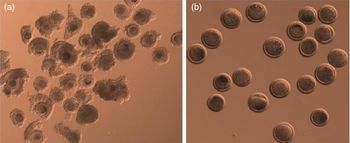
Reproductive tracts of prepubertal goats were obtained from a local abattoir and were transported to the laboratory at 32°C in thermos flasks containing normal saline solution. On arrival in the laboratory, ovaries were cut off from the surrounding tissue and rinsed three times in saline. COCs (Fig. 1a) were recovered in PBS supplemented with 5% FCS using 20 ml disposable syringes fitted with 18-gauge hypodermic needles. COCs were distributed randomly into 4-well culture plates (20–25 COCs/well) containing 500 µl of the maturation medium as described in the following sections and cultured at 39°C in an atmosphere of 5% CO2 in air for 24 h.
(a) Cumulus–oocyte complexes (COCs) harvested from ovaries of prepubertal goats of slaughterhouse origin. (b) Denuded mature oocytes after 24 h of in vitro culture.

Chemical activation of mature oocytes
Oocytes were denuded by gentle pipetting in 4-(2-hydroxyethyl)-1-piperazineethanesulfonic acid (HEPES)-buffered TCM-199 supplemented with 1 mg/ml hyaluronidase. Mature oocytes with a visible polar body (Fig. 1b) were activated by either exposure to 7% ethanol or 5 µM ionomycin in HEPES-buffered TCM-199 supplemented with 1 mg/ml BSA. Oocytes from both treatments were cultured in medium containing 2 mM 6-DMAP for 4 h before transferring them to the embryo culture medium.
Staining and evaluation of chromatin status of activated oocytes
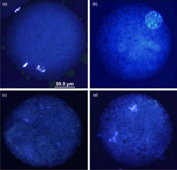
After 18 h of culture, the activated oocytes were washed to remove all the attached cumulus cells. The oocytes were fixed in 2% formaldehyde and 0.25% glutaraldehyde in PBS (supplemented with 0.1% polyvinylpyrrolidone) for 3 min at room temperature. Oocytes were then stained with 5 µg/ml Hoechst 33342 for 10 min. They were examined under a phase contrast microscope (Olympus, Japan) to evaluate the nuclear state under UV light. Oocytes were classified as activated [with pronucleus/i, metaphase III (oocyte chromatin and two polar bodies)], non-activated (with oocyte chromatin in the metaphase II stage) and degenerated (Fig. 2a–d).
Representative oocytes stained with Hoechst 33342 16–18 h after chemical activation. (a) An activated oocyte with an oocyte chromatin and two polar bodies. (b) An activated oocyte with a single pronucleus. (c, d) Oocytes with abnormal or degenerative nuclear chromatin.

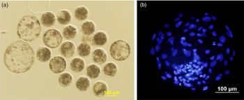
(a) Morulae and blastocysts produced on day 8 of culture. (b) Blastocyst stained with Hoechst 33342 to show the cell numbers.

In vitro culture of activated oocytes
Activated oocytes were cultured in 4-well culture plates (20–25 embryos/well) containing 500 µl of the embryo culture medium supplemented with 3.0 µg/ml BSA for the first 3 days and 10% FCS for subsequent 5 days at 39ºC under 5% CO2 in humidified air.
Experimental design
Experiment 1
The aim of this experiment was to study the effect of LH and cysteine on maturation of prepubertal goat oocytes. COCs were randomly distributed to wells containing basic culture medium (TCM-199 supplemented with 0.15 µg/ml l-glutamine, 0.25 mM sodium pyruvate, 20 ng/ml epidermal growth factor, 10 µg/ml FSH, 1 µg/ml estradiol, 10% FCS and 25 µg/ml gentamycin) or basic medium + 10 µg/ml LH (treatment 1) or basic medium + 10 µg/ml LH + 0.1 mM l-cysteine (treatment 2). Complexes were cultured for 24 h at 39°C under 5% CO2 in humidified air.
Experiment 2
After culturing for 24 h in medium used in treatment 2, mature oocytes were activated by either 5 µM ionomycin or 7% ethanol as described previously. After 18 h of activation, some oocytes were taken randomly, fixed and stained to evaluate the chromatin status from both groups, while all other oocytes were cultured in embryo culture medium to study their further development. The experiment was replicated six times using KSOMaa and SOFaa for embryo culture as described at some point later.
Experiment 3
In this experiment, we used four embryo culture media (CR-1aa, TCM-199, KSOMaa and SOFaa) to evaluate the developmental potential of oocytes activated by 5 µM ionomycin and 2 mM 6-DMAP. All media were supplemented with 3.0 mg/ml BSA for the first 3 days and then supplemented with 10% FCS for the subsequent 5 days of culture.
Statistical analysis
Data were analyzed by dependent sample t-test and one-way analysis of variance with Tukey post-hoc test using SPSS software (SPSS, USA). Data are expressed as mean ± standard error of the mean (SEM).
Results
The maturation rate of prepubertal goat oocytes obtained in control (53.6 ± 2.1), treatment 1 (59.3 ± 4.2) and treatment 2 (58.2 ± 2.7) medium did not differ from each other (P > 0.05). However, the lowest degeneration rate was observed in the oocytes in treatment 2 (P < 0.05) when compared with the other two groups (Table 1).
Effect of LH and cysteine supplementation in maturation media on in vitro maturation of prepubertal goat oocytes

a,b Values within columns with different superscripts differ significantly (P < 0.05).
LH, luteinizing hormone; SEM, standard error of the mean.
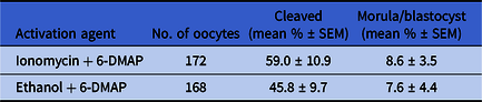
As seen in Table 2, the proportion of oocytes activated was higher (P < 0.05) in the ionomycin group, when compared with the group activated with ethanol (83.2 ± 8.1 vs. 69.3 ± 4.4). Also, non-activated oocytes were significantly less in the ionomycin group than the ethanol group (P < 0.05). However, no significant difference was observed in the cleavage (59.0 ± 10.9 vs. 45.8 ± 9.7) and morula/blastocyst production rates (8.6 ± 3.5 vs. 7.6 ± 4.4) between the groups (Table 3).
Activation of prepubertal goat oocytes following exposure to sequential treatments of ionomycin or ethanol combined with 6-DMAP

a,b Values within column with different superscripts differ significantly (P < 0.05).
6-DMAP, 6-dimethylaminopurine; SEM, standard error of the mean
Embryo development from in vitro matured prepubertal goat oocytes after activation with sequential treatments of ionomycin or ethanol followed by 6-DMAP

6-DMAP, 6-dimethylaminopurine; SEM, standard error of the mean
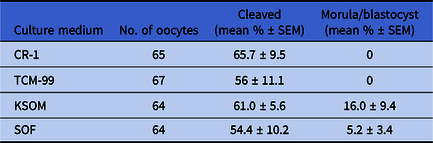
As shown in Table 4, the proportions of oocytes cleaved were 65.7 ± 9.5, 56.8 ± 11.1, 61.0 ± 5.6, 54.4 ± 10.2 in CR-1, TCM-199, KSOM and SOF media, respectively, with no significant difference. However, further development of cleaved oocytes (Fig. 3a, b) was better in KSOM (16.0 ± 9.4), followed by SOF (5.2 ±3.4) media.
Effect of culture medium on embryo development from in vitro matured prepubertal goat oocytes after activation with 5 µM ionomycin and 6-DMAP

CR-1: Charles Rosenkrans medium; TCM-199: Tissue culture medium-199; KSOM: potassium simplex optimization medium; SEM, standard error of the mean; SOF: synthetic oviductal fluid
Discussion
To the best of our knowledge, there have been no earlier reports on the chemical activation of in vitro matured oocytes collected from prepubertal goats. The objective of this study in our laboratory was to optimize in vitro maturation, chemical activation and in vitro culture for prepubertal goat oocytes. Even though supplementation of 10 µg/ml of LH has been reported to have beneficial effects on the maturation rates of bovine (Zuelke and Brackett, Reference Zuelke and Brackett1990) and prepubertal goat (Izquierdo et al., Reference Izquierdo, Villamediana and Paramio1999) oocytes, we did not find any difference in maturation rate of these oocytes at the mentioned concentration when compared with the control group. LH receptors (LHRs) of cumulus cells may not be fully developed in prepubertal goat oocytes, as has been reported in a study on porcine oocytes (Shimada et al., Reference Shimada, Nishibori, Isobe, Kawano and Terada2003). Fully developed LHRs on cumulus cells are responsible for the activation of cAMP production, which stimulates cumulus expansion and oocytes maturation (Shimada et al., Reference Shimada, Nishibori, Isobe, Kawano and Terada2003). LH has been reported to increase IVM rate in oocytes collected from the ovaries of adult animals in many species (Eppig, Reference Eppig1996; Junk et al., Reference Junk, Dharmarajan and Yovich2003; Phillips and Dekel, Reference Phillips and Dekel1982; Zuelke and Brackett, Reference Zuelke and Brackett1990). Our results are similar and in agreement with another study (Khatun et al., Reference Khatun, Bhuiyan, Ahmed, Haque, Rahman and Shamsuddin2010) that also reported a similar maturation rate of 60% in black Bengal goat oocytes.
In our study, the addition of cysteine and LH in IVM medium showed improved maturation rate even though non-significant, but the proportion of degenerated oocytes decreased significantly (P < 0.05). Our results are in agreement with an earlier study in which it was suggested that increased glutathione levels might improve oocyte maturation (Rodríguez-González et al., Reference Rodríguez-González, López-Bejar, Mertens and Paramio2003) and prevent degeneration of prepubertal goat oocytes. The effect of adding cysteine (thiol compound) to the IVM medium is known to promote male pronucleus (MPN) formation, fertilization, and embryo development. Cysteine or cysteamine increases GSH levels in oocytes without cumulus cells in IVM medium (De Matos et al., Reference De Matos, Furnus and Moses1997).Therefore, the addition of thiol compounds in IVM medium has advantages on maturation of prepubertal goat oocytes that do not have fully grown cumulus cells or denuded oocytes.
In the second experiment, we compared two chemical agents, ionomycin and ethanol, followed by culture in 6-DMAP for activation of in vitro matured oocytes. Not only was the proportion of oocytes activated higher (P < 0.05) in ionomycin group, when compared with the group activated with ethanol, but the non-activated oocytes were also significantly less in this group, P < 0.05). Our results are similar and in agreement with earlier studies in which ionomycin in combination with 6-DMAP has been shown to induce a higher activation rate in bovine (Susko-Parrish et al., Reference Susko-Parrish, Leibfried-Rutledge, Northey, Schutzkus and First1994) and camel (Wani, Reference Wani2008) oocytes. Both of these activating agents used in our study were proven to be very effective for activation of bovine (Rho et al., Reference Rho, Kawarsky, Johnson, Kochhar and Betteridge1998), caprine (Ongeri et al., Reference Ongeri, Bormann, Butler, Melican, Gavin, Echelard, Krisher and Behboodi2001), camel (Wani, Reference Wani2008) and buffalo (Gasparrini et al., Reference Gasparrini, Boccia, Rosa, Palo, Campanile and Zicarelli2004) oocytes. They induce an intracellular Ca2+ increase, mimicking the process of penetration by sperm during the fertilization process. Higher activation from ionomycin in the present study could be attributed to induced increase in intracellular Ca2+ in oocytes from both external and internal stores (Kline and Kline, Reference Kline and Kline1992). Whereas, ethanol induced a Ca2+ rise mainly from external sources, although a minor intracellular release was also involved (Shiina et al., Reference Shiina, Kaneda, Matsuyama, Tanaka, Hiroi and Doi1993). The increased Ca2+ levels induced by ionomycin or ethanol led to inactivation of maturation promoting factor (MPF) and cytostatic factor, therefore releasing the oocytes from its meiotic arrest (Lorca et al., Reference Lorca, Cruzalegui, Fesquet, Cavadore, Méry, Means and Dorée1993). Both of these chemicals induce a single Ca2+ hike in oocytes, whereas, during the fertilization process, the initial Ca2+ rise was followed by many oscillations. Therefore, sequential approaches involving persistent inhibition of MPF by addition of protein synthesis, protein phosphorylation or specific MPF inhibitors like 6-DMAP (Susko-Parrish et al., Reference Susko-Parrish, Leibfried-Rutledge, Northey, Schutzkus and First1994) have been used.
We compared media such as CR-1, KSOM, SOF, and TCM-199 for embryo development and observed that KSOM and SOF were better suited for in vitro embryo culture of chemically activated prepubertal goat oocytes. Even though all media used supported the initial cleavage and development to the 4-cell stage, further development was restricted in all media used in the present study. We obtained the lowest embryo development in CR-1 medium, which lacks glucose, suggesting that glucose might be needed for further development of cleaved embryos. Therefore, further studies are needed to evaluate the use of glucose/lactate for culture of embryos obtained from prepubertal goat oocytes. It has, however, been reported that glucose does not support the development of the preimplantation mouse embryos before the 8-cell stage and that lactate does not support development before the 2-cell stage (Biggers and Summers, Reference Biggers and Summers2008). We achieved the highest morula/ blastocyst rate when embryos were cultured in KSOM. This medium contains ethylenediaminetetraacetic acid (EDTA), which has been reported to overcome the 2-cell block in mouse embryos (Abramczuk et al., Reference Abramczuk, Solter and Koprowski1977) and acts as a chelator for transition of metal ions to prevent the extracellular generation of oxygen radicals (Matsukawa et al., Reference Matsukawa, Ikeda, Imai and Yamada2002). The percentage of morulae/blastocysts was lower in our study when compared with the results of an earlier study (Izquierdo et al., Reference Izquierdo, Villamediana and Paramio1999) in which authors used a co-culture system and achieved about a 21% developmental rate in TCM-199 supplemented with oviduct cells. The developmental rates in TCM-199 without cells and conditioned medium were, however, similar to our results in the present study.
In conclusion, supplementation of LH and cysteine in IVM medium did not increases maturation rate but reduced the degeneration rate of prepubertal goat oocytes. Both ionomycin and ethanol could be used to activate in vitro matured oocytes obtained from prepubertal goats, however activation with 5 µM ionomycin followed by 6-DMAP tended to have better activation results and embryonic development. KSOM seemed to be a better embryo culture medium for activated in vitro matured oocytes from prepubertal goats. Further studies are needed to optimize embryo culture media for prepubertal goat oocytes to enhance their development up to the blastocyst stage.
Acknowledgements
This work would not have been possible without the moral, financial and administrative support of Dr Ali Redha, Administrative Director of our centre. This study was kindly sponsored by H.H. Sheikh Hamadan bin Mohammed bin Rashid al Makhtoum, Crown Prince of Dubai, UAE.
Conflict of Interest
We declare that there is no conflict of interest that could be perceived as prejudicing the impartiality of the research reported.
Financial Support
This study was kindly sponsored by H.H. Sheikh Hamadan bin Mohammed bin Rashid Al Maktoum, Crown Prince of Dubai, UAE.
Ethical Standards
Not applicable.