Introduction
The membranous portion of the human vocal fold mucosa is connected to the thyroid cartilage anteriorly via the intervening anterior macula flava and anterior commissure tendon.Reference Hirano and Sato1 Posteriorly, it is joined to the vocal process of the arytenoid cartilage via the intervening posterior macula flava.Reference Hirano and Sato1 The vocal ligament runs between the anterior and posterior maculae flavae.Reference Hirano and Sato1 Many vocal fold stellate cells, which are stellate in shape and store vitamin A in their lipid droplets, are distributed in the maculae flavae.Reference Sato, Hirano and Nakashima2, Reference Sato, Hirano and Nakashima3
Human maculae flavae located at both ends of the vocal fold mucosa are inferred to be involved in the metabolism of extracellular matrices, which are essential for the viscoelastic properties of the lamina propria of the human vocal fold.Reference Sato, Umeno and Nakashima4 Human adult maculae flavae are thought to be responsible for maintaining the characteristic layered structure of the human vocal fold mucosa.Reference Sato, Umeno and Nakashima4 Human newborn, infant and child maculae flavae are inferred to be responsible for forming the characteristic layered structure of the human vocal fold mucosa.Reference Sato, Hirano and Nakashima5–Reference Sato, Umeno and Nakashima7 Human maculae flavae are considered to be an important structure in the growth, development and ageing of the human vocal fold mucosa.Reference Sato, Umeno and Nakashima4–Reference Sato, Hirano and Nakashima8
Vocal fold stellate cells in the maculae flavae are vitamin A storing cells and a member of the ‘diffuse stellate cell system’.Reference Sato, Umeno and Nakashima9 Vocal fold stellate cells are considered a new category of cells in the human vocal fold.
Adult tissue-specific stem cells (tissue stem cells) have the capacity to self-renew and to generate functionally differentiated cells that replenish lost cells throughout an organism's lifetime. There is growing evidence to suggest that the vocal fold stellate cells in the human maculae flavae are tissue stem cells or progenitor cells.Reference Sato, Umeno and Nakashima10, Reference Kurita, Sato, Chitose, Fukahori, Sueyoshi and Umeno11 The human maculae flavae are a candidate for a stem cell niche, which is a microenvironment nurturing a pool of stem cells which, in this case, are vocal fold stellate cells.Reference Sato, Umeno and Nakashima10, Reference Kurita, Sato, Chitose, Fukahori, Sueyoshi and Umeno11
As a result of this heterogeneity, it is uncertain whether the cells including vocal fold stellate cells derive from the same embryonic source as conventional fibroblasts in the human vocal fold mucosa.
This study aimed to investigate the origin of the cells including vocal fold stellate cells in the human newborn maculae flavae, focusing on their relationship with bone marrow derived cells.
Materials and methods
Three normal human newborn larynges from autopsy cases were used. Any larynges that had diseases which could possibly affect the tissue of the vocal fold were excluded from the study.
For light microscopy, specimens were fixed in 10 per cent formalin, dehydrated in graded concentrations of ethanol and embedded in paraffin. Haematoxylin and eosin stain was used for each section, and immunohistochemical staining was carried out.
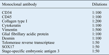
The expression of cluster of differentiation 34, cluster of differentiation 45, collagen type I, cytokeratin, vimentin, glial fibrillary acidic protein, desmin, telomerase reverse transcriptase, SOX17 and stage-specific embryonic antigen 3 (monoclonal antibody; Abcam, Cambridge, UK) (Table I) was determined histologically in formalin-fixed and paraffin-embedded tissue by immunohistochemistry, for which a universal immuno-enzyme polymer method staining kit (Histofine Simple Stain Max-PO; Nichirei, Tokyo, Japan) was used.
Dilutions of antibodies

CD = cluster of differentiation
All specimens were sectioned to a thickness of 5–6 µm and mounted on glass slides. Deparaffinised and hydrated sections were rinsed with 0.01 mol/l phosphate-buffered saline at pH 7.4. The specimens were covered with 3 per cent hydrogen peroxide for 10 minutes and rinsed with 0.01 mol/l phosphate-buffered saline. The specimens were then incubated with the primary antibody overnight at 4°C.
After rinsing with phosphate-buffered saline and labelling with the universal immuno-enzyme polymer method staining kit, a colour reaction was developed with 3,3’-diaminobenzidine tetrahydrochloride for 3–5 minutes at room temperature. The specimens were counterstained with haematoxylin. Immunoreactivity was examined by light microscopy.
Results
Cytoplasmic cytokeratin (epithelium-associated protein), vimentin (mesenchymal cell associated protein), glial fibrillary acidic protein (neural-associated protein), desmin (muscle-associated protein) and SOX17 (endodermal marker) immunoreactivity were present in the cells including vocal fold stellate cells in the human newborn maculae flavae (Figure 1), indicating that these cells were undifferentiated and expressed proteins of all three germ layers.
Immunohistochemical staining of the cells in the macula flava of the human newborn vocal fold. Cytoplasmic cytokeratin (a), vimentin (b), glial fibrillary acidic protein (c), desmin (d), and SOX17 (e) immunoreactivity were present.
The cells including vocal fold stellate cells in the human newborn maculae flavae expressed cluster of differentiation 34 (haematopoietic progenitor cell marker), cluster of differentiation 45 (leukocyte common antigen) and collagen type I (Figure 2), which are the major makers of bone marrow derived circulating fibrocytes.
Immunohistochemical staining of the cells in the macula flava of the human newborn vocal fold. The cells in the human newborn maculae flavae expressed: cluster of differentiation 34 (a), cluster of differentiation 45 (b) and collagen type I (c), which are the major makers of bone marrow derived circulating fibrocytes; telomerase reverse transcriptase (d), indicating that a special DNA polymerase called telomerase resides in the cells in the human newborn maculae flavae; and cytoplasmic stage-specific embryonic antigen 3 (e).
The cells including vocal fold stellate cells expressed telomerase reverse transcriptase (Figure 2), indicating that a special DNA polymerase called telomerase resides in these cells in the human newborn maculae flavae. The cells also expressed stage-specific embryonic antigen 3 (Figure 2). This suggests that the cells in the human newborn maculae flavae are stem cell related.
Discussion
Stem cells are divided into two main groups, pluripotent and multipotent, based on their potential to differentiate. Pluripotent (embryonic) stem cells can differentiate into every kind of cell in the body and multipotent (adult) stem cells can differentiate into multiple, but not all, cell lineages.Reference Xie and Li12
There is growing evidence to suggest that the cells including vocal fold stellate cells in the human maculae flavae are adult multipotent stem cells, tissue stem cells or progenitor cells, and that the human maculae flavae are a candidate for a stem cell niche, which is a microenvironment nurturing a pool of stem cells which, in this case, are vocal fold stellate cells.Reference Sato, Umeno and Nakashima10, Reference Kurita, Sato, Chitose, Fukahori, Sueyoshi and Umeno11
Our previous investigation revealed that the cells in the human adult maculae flavae are undifferentiated cells derived from the differentiation of bone marrow cells.Reference Kurita, Sato, Chitose, Fukahori, Sueyoshi and Umeno11 The results of this study are consistent with the hypothesis that the cells including the vocal fold stellate cells are tissue stem cells or progenitor cells of the human vocal fold mucosa.Reference Kurita, Sato, Chitose, Fukahori, Sueyoshi and Umeno11
In the present study, the origin of cells including the vocal fold stellate cells in the human newborn maculae flavae, especially their relationship with bone marrow derived cells, was investigated.
Intermediate filaments
The expression of proteins in the intermediate filaments of cytoplasm is specific to cell type and differentiation.Reference Becker, Kleinsmith and Hardin13 Because of the tissue specificity of intermediate filaments, cells from different tissues can be distinguished on the basis of the intermediate filament protein present.Reference Becker, Kleinsmith and Hardin13
Our previous studies revealed that the intermediate filaments, including cytokeratin, vimentin, glial fibrillary acidic protein and desmin, are distributed in the cytoplasm of the cells including the vocal fold stellate cells in the adult maculae flavae.Reference Sato, Umeno and Nakashima9, Reference Kurita, Sato, Chitose, Fukahori, Sueyoshi and Umeno11 The cells in the adult maculae flavae expressed epithelium-associated, mesenchymal cell associated, neural-associated and muscle-associated proteins, indicating that the cells in the adult maculae flavae are undifferentiated and multipotent.Reference Sato, Umeno and Nakashima10, Reference Kurita, Sato, Chitose, Fukahori, Sueyoshi and Umeno11
In the present study, cytoplasmic cytokeratin, vimentin, glial fibrillary acidic protein and desmin immunoreactivity were present in the cells including the vocal fold stellate cells in the newborn maculae flavae. Additionally, the vocal fold stellate cells expressed SOX17, which is an endodermal cell marker. Cytokeratin is the protein of the intermediate filaments of epithelial cells, and vimentin is a major subunit protein of the intermediate filaments of mesenchymal cells. Glial fibrillary acidic protein, a member of the intermediate filament protein family and characteristic of neural crest cells, is heavily and specifically expressed in astrocytes and certain other astroglia in the central nervous system. In addition, neural stem cells frequently and strongly express glial fibrillary acidic protein. Desmin is the protein of the intermediate filament; it is characteristic of myogenic crest cells and is found in muscle cells. Consequently, the cells in the human newborn maculae flavae express proteins of all three germ layers. This suggests that they are undifferentiated and multipotent.
Telomerase
A special DNA polymerase called telomerase can catalyse the formation of additional copies of the telomeric repeat sequence, thereby compensating for the gradual shortening that occurs at both ends of the chromosome during DNA replication.Reference Becker, Kleinsmith and Hardin14 In multicellular organisms, telomerase resides mainly in the germ cells that give rise to sperm and eggs, and in a few other kinds of proliferating normal cells such as stem cells.Reference Becker, Kleinsmith and Hardin14 Because telomerase is not found in most cells, their chromosomal telomeres get shorter and shorter with each cell division.Reference Becker, Kleinsmith and Hardin14 The presence of telomerase allows cells to divide indefinitely, without telomere shortening.Reference Becker, Kleinsmith and Hardin14
Our present investigation revealed that cells in the newborn maculae flavae express telomerase reverse transcriptase, indicating that telomerase resides in these cells.
As mentioned above, these findings raise the possibility that the cells including vocal fold stellate cells are tissue stem cells (tissue-specific resident stem cells) such as mesenchymal stem cells or multipotent mesenchymal stromal cells.
Relationship with marrow-derived cells
Colony formation in vitro and asymmetric cell division are one of the characteristics of stem cells.Reference Friedenstein, Deriglasova, Kulagina, Panasuk, Rudakowa and Luria15, Reference Deasy and Rajasekhar16 Our past investigation revealed that the cultured cells from the human adult maculae flavae formed a colony-forming unit and cell division was an asymmetric self-renewal, indicating that these cells are mesenchymal stem cells or stromal stem cells derived from the bone marrow.Reference Kurita, Sato, Chitose, Fukahori, Sueyoshi and Umeno11, Reference Sato, Chitose, Kurita and Umeno17
Bone marrow has two major components: the haematopoietic component and the mesenchymal component.Reference Abedi and Bucala18 In contrast to its haematopoietic component, the mesenchymal component of the haematopoietic organs includes fibroblast-like cells (stromal cells), myofibroblasts (also known as adventitial reticular cells), adipocytes and endothelial cells.Reference Abedi and Bucala18 Some marrow-derived cells, such as circulating fibrocytes and pericytes, have been suggested to contribute to tissue fibroblasts.Reference Abedi and Bucala18 The fibroblast-related cells, such as hepatic stellate cellsReference Forbes, Russo, Rey, Burra, Wright and Alison19 and myofibroblasts in wounded skin,Reference Brittan, Hunt, Jeffery, Poulsom, Forbes and Hodivala-Dilke20 are also derived from bone marrow. It is interesting that the morphological features of the vocal fold stellate cells in the human maculae flavae are similar to the hepatic stellate cells and included in the proposed diffuse stellate cell system.Reference Sato, Umeno and Nakashima9
Marrow-derived circulating fibroblast precursors have been suggested to originate from marrow cells, circulate into blood cells and, after homing to the tissue, differentiate into fibroblasts.Reference Abedi and Bucala18 Circulating fibrocytes were first identified by Bucala et al. in 1994.Reference Bucala, Spiegel, Chesney, Hogan and Cerami21 They were found to be unique cells because they co-express haematopoietic markers, and collagen type I and other matrix proteins (mesenchymal markers). Circulating fibrocytes are specifically defined by the expression of cluster of differentiation 34, cluster of differentiation 45 and collagen type I.Reference Abedi and Bucala18
In the present study, the cells in the human newborn maculae flavae expressed haematopoietic markers (cluster of differentiation 34, cluster of differentiation 45) and collagen type I, which are the major makers of bone marrow derived circulating fibrocytes.
These observations are consistent with the hypothesis that the cells including vocal fold stellate cells in the human maculae flavae arise not from resident interstitial cells but from the differentiation of bone marrow cells.
Pluripotency
Stage-specific embryonic antigen 3 is present on cell surface glycolipids and glycopeptides of human teratocarcinoma cells, on embryonic germ cells and on embryonic stem cells. Stage-specific embryonic antigen 3 expression decreases as those cells differentiate.
In the present study, the cells in the human newborn maculae flavae expressed stage-specific embryonic antigen 3. This suggests that the cells including vocal fold stellate cells in the human newborn maculae flavae are stem cell related cells and possibly have pluripotency.
Vocal fold regeneration
The identification of tissue stem cells and their stem cell niche in various organs has become a focus of recent research, as they represent a cell source for organ regeneration or tissue repair.
Side population cells have been considered to contain a high number of stem cells or progenitor cells. Side population cells have been identified in the anterior and posterior maculae flavae in the human vocal fold.Reference Yamashita, Hirano, Manemaru, Tsuji, Suehiro and Ito22 This investigation is consistent with the hypothesis that the cells including the vocal fold stellate cells in the maculae flavae are tissue stem cells or progenitor cells.
Side population cells in the anterior and posterior maculae flavae participate in the early stages of wound healing of the rat vocal fold.Reference Gugatschka, Kojima, Ohno, Kanemaru and Hirano23
The regeneration of vocal folds requires three important elements: cell therapy, the development and implementation of a scaffold, and the use of growth factors.Reference Fishman, Long, Gugatschka, De Coppi, Hirano and Hertegard24 The results of this study are consistent with the hypothesis that the cells in the maculae flavae are tissue stem cells and are therefore a potential endogenous cell source for vocal fold regeneration.
• Cells in human newborn maculae flavae are undifferentiated, and arise from bone marrow cell differentiation not resident interstitial cells
• The study findings suggest that the macula flava cells are tissue stem cells or progenitor cells of vocal fold mucosa
• At birth, these cells have already been supplied from the bone marrow into the newborn vocal fold maculae flavae
• From there, these cells are ready to start the growth and development of the human vocal fold mucosa as a vibrating tissue
Understanding the mechanism responsible for the regulation of cells in the human maculae flavae will provide the tools needed to manipulate them for the development of therapeutic approaches to diseases and tissue injuries.
Conclusion
The cells including the vocal fold stellate cells in the human newborn maculae flavae are undifferentiated cells which arise not from resident interstitial cells but from the differentiation of bone marrow cells.
The results of this study are consistent with the hypothesis that the cells including the vocal fold stellate cells are tissue (mesenchymal) stem cells or progenitor cells of the human vocal fold mucosa.
At birth, these cells have already been supplied from the bone marrow into the maculae flavae in the newborn vocal fold, and are ready to start the growth and development of the human vocal fold mucosa as a vibrating tissue.
Acknowledgement
This investigation was supported by a Grant-in-Aid for Scientific Research (number 24592612) from the Japanese Ministry of Education, Culture, Sports, Science and Technology.