Introduction
Copper(II) hydroxychloride minerals are typical oxidation products of other copper minerals under arid conditions. This paper describes the first mixed-valency copper hydroxychloride mineral bounahasite, Cu+Cu2+2(OH)3Cl2. Only two other minerals with both univalent and divalent copper cations are known – a rare secondary mineral paramelaconite Cu+2Cu2+2O3 (O'Keeffe and Bovin, Reference O'Keeffe and Bovin1987) and a rare fumarolic mineral allochalcoselite Cu+Cu2+5PbO2(SeO3)2Cl5 (Vergasova et al., Reference Vergasova, Krivovichev, Britvin, Filatov, Burns and Ananyev2005; Krivovichev et al., Reference Krivovichev, Filatov, Burns and Vergasova2006).
Bounahasite was named after its type locality – Bou Nahas Mine, Morocco. Both the new mineral and the name (symbol Bnhs) have been approved by the Commission on New Minerals, Nomenclature and Classification of the International Mineralogical Association, proposal IMA2021-114 (Lykova et al., Reference Lykova, Rowe, Poirier, Friis and Helwig2022). The holotype has been deposited in the collection of the Canadian Museum of Nature, Ottawa, Canada. The catalogue number is CMNMC 89874. A part of the holotype used for structure determination has been kept at the Natural History Museum in Oslo under catalogue number KNR 44404.
Occurrence and general appearance
Bounahasite occurs at the Bou Nahas (Bou N'hass) Cu Mine, Oumjrane mining area, Anti-Atlas Mountains, Morocco. It is a supergene mineral found in the oxidation zone of hydrothermal polymetallic veins. The veins were formed along faults and fractures in Upper Ordovician marlstones, shales, sandstones and conglomerates during hydrothermal activity related to late Triassic volcanism. The main minerals of the polymetallic veins are pyrite, chalcopyrite, galena and baryte. The secondary supergene mineralisation developed later, probably between Jurassic and Neogene periods (Praszkier, Reference Praszkier2015).
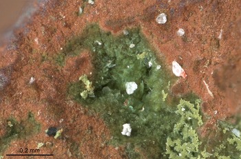
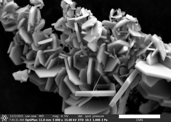
The new mineral forms very small pseudo-hexagonal plates up to 3 × 30 × 40 μm in size. The dominant form is pinacoid {110} with minor rhombic prism and another pinacoid. The crystals are combined into loose clusters (Figs 1–2) on native copper together in association with paratacamite.
Green platy crystals of bounahasite on native copper. FOV 1 mm. Specimen CMNMC 89874. Photo: François Génier.

Bounahasite crystals. Scanning electron microscopy image, scale bar = 10 μm.

Physical and optical properties
Bounahasite is green with pale green powder colour and vitreous lustre (Fig. 1). The cleavage is parallel to {110}, perfect. The fracture is uneven, stepped. The Mohs hardness could not be determined as the crystals are very small. The mineral is non-fluorescent under ultraviolet light. The density calculated using the empirical formula and unit-cell volume refined from the single-crystal X-ray diffraction data is 3.90 g/cm3.
Bounahasite is optically biaxial, 2V (meas.) = 60(5)° (from a spindle-stage extinction curve). The refractive indices were not measured due to the lack of available high refractive index (>1.8) immersion fluids. The mean refractive index obtained from the Gladstone–Dale relationship (Mandarino, Reference Mandarino1981) is 1.961.
Experimental methods
Electron microprobe analyses of bounahasite were obtained using a JEOL 8230 SuperProbe electron microscope equipped with five wavelength dispersive spectrometers (University of Ottawa – Canadian Museum of Nature MicroAnalysis Laboratory, Canada) with an acceleration voltage of 10 kV, a beam current of 10 nA and a beam diameter of 5 μm. The following reference materials were used: cuprite (CuLα) and tugtupite (ClKα). The intensity data were corrected for Time Dependent Intensity (TDI) loss (or gain) using a self-calibrated correction for ClKα.
The Fourier-transform infrared spectrum of bounahasite was obtained at the Canadian Conservation Institute, Canada using a Bruker Hyperion 2000 microscope interfaced to a Tensor 27 spectrometer with a wide-band mercury cadmium telluride (MCT) detector. A crystal of bounahasite ~20 μm in size was mounted on a low-pressure diamond anvil microsample cell and analysed in transmission mode. The spectrum was collected between 400–4000 cm–1 with the co-addition of 150 scans at a 4 cm–1 resolution.
Powder X-ray diffraction data were collected at the Canadian Museum of Nature, Canada using a Bruker D8 Discover microdiffractometer equipped with a DECTRIS EIGER2 R 500K detector and IμS microfocus X-ray source (λCuKα1 = 1.54060Å) with the Kα2 contribution removed using the ‘Strip Kα2’ tool in Bruker Diffrac.EVA V4.3. The instrument was calibrated using a statistical calibration method (Rowe, Reference Rowe2009). A powder ball 200 μm in diameter, mounted on a fibre pin mount, was analysed with continuous Phi rotation and 10° rocking motion along the Psi axis of the Centric Eulerian Cradle stage.
Single-crystal X-ray studies were carried out at room temperature on a Rigaku XtaLAB Synergy-S diffractometer equipped with a HyPix 6000HE detector (λCuKα = 1.54184) operating at 50 kV and 1 mA, housed at the Natural History Museum, University of Oslo, Norway. The data were collected and processed using Rigaku's CrysAlis Pro software.
Results
Chemical data
Chemical data for bounahasite are given in Table 1. Obtaining good data proved to be very challenging due to the small size of the crystals, especially their thickness. We were able to collect only a few reliable analyses. The empirical formula calculated on the basis of 3 Cu atoms per formula unit is: Cu+Cu2+2(OH)2.97Cl2.03. The content of Zn is below the detection limit. The ideal formula for bounahasite is Cu+Cu2+2(OH)3Cl2, which requires Cu2O 22.90, CuO 50.90, H2O 8.64, Cl 22.68, –O = Cl2 –5.12, total 100 wt.%.
Chemical data (in wt.%, average of 3 analyses) for bounahasite.

*Calculated from the stoichiometry.
S.D. – standard deviation
Bounahasite is readily soluble in acids at room temperature.
Infrared spectroscopy
The infrared (IR) spectrum of bounahasite (Fig. 3) shows IR bands of O–H-stretching (in the range from 3470 to 3350 cm–1) vibrations of OH groups and absorbed H2O molecules (the wider band at 3350 cm–1), as well as multiple bands in the range 400–1150 cm–1. A weak broad band at 1630 cm–1 could be assigned to H–O–H bending vibrations of absorbed H2O molecules. Similar weak bands are often observed in the IR spectra of copper(II) hydroxychloride minerals of the atacamite group (Chukanov, Reference Chukanov2014). The latter are also characterised by multiple IR bands in the 400–600 and 750–1150 cm–1 ranges but the lack of bands in the range 600–750 cm–1, unlike the spectrum of bounahasite with its distinct band at 684 cm–1. Direct attribution of the bands in the range 400–1150 cm–1 is very challenging due to the paucity of compounds with comparable chemistry, and thus the lack of theoretical works on vibrations in them.
Infrared spectrum of bounahasite

X-ray diffraction data
The indexed powder X-ray diffraction data are given in Table 2. Parameters of the monoclinic unit cell refined from the powder data are as follows: a = 8.5906(2) Å, b = 6.4203(1) Å, c = 10.406(1) Å, β = 111.783(1)° and V = 532.98(2) Å3.
Powder X-ray diffraction data of bounahasite.

The strongest lines are given in bold.
1 Calculated from the crystal structure determination, only reflections with intensities >1 are given.
2 Calculated from powder XRD Rietveld unit-cell refinement with a = 8.5906(2), b = 6.4203(1), c = 10.406(1) Å, β = 111.783(1)° and V = 532.98(2) Å3.
The single-crystal X-ray diffraction data were indexed in the P21/n space group with the following unit-cell parameters: a = 8.5925(1), b = 6.4189(1), c = 10.4118(2) Å, β = 111.804(2)° and V = 533.17(2) Å3. The structure was solved and refined to R1 = 0.028 on the basis of 1054 independent reflections with I > 2σ(I) using the SHELXL-2018/3 program package (Sheldrick, Reference Sheldrick2015). Crystal data, data collection and structure refinement details are given in Table 3, atom coordinates, equivalent displacement parameters, site occupancy factors in Table 4, selected interatomic distances in Table 5 and bond valence sums (BVS) in Table 6. The crystallographic information file has been deposited with the Principal Editor of Mineralogical Magazine and is available as Supplementary material (see below). In addition, it has been deposited in the Inorganic Crystal Structure Database (ICSD; #CSD 2189172).
Crystal data, data collection information and structure refinement details for bounahasite.

*Located 0.64 Å away from the Cu4a site.
Coordinates, equivalent (U eq) and anisotropic displacement parameters (in Å2) of atoms, and site-occupancy factors# for bounahasite.

# Site-occupancy factor for Cu4a is Cu0.9596(17) and Cu4b is Cu0.0404(17), all other sites = 1
*The anisotropic displacement parameters for Cu4a and Cu4b were refined constraining site movements and occupancies to each other (the EADP command).
**U iso; the values were fixed during refinement.
Selected interatomic distances (Å) in the structure of bounahasite.

Bond-valence calculations* for bounahasite.

*Bond-valence parameters were taken from Gagné and Hawthorne (Reference Gagné and Hawthorne2015) for Cu2+–O bonds and Brese and O'Keeffe (Reference Brese and O`Keeffe1991) for Cu2+–Cl and Cu+–Cl bonds.
Description and discussion of the crystal structure
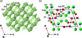
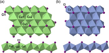
The crystal structure of bounahasite is unique. It is based on two alternating sheets – octahedral and tetrahedral – coplanar to (110) (Fig. 4a,b). The octahedral sheet consists of alternating edge-sharing slightly Jahn–Teller-distorted Cu(2)2+(OH)6 octahedra with a <Cu–O> distance of 2.133 and two octahedra coordinated by oxygen and chlorine: Cu(1)2+(OH)4Cl2 and Cu(3)2+(OH)4Cl2. Both Cu(1)- and Cu(3)-centred octahedra are strongly distorted with short equatorial Cu–O bonds with <Cu–O> distances of 1.960 and 1.974 Å, respectively, and elongated axial Cu–Cl bonds (Fig. 5a; Table 5). The distortion is attributed to the pseudo Jahn–Teller effect observed in Cu2+-centred octahedra with mixed ligands (Hathaway, Reference Hathaway1984). Ignoring the long Cu–Cl bonds, the sheet can be described as consisting of alternating edge-sharing Cu2+(OH)6 octahedra and two Cu2+(OH)4 squares.
General view of the crystal structure of bounahasite: (a) polyhedral and (b) ball-and-stick models. The unit cell is outlined. Projection on (001).

(a) Octahedral sheet in the crystal structure of bounahasite and (b) a similar sheet in the crystal structure of botallackite (after Hawthorne, Reference Hawthorne1985).

A similar sheet formed by alternating edge-sharing Cu(OH)4Cl2 and Cu(OH)5Cl octahedra was found in the structure of botallackite, Cu2(OH)3Cl (Fig. 5b; Hawthorne, Reference Hawthorne1985).
The tetrahedral sheet is based on Cu(4)+Cl4 tetrahedra (Fig. 6). Two edge-sharing Cu+Cl4 tetrahedra form Cu+2Cl6 dimers connected with each other via shared vertices (Fig. 7). The Cu4 site is split onto the Cu4a and Cu4b subsites (Fig. 8) with the occupancy factors 0.9596(17) and 0.0404(17) and <Cu–Cl> distances of 2.364 and 2.449 Å, respectively. The Cu4a–Cu4b distance is 1.24 Å. There are several peaks and holes in the electron density map between the Cu4a and Cu4b sites, which indicates further splitting with multiple low-occupancy Cu4 subsites located near the Cu4a site.
Tetrahedral sheet in the crystal structure of bounahasite.

Cu+2Cl6 dimer connected with other dimers via shared vertices in the crystal structure of bounahasite.

Splitting of the Cu4 site into two subsites in the crystal structure of bounahasite.

The two sheets are connected via Cl1 atoms. The weak bonding between the sheets explains the perfect cleavage in one direction and the platy habit of bounahasite.
Structural analysis was crucial in determining both the number and the nature of the anions in the formula, as well as the valence state of copper. The bond-valence sums (BVS) at the O1 [1.23 valence units (vu)], O2 [1.26 vu], and O3 [1.29 vu; Table 6] sites indicate that all three sites are occupied by hydroxyl groups. H atoms were tentatively localised in the structure (Fig. 4b, Table 7). The distances between H and O atoms were restrained during refinement using a soft restraint (0.85 Å using the DFIX command). Hydrogen bonding between O atoms of the octahedral sheet and Cl2 atoms of the tetrahedral sheet (O–H⋅⋅⋅Cl) connects the sheets. Each Cl2 atom is involved in three hydrogen bonds (Table 7). The low BVS at the Cl2 site [0.53 vu; Table 6] is due to the unaccounted hydrogen bonds contribution.
Hydrogen bonds in the structure of bounahasite.*

*D = donor; A = acceptor
The BVS at the Cu1, Cu2 and Cu3 sites [2.16, 1.96 and 2.10 vu, respectively; Table 6] confirm the bivalent state of copper at those sites. Although, as discussed above, the splitting of the Cu4 site into multiple subsites complicates bond-valence calculations for both Cu and Cl sites, the results are unambiguous. The BVS calculated using bond-valence parameters taken for Cu2+–Cl bonds (Brese and O'Keeffe, Reference Brese and O`Keeffe1991) is 1.44 vu, whereas the parameters were taken for Cu+–Cl bonds give 0.96 vu, indicating that Cu+ is the dominant valence state of copper at the Cu4 site.
The resulting structural formula of bounahasite is Cu+Cu2+2Cl2(OH)3.
No minerals or synthetic compounds structurally related to bounahasite have been found in the literature or databases. All five copper hydroxychlorides that have been found in Nature so far (atacamite Cu2Cl(OH)3, belloite Cu(OH)Cl, botallackite Cu2Cl(OH)3, clinoatacamite Cu2Cl(OH)3 and paratacamite Cu3(Cu,Zn)Cl2(OH)6 [IMA-CNMNC List of Mineral Names, Pasero, Reference Pasero2022] contain only bivalent copper, and their structures are based on Cu-centred polyhedra with mixed Cl– and OH– ligands (Wells, Reference Wells1949; Fleet, Reference Fleet1975; Hawthorne, Reference Hawthorne1985; Grice et al., Reference Grice, Szymanski and Jambor1996; Pollard et al., Reference Pollard, Thomas and Williams1989; Krivovichev et al., Reference Krivovichev, Hawthorne and Williams2017; Zheng et al., Reference Zheng, Yamauchi, Kitajima, Fujihala, Maki, Lee, Hagihala, Torii, Kamiyama and Kawae2018).
It is rare to find Cu–Cl polyhedra in minerals and occurrences are usually structural types unique to each mineral, for example vertex-sharing Cu+Cl4 tetrahedra in nantokite CuCl, which is a representative of the sphalerite structural type (Wyckoff and Posnjak, Reference Wyckoff and Posnjak1922), distorted octahedra Cu2+Cl6 in tolbachite CuCl2 (Burns and Hawthorne, Reference Burns and Hawthorne1993) or discrete planar dimers Cu2+2Cl6 in sanguite KCuCl3 (Pekov et al., Reference Pekov, Zubkova, Belakovskiy, Lykova, Yapaskurt, Vigasina, Sidorov and Pushcharovsky2015). In this study, the tetrahedral sheet of bounahasite is no exception, as no sheet of a similar topology has been described before in either minerals or synthetic compounds. This implies that there is no preferential stable configuration of Cu–Cl polyhedra. All three minerals mentioned above – nantokite, tolbachite and sanguite – are unstable in air and alter into secondary minerals. Bounahasite has shown no signs of alteration so far, possibly due to the octahedral sheet stabilising the structure. The splitting of the Cu4 site into multiple subsites may play a similar role by relaxing the bonds and thus satisfying the local charge balance within the tetrahedral sheet.
Acknowledgements
We thank anonymous referees for their valuable comments. We thank Donald Doell Jr. for donating a suite of specimens, one of which became the holotype of bounahasite, to the Canadian Museum of Nature. We are grateful to François Génier for taking the colour photo.
Supplementary material
To view supplementary material for this article, please visit https://doi.org/10.1180/mgm.2022.133
Competing interests
The authors declare none.